. 2020 Jul 27:9:115-130.
doi: 10.2147/ITT.S257443. eCollection 2020.

Targeting Prostate Cancer Using Intratumoral Cytotopically Modified Interleukin-15 Immunotherapy in a Syngeneic Murine Model

Affiliations

Targeting Prostate Cancer Using Intratumoral Cytotopically Modified Interleukin-15 Immunotherapy in a Syngeneic Murine Model

Efthymia Papaevangelou et al. Immunotargets Ther. .

Abstract

Background: The prostate cancer microenvironment is highly immunosuppressive; immune cells stimulated in the periphery by systemic immunotherapies will be rendered inactive once entering this environment. Immunotherapies for prostate cancer need to break this immune tolerance. We have previously identified interleukin-15 (IL-15) as the only cytokine tested that activates and expands immune cells in the presence of prostate cancer cells. In the current study, we aimed to identify a method of boosting the efficacy of IL-15 in prostate cancer.

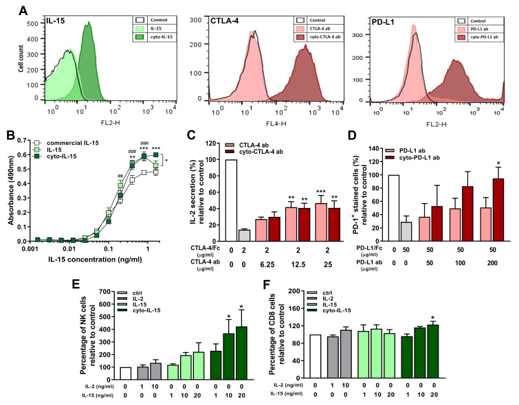
Methods: We engineered, by conjugation to a myristoylated peptide, a membrane-localising form of IL-15 (cyto-IL-15) and the checkpoint inhibitor antibodies cytotoxic T lymphocyte antigen 4 (CTLA-4) and programmed death ligand 1 (PD-L1) (cyto-abs) to enable them to bind to cell surfaces by non-specific anchoring to the phospholipid bilayer. The efficacy of these agents was investigated by intratumoral administration either alone (cyto-IL-15 or cyto-abs) or in combination (cyto-combo) in subcutaneous TRAMP-C2 prostate tumors in C57BL/6J mice and compared with their non-modified equivalents in vivo. Following the survival endpoint, histological analyses and RNA sequencing were performed on the tumors.

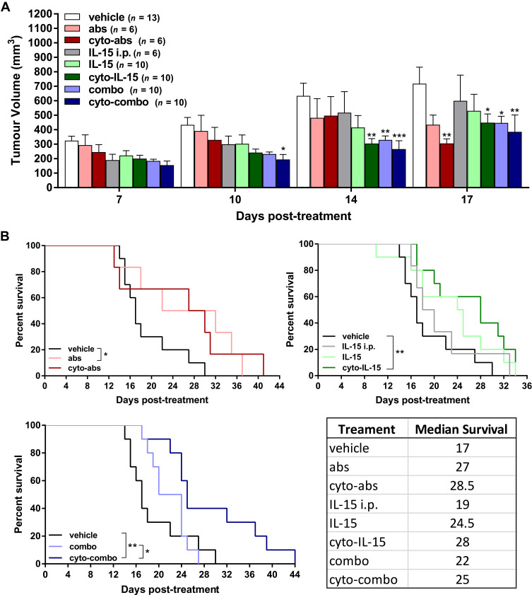
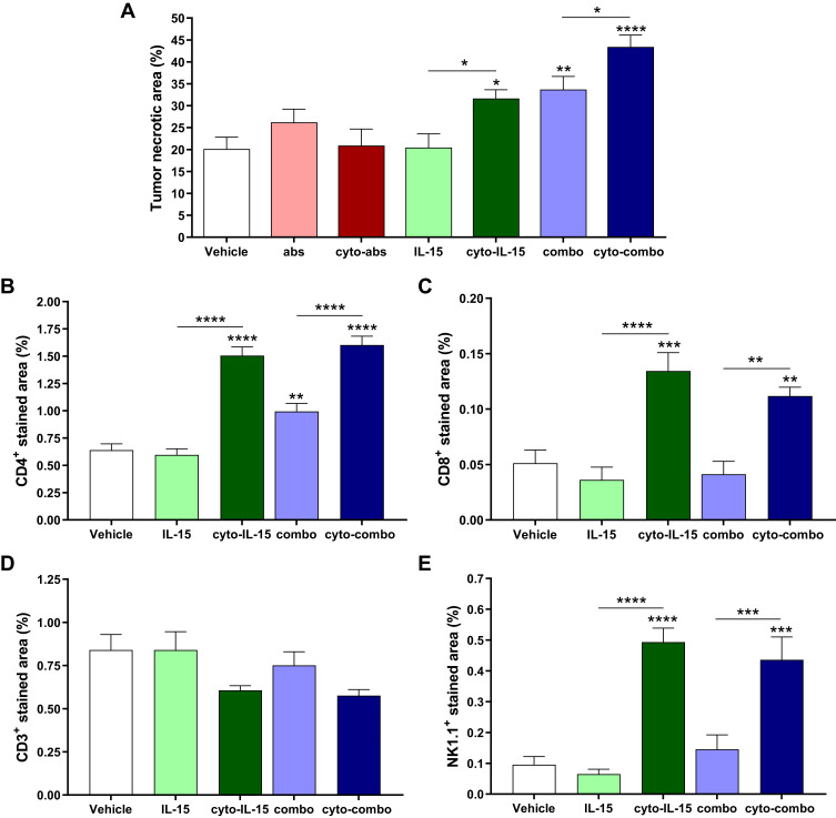
Results: Intratumoral injection of cyto-IL-15 or cyto-combo delayed tumor growth by 50% and increased median survival to 28 and 25 days, respectively, compared with vehicle (17 days), whereas non-modified IL-15 or antibodies alone had no significant effects on tumor growth or survival. Histological analysis showed that cyto-IL-15 and cyto-combo increased necrosis and infiltration of natural killer (NK) cells and CD8 T cells in the tumors compared with vehicle and non-modified agents. Overall, the efficacy of cyto-combo was not superior to that of cyto-IL-15 alone.

Conclusion: We have demonstrated that intratumoral injection of cyto-IL-15 leads to prostate cancer growth delay, induces tumor necrosis and increases survival. Hence, cytotopic modification in combination with intratumoral injection appears to be a promising novel approach for prostate cancer immunotherapy.

Keywords: IL-15; NK cells; checkpoint blockade; cytotopic modification; prostate cancer.


Conflict of interest statement

Efthymia Papaevangelou, Dorota Smolarek, Richard A Smith, Prokar Dasgupta, and Christine Galustian report a patent, GB 1,913,804.9, pending to King's College London and the Prostate Cancer Research Centre Charity. The authors report no other possible conflicts of interest in this work.

Figures

Figure 1
Figure 1
In vitro characterization of cytotopically-modified proteins. (A) Cell membrane binding of cytotopically-modified IL-15 on RBCs, and cytotopically-modified CTLA-4 or PD-L1 antibodies on naïve Jurkat cells, detected by flow cytometric analysis using fluorescent-labelled antibodies to these proteins. (B) Proliferation of CTLL-2 cells treated with different forms of IL-15 (n = 3 independent experiments performed in quadruplicates). Results are means ±1 SEM (*cyto-IL-15 versus commercial IL-15 or cyto-IL-15 versus IL-15, #IL-15 versus commercial IL-15, *p <0.05, **or ##p <0.01, ***or ###p <0.001 two-way ANOVA with Tukey multiple comparisons post-test). (C) IL-2 secretion by activated Jurkat cells inhibited with CTLA-4/Fc protein and reconstituted with varying concentrations of CTLA-4 or cyto-CTLA-4 antibodies. Results are expressed relative to IL-2 secretion by activated control cells (n = 4). (D) PD-1 binding in EL4 cells inhibited with PD-L1/Fc protein and reconstituted with varying concentrations of PD-L1 or cyto-PD-L1 antibodies. Results are expressed relative to PD-1 expression in control cells (n = 4). (E, F) NK and CD8 T cell expansion in a human non-adherent PBMC population treated with varying concentrations of IL-2, IL-15 or cyto-IL-15 (n = 3). (CF) Results are means +1 SEM (*p <0.05, **p <0.01, ***p <0.001 one-way ANOVA with Sidak’s multiple comparisons post-test).
Figure 2
Figure 2
Effect of modified IL-15 and antibodies on growth of TRAMP-C2 subcutaneous prostate tumors. (A) Tumor volumes up to day 17 post-treatment. Data are means + 1 SEM for all the tumors per group and comparisons are relative to vehicle (*p <0.05, **p <0.01, ***p <0.001 two-way ANOVA with Dunnett’s multiple comparisons post-test). (B) Survival curves of mice post-treatment (*p <0.05, **p <0.01, comparisons of equality of two survival curves using Log-rank (Mantel-Cox) test). Table shows the median survival of each group.
Figure 3
Figure 3
Assessment of cytokines in TRAMP-C2 prostate tumor homogenates. CCL2 (A), IL-1β (B), GM-CSF (C), IL-10 (D) and IFN-α (E) concentration in tumor homogenates derived from tumors treated with vehicle, IL-15, cyto-IL15, combo or cyto-combo. Results are means +1 SEM of duplicate measurements made from all tumors in each cohort (n = 10) corrected for protein concentration. Comparisons are relative to vehicle (*p <0.05, **p <0.01, one-way ANOVA with Dunnett’s multiple comparisons post-test).
Figure 4
Figure 4
Histological assessment of TRAMP-C2 prostate tumors. (A) Composite images and magnified images of H&E-stained sections indicating necrotic regions. (B, C) RGB images from tumor sections stained with (B) CD4 (red) and CD8 (green), and (C) CD3 (red) and NK1.1 (green) antibodies. Nuclei were stained with DAPI (blue).
Figure 5
Figure 5
Quantification of necrotic area and immune cell infiltration in TRAMP-C2 tumors. (A) Necrotic area. Results are means +1 SEM of two sections per tumor for all tumors. (B) CD4 positive-stained area, (C) CD8 positive area, (D) CD3 positive area, and (E) NK1.1 positive-stained area. Results are means +1 SEM of 10 images per tumor for n = 5 per group. Comparisons are relative to vehicle unless otherwise indicated (*p <0.05, **p <0.01, ***p <0.001, ****p <0.0001 one-way ANOVA with Dunnett’s multiple comparisons post-test).
Figure 6
Figure 6
Effects of cyto-IL15 treatment on gene expression in TRAMP-C2 tumors. Differential gene expression analysis followed by gene ontology analysis were performed between tumors treated with vehicle or IL-15 (top panel), with vehicle or cyto-IL-15 (middle panel), and with IL-15 or cyto-IL-15 (bottom panel) (n = 3/cohort). (A) Bi-clustering heatmaps of the log2 transformed expression values in each sample showing the expression profiles of the top 30 differentially expressed genes (all 8 differentially expressed genes are shown in the vehicle versus IL-15 comparison). (B) Gene ontology (GO) enrichments of all significantly differentially expressed genes (adjusted p-value < 0.05) for each of the three comparison sets. The numbers next to the bars indicate the number of significantly differentially expressed genes involved in each biological process.

Similar articles

Cited by

References

    1. Siegel RL, Miller KD, Jemal A. Cancer statistics, 2018. CA Cancer J Clin. 2018;68(1):7–30. doi:10.3322/caac.21442 - DOI - PubMed
    1. Cheng HH, Lin DW, Yu EY. Advanced clinical states in prostate cancer. Urol Clin North Am. 2012;39(4):561–571. doi:10.1016/j.ucl.2012.07.011 - DOI - PubMed
    1. Bruno TC, French JD, Jordan KR, et al. Influence of human immune cells on cancer: studies at the University of Colorado. Immunol Res. 2013;55(1–3):22–33. doi:10.1007/s12026-012-8346-y - DOI - PMC - PubMed
    1. Sfanos KS, De Marzo AM. Prostate cancer and inflammation: the evidence. Histopathology. 2012;60(1):199–215. doi:10.1111/j.1365-2559.2011.04033.x - DOI - PMC - PubMed
    1. Westdorp H, Skold AE, Snijer BA, et al. Immunotherapy for prostate cancer: lessons from responses to tumor-associated antigens. Front Immunol. 2014;5:191. doi:10.3389/fimmu.2014.00191 - DOI - PMC - PubMed