. 2020 Jul 28;6(5):e498.
doi: 10.1212/NXG.0000000000000498. eCollection 2020 Oct.

Integrated sequencing and array comparative genomic hybridization in familial Parkinson disease

Affiliations

Integrated sequencing and array comparative genomic hybridization in familial Parkinson disease

Laurie A Robak et al. Neurol Genet. .

Abstract

Objective: To determine how single nucleotide variants (SNVs) and copy number variants (CNVs) contribute to molecular diagnosis in familial Parkinson disease (PD), we integrated exome sequencing (ES) and genome-wide array-based comparative genomic hybridization (aCGH) and further probed CNV structure to reveal mutational mechanisms.

Methods: We performed ES on 110 subjects with PD and a positive family history; 99 subjects were also evaluated using genome-wide aCGH. We interrogated ES and aCGH data for pathogenic SNVs and CNVs at Mendelian PD gene loci. We confirmed SNVs via Sanger sequencing and further characterized CNVs with custom-designed high-density aCGH, droplet digital PCR, and breakpoint sequencing.

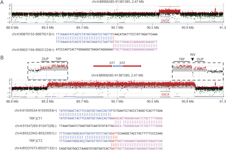
Results: Using ES, we discovered individuals with known pathogenic SNVs in GBA (p.Glu365Lys, p.Thr408Met, p.Asn409Ser, and p.Leu483Pro) and LRRK2 (p.Arg1441Gly and p.Gly2019Ser). Two subjects were each double heterozygotes for variants in GBA and LRRK2. Based on aCGH, we additionally discovered cases with an SNCA duplication and heterozygous intragenic GBA deletion. Five additional subjects harbored both SNVs (p.Asn52Metfs*29, p.Thr240Met, p.Pro437Leu, and p.Trp453*) and likely disrupting CNVs at the PRKN locus, consistent with compound heterozygosity. In nearly all cases, breakpoint sequencing revealed microhomology, a mutational signature consistent with CNV formation due to DNA replication errors.

Conclusions: Integrated ES and aCGH yielded a genetic diagnosis in 19.3% of our familial PD cohort. Our analyses highlight potential mechanisms for SNCA and PRKN CNV formation, uncover multilocus pathogenic variation, and identify novel SNVs and CNVs for further investigation as potential PD risk alleles.


Figures

Figure 1
Figure 1. aCGH plots and breakpoint junction sequences of 2 CNVs involving SNCA
(A) In subject 3, a 248-kb duplication was identified. In this case, the whole SNCA gene was duplicated. The junction sequence (bottom) is aligned with upstream and downstream reference sequences, with the blue and pink colors indicating their different origins and the red indicating inserted nucleotides and microhomology. (B) A 1.7-Mb DUP-TRP/INV-DUP rearrangement was identified in the index subject with a known SNCA multiplication. The x-axis indicates the chromosomal regions surrounding SNCA. The y-axis indicates the subject vs control log2 ratio of the aCGH results, with duplications at 0.58, triplications at 1, and heterozygous deletions at −1 based on theoretical calculations. Red dots in the graph represent probes with log2 ratio >0.25, black dots with log2 ratio from 0.25 to −0.25, and green dots with log2 ratio <−0.25. The normal-duplication-triplication transition regions are magnified in boxes above the plot. The entire SNCA gene is triplicated. In addition, an SNP (rs12651181, underlined) was detected close to JCT2. aCGH = array-based comparative genomic hybridization; CNV = copy number variant; DUP-TRP/INV-DUP; duplication-triplication inverted-duplication; SNP = single nucleotide polymorphism.
Figure 2
Figure 2. aCGH plot and breakpoint junction sequence of GBA deletion
(A) aCGH plot and (B) junction sequence of a 4.7-kb deletion identified involving GBA in subject 1. The deletion (shadowed) encompasses 7 exons of GBA (from exon 2 to exon 8). (C) By agarose gel electrophoresis, the amplification of the deleted region (Del) in subject 1 showed a ∼5 kb discrepancy compared with a control (Ctl), consistent with aCGH findings. PCR showed preferential amplification of the shorter fragment in the Del lane. aCGH = array-based comparative genomic hybridization.
Figure 3
Figure 3. aCGH plots and breakpoint junction sequences of PRKN CNVs
aCGH plots (left panel) and breakpoint junction sequences (right panel) of CNVs identified involving the PRKN gene in the cohort. At the top, a schematic gene structure demonstrates the 12 exons of PRKN. (A) In subject 20, a 222-kb deletion covering exons 8 and 9 was accompanied by a known pathogenic nonsense mutation c.1358G>A:p.Trp453* (gnomAD frequency = 0) in exon 12. (B) In subject 11, in addition to a missense variant c.1310C>T:p.Pro437Leu (exon 12), a duplication-normal-duplication (DUP-NML-DUP) was identified. (C) Siblings 21 and 22 share a pathogenic missense variant c.719C>T:p.Thr240Met (in exon 6) and a 178-kb deletion (disrupting exons 5 and 6). (D) In subject 6, a 364-kb duplication encompassed exons 4 to 6. A known pathogenic frameshift variant c.155delA:p.Asn52Metfs*29 was identified in exon 2 (gnomAD frequency = 2.5 × 10⁻⁴). Breakpoint sequencing was not successful in this sample. aCGH = array-based comparative genomic hybridization; CNV = copy number variant; gnomAD = Genome Aggregation Database.
Figure 4
Figure 4. PRKN and GBA ddPCR results of representative subjects
(A) Positive droplet concentrations in 8 subjects. Primer pairs for the 12 exons of PRKN and 2 control genes, RPPH1 and TERT, were used to obtain positive droplet concentrations from PCR in each individual (e-Methods and figure e-4A, links.lww.com/NXG/A305). The y-axis shows exon-by-exon results in 13 columns with different colors, showing comparable results to the average value of RPPH1 and TERT. A y-axis value of 0.5 indicates a deletion, 1 copy neutral (no deletion, no duplication), and 1.5 a duplication. In subject 6, a duplication involving exons 4 to 6 was identified as shown by aCGH; in subject 11, exons 2, 4, 5, and 6 demonstrated copy number gains; in subject 20, there is a copy number loss involving exons 8 and 9; similarly, in subjects 21 and 22 a copy number loss of exons 5 and 6 is detected. In subjects 1, 23, and HapMap NA10851, no amplicons showed altered copy number. See also figure e-2 (links.lww.com/NXG/A305). Copy number variants are denoted with asterisks (*). (B) GBA and its nearby pseudogene, GBAP1, share a high degree of sequence homology, with ddPCR primer pairs for 6 of the 12 exons of GBA producing amplicons concurrently from GBA and GBAP1. GBA exons 3, 5, 6, 8, 11, and 12 are color coded to demonstrate their homologous regions within GBAP1, which result in a doubling of the apparent copy number identified by ddPCR: 4 instead of 2 copies (ratio = 2), indicate copy number neutrality for these exons. GBA exon 5 is homologous with an intragenic region between exons 4 and 5 of GBAP1. (C) ddPCR detected potential exonic CNVs in GBA. Here, we demonstrate a deletion identified in subject 1, compared with HapMap subject NA10581 and other 2 subjects, ratios of exons 2 to 8 were each reduced by 0.5-fold, consistent with a deletion involving these exons. Deleted exons are denoted with an asterisk (*); deleted exons with a droplet ratio of 1.5 due to GBAP1 amplification are denoted with an arrowhead. See also figure e-3 (links.lww.com/NXG/A305). aCGH = array-based comparative genomic hybridization; CNV = copy number variant; ddPCR = droplet digital PCR.

Similar articles

Cited by

References

    1. Elbaz A, Grigoletto F, Baldereschi M, et al. . Familial aggregation of Parkinson's disease: a population-based case-control study in Europe. EUROPARKINSON Study Group. Neurology 1999;52:1876–1882. - PubMed
    1. Karimi-Moghadam A, Charsouei S, Bell B, Jabalameli MR. Parkinson disease from Mendelian forms to genetic susceptibility: new molecular insights into the neurodegeneration process. Cell Mol Neurobiol 2018;38:1153–1178. - PMC - PubMed
    1. Espay AJ, Brundin P, Lang AE. Precision medicine for disease modification in Parkinson disease. Nat Rev Neurol 2017;13:119–126. - PubMed
    1. Falcone DC, Wood EM, Xie SX, Siderowf A, Van Deerlin VM. Genetic testing and Parkinson disease: assessment of patient knowledge, attitudes, and interest. J Genet Couns 2011;20:384–395. - PMC - PubMed
    1. Brás J, Guerreiro R, Hardy J. SnapShot: genetics of Parkinson's disease. Cell 2015;160:570–570.e1. - PubMed