

Artificial Intelligence: A Primer for Breast Imaging Radiologists
- PMID: 32803154
- PMCID: PMC7418877
- DOI: 10.1093/jbi/wbaa033
Artificial Intelligence: A Primer for Breast Imaging Radiologists
Abstract
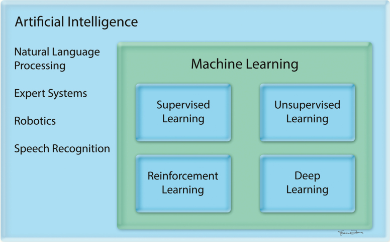
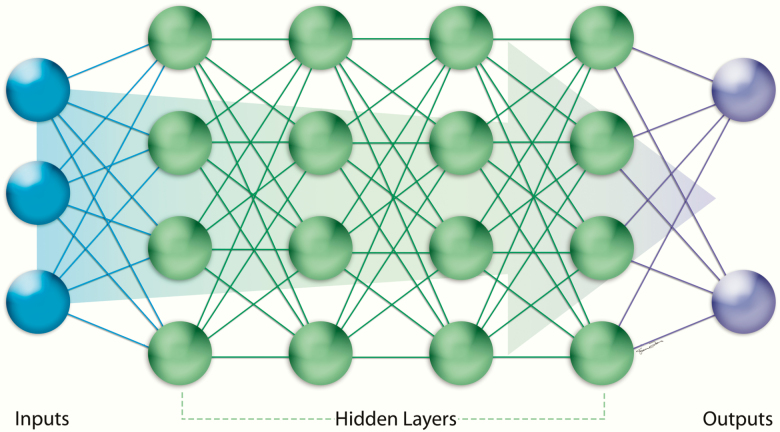
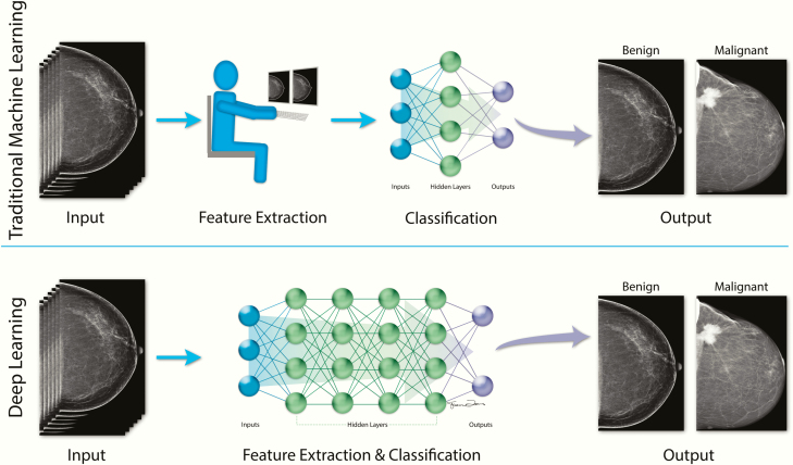
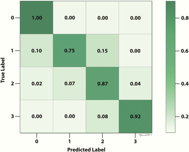
Artificial intelligence (AI) is a branch of computer science dedicated to developing computer algorithms that emulate intelligent human behavior. Subfields of AI include machine learning and deep learning. Advances in AI technologies have led to techniques that could increase breast cancer detection, improve clinical efficiency in breast imaging practices, and guide decision-making regarding screening and prevention strategies. This article reviews key terminology and concepts, discusses common AI models and methods to validate and evaluate these models, describes emerging AI applications in breast imaging, and outlines challenges and future directions. Familiarity with AI terminology, concepts, methods, and applications is essential for breast imaging radiologists to critically evaluate these emerging technologies, recognize their strengths and limitations, and ultimately ensure optimal patient care.
Keywords: artificial intelligence; breast imaging; deep learning; machine learning; mammography.
© Society of Breast Imaging 2020. All rights reserved. For permissions, please e-mail: journals.permissions@oup.com.
Figures
References
-
- Chartrand G, Cheng PM, Vorontsov E, et al. . Deep learning: a primer for radiologists. Radiographics 2017;37(7):2113–2131. - PubMed
-
- Tang A, Tam R, Cadrin-Chênevert A, et al. ; Canadian Association of Radiologists (CAR) Artificial Intelligence Working Group Canadian Association of Radiologists white paper on artificial intelligence in radiology. Can Assoc Radiol J 2018;69(2):120–135. - PubMed
-
- Kohli M, Prevedello LM, Filice RW, Geis JR. Implementing machine learning in radiology practice and research. AJR Am J Roentgenol 2017;208(4):754–760. - PubMed
-
- Fuchsjäger M. Is the future of breast imaging with AI? Eur Radiol 2019;29(9):4822–4824. - PubMed
-
- Soffer S, Ben-Cohen A, Shimon O, Amitai MM, Greenspan H, Klang E. Convolutional neural networks for radiologic images: a radiologist’s guide. Radiology 2019;290(3):590–606. - PubMed
Publication types
Grants and funding
LinkOut - more resources
Full Text Sources