. 2020 Aug 16;20(3):12.
doi: 10.1007/s10158-020-00247-1.

Adapting techniques for calcium imaging in muscles of adult Brugia malayi

Affiliations

Adapting techniques for calcium imaging in muscles of adult Brugia malayi

Paul D E Williams et al. Invert Neurosci. .

Abstract

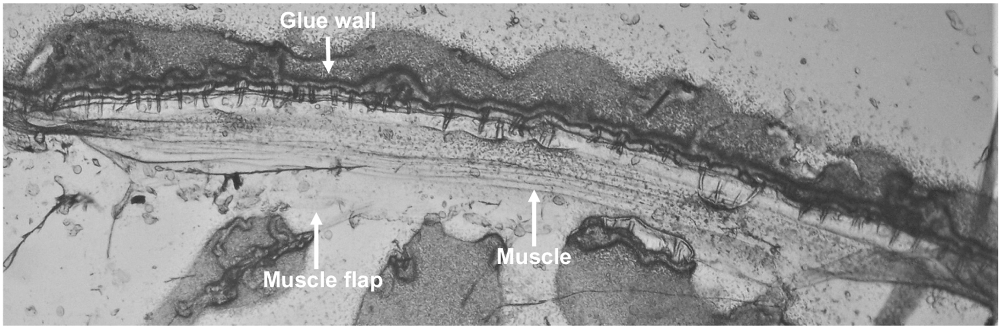
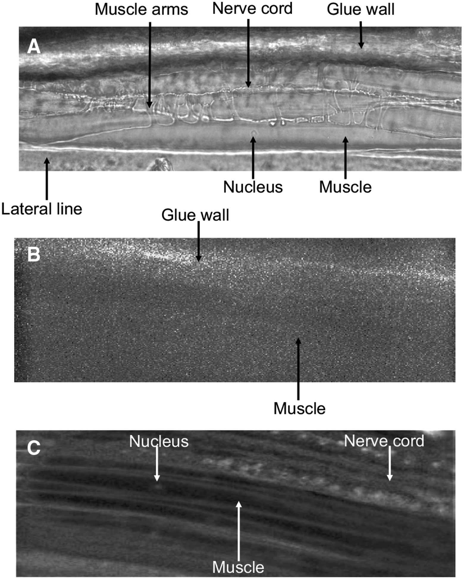
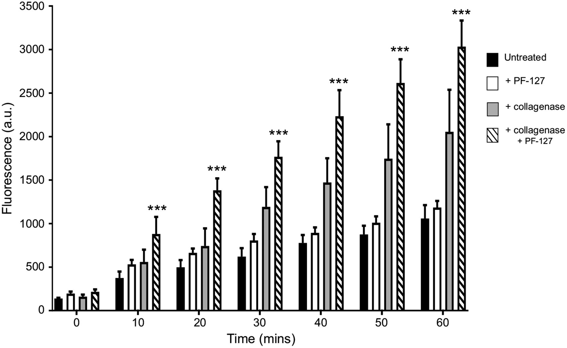
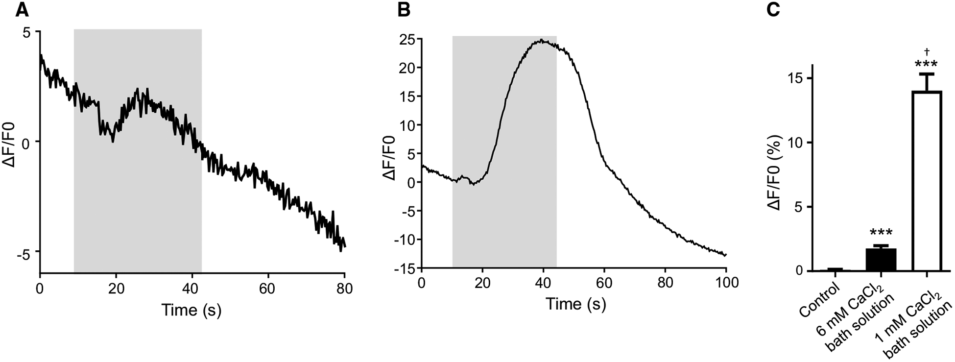
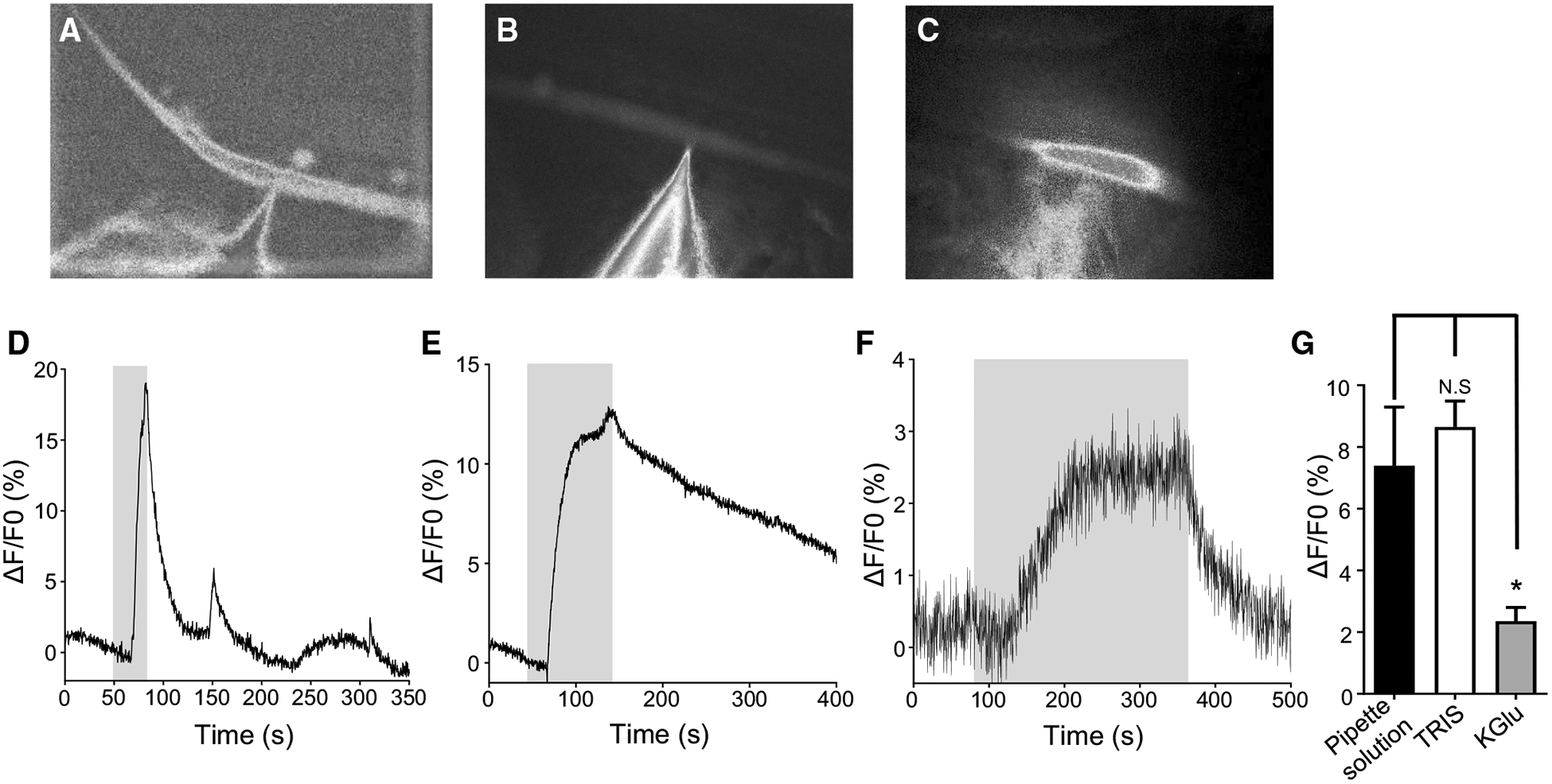
Brugia malayi is a human filarial nematode parasite that causes lymphatic filariasis or 'elephantiasis' a disfiguring neglected tropical disease. This parasite is a more tractable nematode parasite for the experimental study of anthelmintic drugs and has been studied with patch-clamp and RNAi techniques. Unlike in C. elegans however, calcium signaling in B. malayi or other nematode parasites has not been achieved, limiting the studies of the mode of action of anthelmintic drugs. We describe here the development of calcium imaging methods that allow us to characterize changes in cellular calcium in the muscles of B. malayi. This is a powerful technique that can help in elucidating the mode of action of selected anthelmintics. We developed two approaches that allow the recording of calcium signals in the muscles of adult B. malayi: (a) soaking the muscles with Fluo-3AM, promoting large-scale imaging of multiple cells simultaneously and, (b) direct insertion of Fluo-3 using microinjection, providing the possibility of performing dual calcium and electrophysiological recordings. Here, we describe the techniques used to optimize dye entry into the muscle cells and demonstrate that detectable increases in Fluo-3 fluorescence to elevated calcium concentrations can be achieved in B. malayi using both techniques.

Keywords: B. malayi; Calcium imaging; Fluo-3; Muscle.


Conflict of interest statement

Conflict of interest The authors declare that they have no conflict of interests.

Figures

Fig. 1
Fig. 1
Micrograph of a dissected body piece of B. malayi: micrograph of a suitable dissected B. malayi body piece with intestine and uterus removed used for calcium and electrophysiological experiments. Key structures glue wall, muscle and muscle flap indicated
Fig. 2
Fig. 2
Micrographs of B. malayi muscles: a micrograph of B. malayi muscles under white light. b Untreated muscle exposed to blue light. c Muscles after being treated with 5 μM Fluo-3AM for 60 min at 34–36 °C. Key structures, muscle arms, muscle, nucleus, nerve cord, lateral line and glue wall are highlighted
Fig. 3
Fig. 3
Collagenase and Pluronic F-127 improve Fluo-3AM absorption: 60 min time course of overall Fluo-3 fluorescencein B. malayi muscle cells for tissues treated with Fluo-3AM alone (black bars), Fluo-3AM and 10% (v/v) Pluronic F-127 (white bars), Fluo-3AM and 2 mg/mL collagenase (gray bars) and Fluo-3AM, collagenase and Pluronic F-127 (dashed bars). ***Significantly different to untreated (Fluo-3AM alone) (P = < 0.001; two-way ANOVA). For all treatments, n = 5. All values are represented as mean ± SEM
Fig. 4
Fig. 4
Fluo-3AM treated muscles cells display Ca2+ signals: a representative calcium trace of a muscle exposed to 10 mM CaCl2 that was maintained in regular bath solution before stimulus application. Gray box represents stimulus application. b Representative calcium trace in response to 10 mM CaCl2 in a muscle maintained in low CaCl2 (1 mM) bath solution. Gray box represents stimulus application. c Amplitudes of calcium signals in muscles perfused with low calcium bath solution, 10 mM CaCl2 in regular bath solution and 10 mM CaCl2 in low CaCl2 bath solution. ***Significantly different from untreated (untreated vs. 10 mM CaCl2 regular buffer; P < 0.0001, t = 6.792, df = 25, unpaired t test), (untreated vs. 10 mM CaCl2 low CaCl2 buffer; P < 0.0001, t = 8.273, df = 30, unpaired t test). † = significantly different to 10 mM CaCl2 in regular buffer (10 mM CaCl2 in regular buffer vs. 10 mM CaCl2 in low CaCl2 buffer P = < 0.0001, t = 8.108, df = 33, unpaired t test). Untreated n = 12; 10 mM CaCl2 in regular buffer n = 15, 10 mM CaCl2 in low CaCl2 buffer n = 20. All values are represented as mean ± SEM
Fig. 5
Fig. 5
Microinjection of Fluo-3: a micrograph of muscle under blue light injected with electrophysiology pipette solution containing 5 μM Fluo-3. b Micrograph of Fluo-3 injection using TRIS-HCl. c Micrograph of Fluo-3 injection using K-gluconate solution. d Representative trace of a calcium response to 10 mM CaCl2 in a muscle injected with pipette solution containing Fluo-3. Gray box indicates stimulus application. e Representative trace of TRIS-HCl Fluo-3 inject muscles to 10 mM CaCl2. Gray box indicates stimulus application. f Representative trace of calcium signal in muscle inject with Fluo-3 using K-gluconate solution. Gray box indicates stimulus application. g Quantification of calcium amplitudes to 10 mM CaCl2 in muscles injected using pipette solution (black bar), TRIS-HCl (white bar) and K-gluconate (gray bar). N.S. = not significant pipette solution versus TRIS-HCl (P = 0.5526, t = 0.6031, df = 22, unpaired t test). *Significantly different pipette solutions versus K-Glu (P = 0.0220, t = 2.473, df = 21, unpaired t test). Pipette solution n = 12; TRIS-HCl n = 12, K-Glu n = 11. All values are represented as mean ± SEM

Similar articles

Cited by

References

    1. Badura A, Sun XR, Giovannucci A, Lynch LA, Wang SS (2014) Fast calcium sensor proteins for monitoring neural activity. Neurophotonics 1(2):025008 10.1117/1.NPh.1.2.025008 - DOI - PMC - PubMed
    1. Cobbold PH, Rink TJ (1987) Fluorescence and bioluminescence measurement of cytoplasmic free calcium. Biochem J 248(2):313–328. 10.1042/bj2480313 - DOI - PMC - PubMed
    1. Coghlan A (2005) Nematode genome evolution. WormBook. 10.1895/wormbook.1.15.1 - DOI - PMC - PubMed
    1. Fieseler C, Kunert-Graf J, Kutz JN (2018) The control structure of the nematode Caenorhabditis elegans: neuro-sensory integration and proprioceptive feedback. J Biomech 74:1–8. 10.1016/j.jbiomech.2018.03.046 - DOI - PubMed
    1. Gee KR, Brown KA, Chen WN, Bishop-Stewart J, Gray D, Johnson I (2000) Chemical and physiological characterization of fluo-4 Ca(2 +)-indicator dyes. Cell Calcium 27(2):97–106. 10.1054/ceca.1999.0095 - DOI - PubMed

Publication types

LinkOut - more resources