Review
. 2020 Nov 4;15(21):1958-1973.
doi: 10.1002/cmdc.202000465. Epub 2020 Sep 18.

Purinergic Signaling: Impact of GPCR Structures on Rational Drug Design

Affiliations
Review

Purinergic Signaling: Impact of GPCR Structures on Rational Drug Design

Veronica Salmaso et al. ChemMedChem. .

Abstract

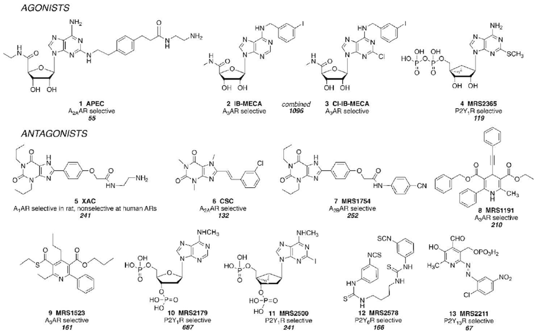
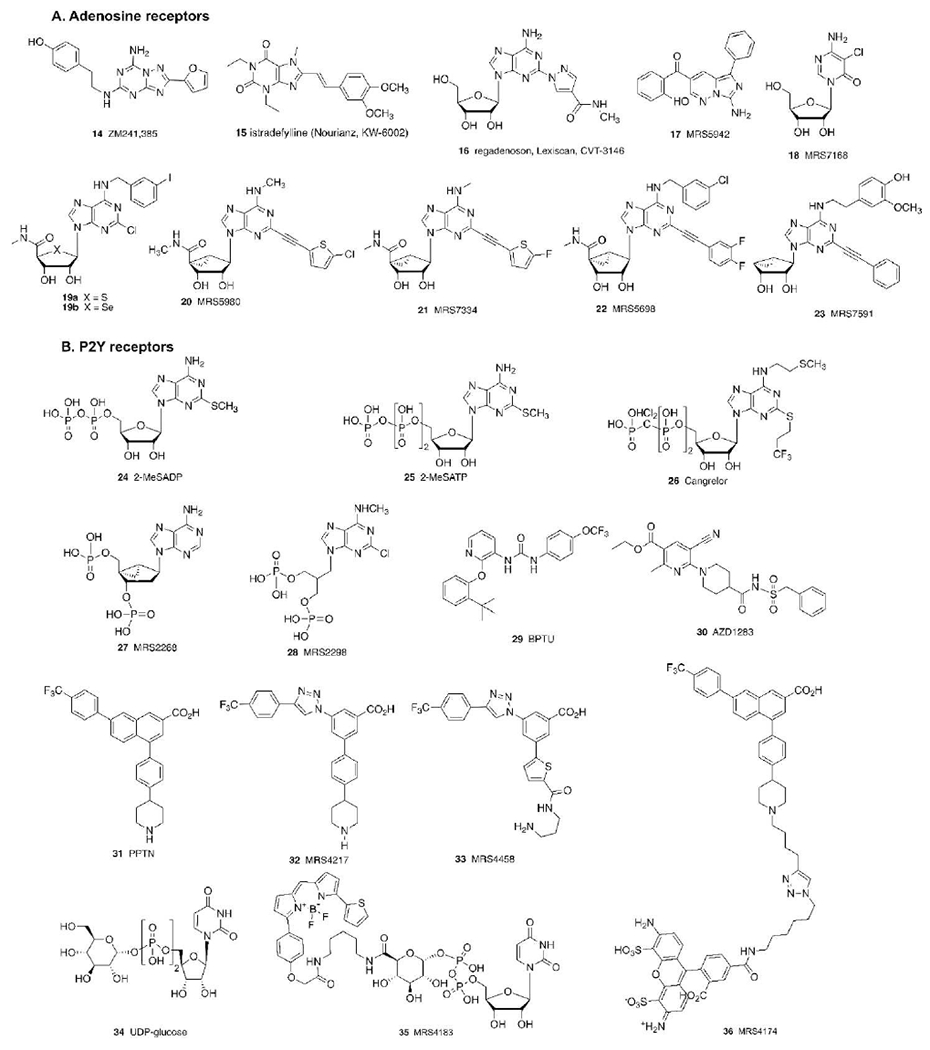
The purinergic signaling system includes membrane-bound receptors for extracellular purines and pyrimidines, and enzymes/transporters that regulate receptor activation by endogenous agonists. Receptors include: adenosine (A1 , A2A , A2B, and A3 ) and P2Y (P2Y1 , P2Y2 , P2Y4 , P2Y6 , P2Y11 , P2Y12 , P2Y13 , and P2Y14 ) receptors (all GPCRs), as well as P2X receptors (ion channels). Receptor activation, especially accompanying physiological stress or damage, creates a temporal sequence of signaling to counteract this stress and either mobilize (P2Rs) or suppress (ARs) immune responses. Thus, modulation of this large signaling family has broad potential for treating chronic diseases. Experimentally determined structures represent each of the three receptor families. We focus on selective purinergic agonists (A1 , A3 ), antagonists (A3 , P2Y14 ), and allosteric modulators (P2Y1 , A3 ). Examples of applying structure-based design, including the rational modification of known ligands, are presented for antithrombotic P2Y1 R antagonists and anti-inflammatory P2Y14 R antagonists and A3 AR agonists. A3 AR agonists are a potential, nonaddictive treatment for chronic neuropathic pain.

Keywords: drug discovery; molecular modeling; nucleosides; nucleotides; receptors.


Conflict of interest statement

Conflict of Interest

The authors declare no conflict of interest.

Figures

Figure 1.
Figure 1.
Early signal transduction by purinergic GPCRs that respond to extracellular nucleotides (P2YRs) and to adenosine (ARs). Enzymes, transporters, channels and cell damage control the level of endogenous agonists, which act in an autocrine or paracrine fashion. Pathways downstream of the G proteins leading to various second messengers, kinases and gene transcription are not shown. P2XRs, which respond rapidly to ATP and are ligand-gated cation channels, consisting of 7 monomer subtypes forming active trimers, are not shown. Various sources of extracellular nucleotides are indicated, in addition to synaptic release from neurons and astrocytes. Ectonucleotidase CD39 is an integral membrane protein and hydrolyzes both ATP and ADP to AMP, while CD73 is anchored through glycosylphosphatidyl inositol (GPI) and acts only on AMP to produce adenosine.
Figure 2.
Figure 2.
Representative ligand toolbox of purinergic receptor agonists and antagonists that were developed by Jacobson et al. through empirical probing of GPCR SAR, from 1985 (XAC) to 2004 (MRS2578). The citation numbers are shown below each description of selectivity (according to SciFinder, https://scifinder.cas.org/, accessed May 22, 2020).
Figure 3.
Figure 3.
Various other purine receptor ligands discussed in the text, including both compounds discovered by empirical methods and designed by structure-based approaches. Most of the compounds shown are GPCR antagonists, but some agonists (16, 18, 19-22, 24, 25, 34, 35) are included. Compound 23 is a partial agonist selective for the A3AR. Cangrelor is an approved i.v. antithrombotic drug, while BPTU and AZD1283 are experimental antithrombotics. 35 and 36 are high affinity P2Y14R fluorescent probes.
Figure 4.
Figure 4.
A superposition of separate X-ray structures of the hP2Y1R in complex with MRS2500 11 and BPTU 29.[35] These inactive receptor conformations are nearly identical, even though the ligands bind to disparate sites on the protein. The MRS2500-bound structure (PDB ID: 4XNW) is shown in light gray (ligand in lime) and the BPTU-bound structure (PDB ID: 4XNV) in dark gray (ligand in violet). Specific stabilizing interactions are indicated by dashed lines.
Figure 5.
Figure 5.
A) Docking pose of UDP-glucose 34 (lime) at the hP2Y14R (light gray) structure, obtained by homology modeling based on the P2Y12R X-ray structure.[62] Two subpockets were found in the P2Y14R putative orthosteric binding site, occupied by the UDP (subpocket 1) and glucose (subpocket 2) moieties. B) Docking pose of the antagonist PPTN 31 (violet) at the P2Y14R model (gray).[21]
Figure 6.
Figure 6.
Pose of MRS7591 23 at the A) human and B) mouse A3AR hybrid models, built using hA2AAR and hA1AR X-ray (for TM2) structures.[78] A) Last frame of a 30-ns MD simulation starting from the docking pose of MRS7591 (lime) at the hA3AR model (light gray). B) Last frame of a 30-ns MD simulation starting from the docking pose of MRS7591 (violet) at the mA3AR model (gray).

References

    1. Congreve M, de Graaf C, Swain NA, Tate CG, Cell 2020, 181, 81–91. - PubMed
    1. Fuentes AV, Pineda MD, Nagulapalli Venkata KC, Pharmacy (Basel) 2018, 6, 43. - PMC - PubMed
    1. Novel Drug Approvals for 2019. Accessed: Dec. 29, 2019: https://www.fda.gov/drugs/new-drugs-fda-cders-new-molecular-entities-and....
    1. Chen JF, Cunha R, Purinergic Signal. 2020, 16, 167–174. - PMC - PubMed
    1. Cherezov V, Rosenbaum DM, Hanson MA, Rasmussen SG, Thian FS, Kobilka TS, Choi HJ, Kuhn P, Weis WI, Kobilka BK, Stevens RC, Science 2007, 318, 1258–1265. - PMC - PubMed

Publication types

MeSH terms

Substances