. 2020 Oct;10(10):e01799.
doi: 10.1002/brb3.1799. Epub 2020 Aug 16.

A lab-on-a-chip model of glaucoma

Affiliations

A lab-on-a-chip model of glaucoma

Fatemeh Nafian et al. Brain Behav. 2020 Oct.

Abstract

Aims: We developed a glaucoma-on-a-chip model to evaluate the viability of retinal ganglion cells (RGCs) against high pressure and the potential effect of neuroprotection.

Methods: A three-layered chip consisting of interconnecting microchannels and culture wells was designed and fabricated from poly-methyl methacrylate sheets. The bottom surface of the wells was modified by air plasma and coated with different membranes to provide a suitable extracellular microenvironment. RGCs were purified from postnatal Wistar rats by magnetic assisted cell sorting up to 70% and characterized by flow cytometry and immunocytochemistry. The cultured RGCs were exposed to normal (15 mmHg) or elevated pressure (33 mmHg) for 6, 12, 24, 36, and 48 hr, with and without adding brain-derived neurotrophic factor (BDNF) or a novel BDNF mimetic (RNYK).

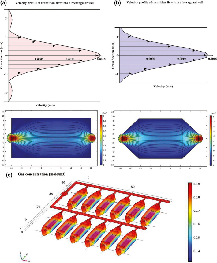
Results: Multiple inlet ports allow culture media and gas into the wells under elevated hydrostatic pressure. PDL/laminin formed the best supporting membrane. RGC survival rates were 85%, 78%, 70%, 67%, and 61% under normal pressure versus 40%, 22%, 18%, 12%, and 10% under high pressure at 6, 12, 24, 36, and 48 hr, respectively. BDNF and RNYK separately reduced RGC death rates about twofold under both normal and elevated pressures.

Conclusion: This model recapitulated the effects of elevated pressure over relatively short time periods and demonstrated the neuroprotective effects of BDNF and RNYK.

Keywords: glaucoma; hydrostatic pressure; lab-on-a-chip; microenvironment; retinal ganglion cell.


Conflict of interest statement

The authors declare that no conflict of interest exists.

Figures

FIGURE 1
FIGURE 1
Schematic of a designed chip in AutoCAD software. (a) Three‐dimensional design of layers a (1), b (2), and c (3) containing 12 wells (4), feeding ports (5), and microchannels (6), 2 main channels (7) and 1 single gas inlet (8). Layer (a) consisted of one feeding port per well and one main gas inlet. Layer B involved 12 hexagonal wells and microchannels. Layer (c) provided a surface area for cell cultures. Layers were fabricated (b) and assembled (c) to establish a complete chip
FIGURE 2
FIGURE 2
Estimated velocity profiles and gas concentration by Comsol Multiphysics software. (a) An entrance region was observed where the upstream flow entered the rectangular well. The boundary layer increased downstream, delayed the axial flow at the wall and accelerated the core flow in the center by maintaining the same flow rate. (b) The velocity profile became more even across the hexagonal well by straitening the walls at the entrance region and reducing the boundary layers at downstream (c) Gas concentration slightly changed throughout the main channels, sub‐channels, and wells
FIGURE 3
FIGURE 3
Phase‐contrast images of SH‐SY5Y cells. (a) Cells phenotypically changed from N‐type to S‐type by ATRA treatment. (b) Relatively greater number of differentiated cells oriented and aligned on different membranes compared to the naked surface as a negative control. PDL/laminin provided a physiologically optimal environment for neuronal adhesion and expansion. Scale bars: 50 µm
FIGURE 4
FIGURE 4
Purification of primary rat RGCs. (a) Retinal cells were isolated and converted into a cell suspension by triturating. The cell suspension was filtered through a mesh filter (>40 µm) to yield a single‐cell suspension. For magnetic assisted cell sorting (MACS), the cell suspension was incubated with CD90.1 microbeads and passed through an LS column to hold the magnetically labeled cells (RGCs) in the magnetic field. The adherent RGCs were rinsed to deplete endothelial cells and microglia. The column was removed from the separator, and the adhered cells were flushed out by firmly pushing the plunger into the column and counted. (b) Flow cytometry results showed that CD90.1+ cells were more purified after positive selection using MACS (orange, 70.4% purity) rather than mesh filtration (blue, 37.4%) and initial retinal single‐cell suspension (red, 19.8%)
FIGURE 5
FIGURE 5
CD90.1 immunostaining of isolated rat RGCs by MACS. (a) After 3‐day normal culture, RGCs continued to express the specific marker CD90.1 (green). Nuclear staining was performed using DAPI (blue). (b) The two‐color merged image was produced by overlaying the original CD90.1 and DAPI images, which indicates colocalization of the axonal connections between multiple RGCs against their nucleus of cell bodies. Scale bars: 100 µm
FIGURE 6
FIGURE 6
Morphological changes of primary RGCs on PMMA surface modified by plasma plus PDL/Laminin compare to nonmodified, before the pressure test. Primary RGCs formed cell clusters contained small cell bodies and few extended fine neurites on nonmodified PMMA surface. On the modified PMMA surface, RGCs exhibited uniform and round cell bodies, with neurites extending to connect to one other. At day 10, the cells developed a complex dendritic network with numerous neurites and branches. Scale bars: 50 µm
FIGURE 7
FIGURE 7
Schematic of the pressure set‐up to simulate glaucomatous condition. (a) A sealed pressure chip inside the incubator was connected to a gauge and regulators placed outside by the polyurethane hoses and pneumatic valves via a side opening in the back of incubator. (b1) There was a significant decrease over time for untreated cells under EHP condition with mean difference (Mean Diff.) = 45.35, 56, 51.83, 55.43, and 51.2 (Two‐way ANOVA, N = 4, graphs display mean ± SD) at 6, 12, 24, 36, and 48 hr, respectively (p < .0001). (b2) BDNF prevented degeneration of treated RGCs under EHP condition with Mean Diff. = 20, 22.52, 23.83, 24, and 26.7 at 6 (p = .0198), 12 (p = .0022), 24 (p = .0004), 36 (p < .0001) and 48 (p < .0001) hr, respectively. (B3) RNYK identically protected RGCs under EHP condition with Mean Diff. = 26.33, 26.67, 25.67, 26.33, and 27.33 at 6, 12, 24, 36, and 48 hr, respectively (p < .0001). *EHP, elevated hydrostatic pressure; NHP, normal hydrostatic pressure

References

    1. Agar, A. , Li, S. , Agarwal, N. , Coroneo, M. T. , & Hill, M. A. (2006). Retinal ganglion cell line apoptosis induced by hydrostatic pressure. Brain Research, 1086(1), 191–200. 10.1016/j.brainres.2006.02.061 - DOI - PubMed
    1. Agar, A. , Yip, S. S. , Hill, M. A. , & Coroneo, M. T. (2000). Pressure related apoptosis in neuronal cell lines. Journal of Neuroscience Research, 60(4), 495–503. 10.1002/(SICI)1097-4547(20000515)60:4<495:AID-JNR8>3.0.CO;2-S - DOI - PubMed
    1. Aires, I. , Ambrósio, A. , & Santiago, A. (2017). Modeling human glaucoma: Lessons from the in vitro models. Ophthalmic Research, 57(2), 77–86. 10.1159/000448480 - DOI - PubMed
    1. Balijepalli, A. , & Sivaramakrishan, V. (2017). Organs‐on‐chips: Research and commercial perspectives. Drug Discovery Today, 22(2), 397–403. 10.1016/j.drudis.2016.11.009 - DOI - PubMed
    1. Bamshad, A. , Nikfarjam, A. , & Khaleghi, H. (2016). A new simple and fast thermally‐solvent assisted method to bond PMMA–PMMA in micro‐fluidics devices. Journal of Micromechanics and Microengineering, 26(6), 1–12. 10.1088/0960-1317/26/6/065017 - DOI

Substances