. 2020 Aug 17:9:e60371.
doi: 10.7554/eLife.60371.

CMG helicase disassembly is controlled by replication fork DNA, replisome components and a ubiquitin threshold

Affiliations

CMG helicase disassembly is controlled by replication fork DNA, replisome components and a ubiquitin threshold

Tom D Deegan et al. Elife. .

Abstract

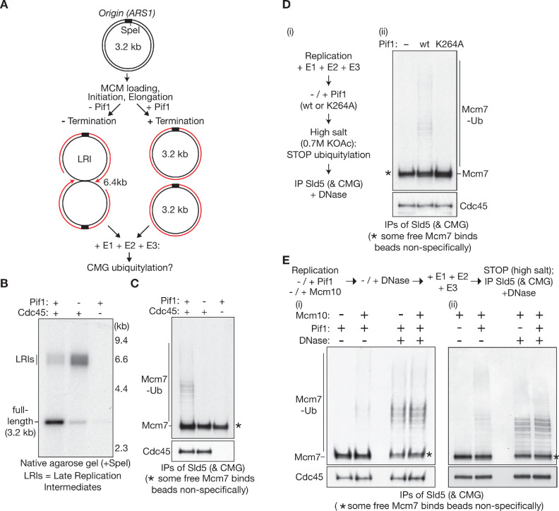
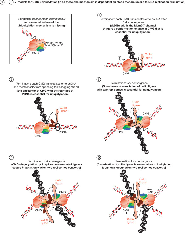
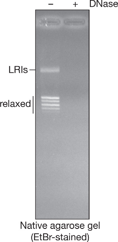
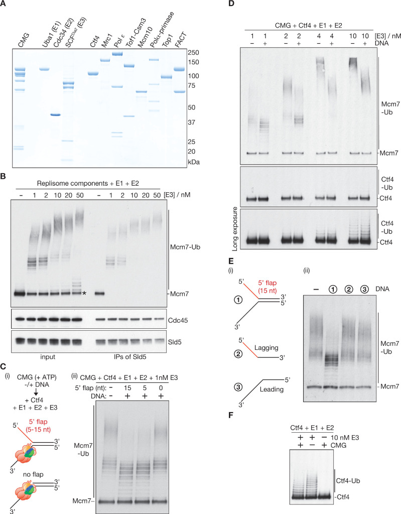
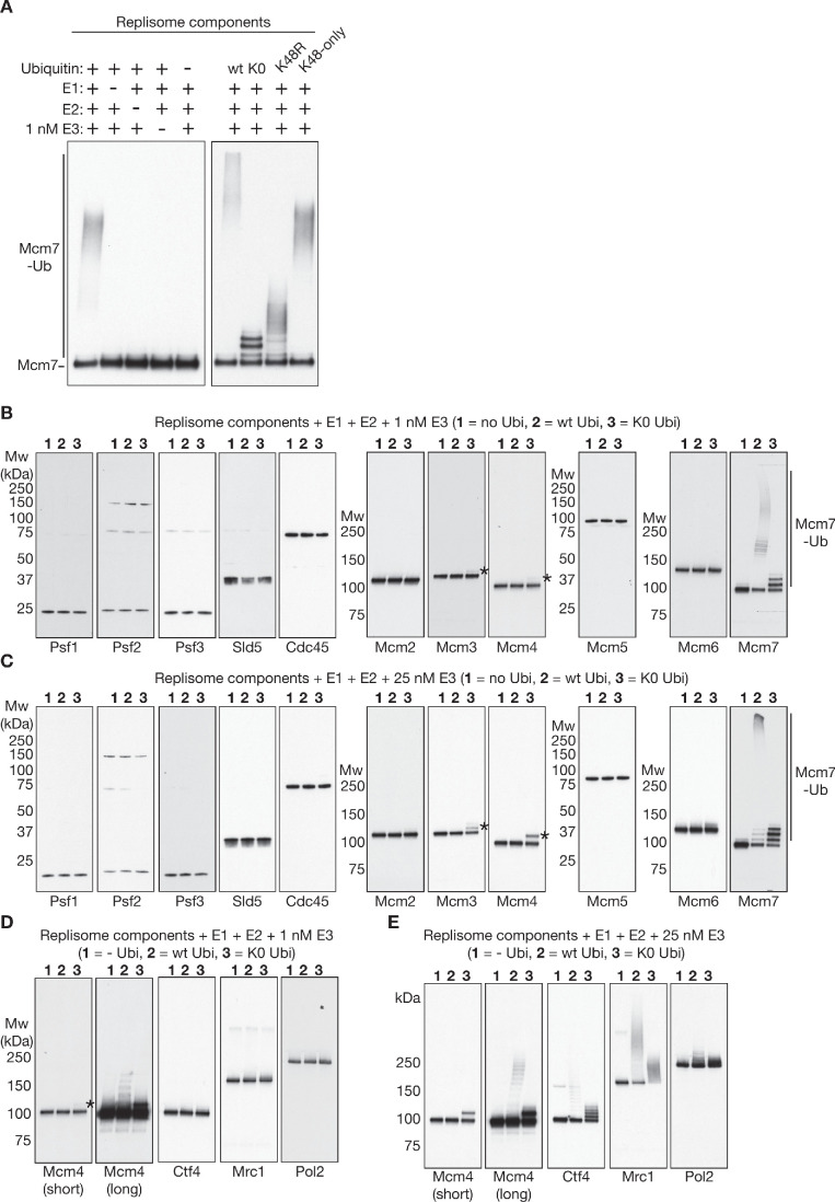
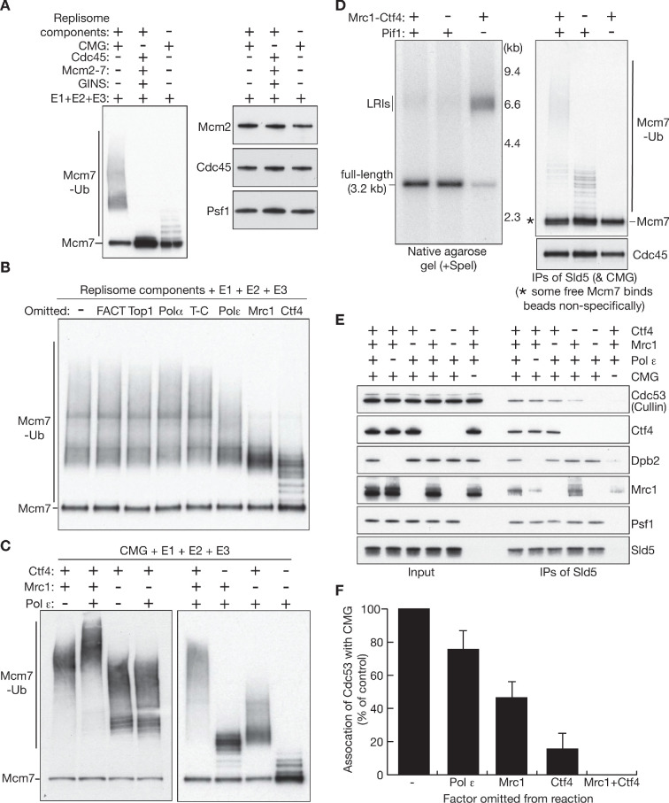
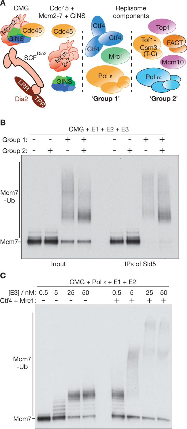
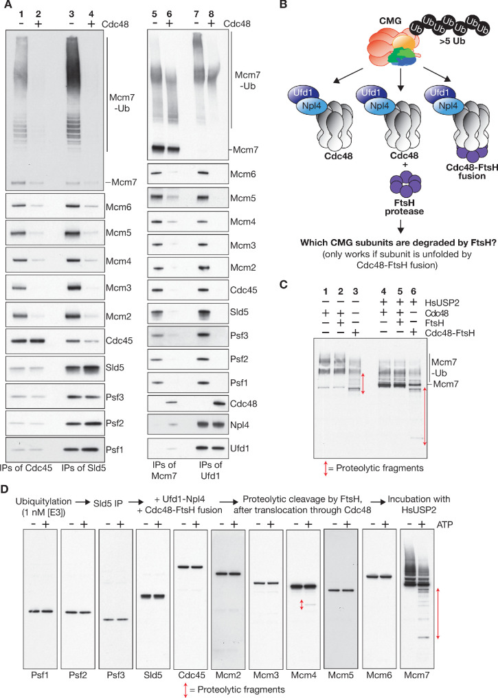
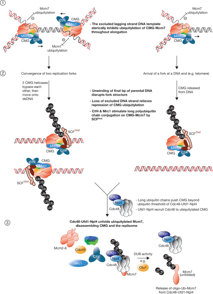
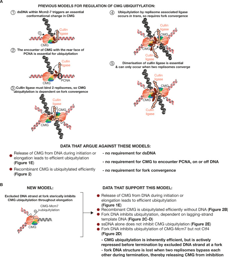
The eukaryotic replisome assembles around the CMG helicase, which stably associates with DNA replication forks throughout elongation. When replication terminates, CMG is ubiquitylated on its Mcm7 subunit and disassembled by the Cdc48/p97 ATPase. Until now, the regulation that restricts CMG ubiquitylation to termination was unknown, as was the mechanism of disassembly. By reconstituting these processes with purified budding yeast proteins, we show that ubiquitylation is tightly repressed throughout elongation by the Y-shaped DNA structure of replication forks. Termination removes the repressive DNA structure, whereupon long K48-linked ubiquitin chains are conjugated to CMG-Mcm7, dependent on multiple replisome components that bind to the ubiquitin ligase SCFDia2. This mechanism pushes CMG beyond a '5-ubiquitin threshold' that is inherent to Cdc48, which specifically unfolds ubiquitylated Mcm7 and thereby disassembles CMG. These findings explain the exquisite regulation of CMG disassembly and provide a general model for the disassembly of ubiquitylated protein complexes by Cdc48.

Keywords: CMG helicase; DNA replication; S. cerevisiae; chromosomes; gene expression; termination.


Conflict of interest statement

TD, PM, RF, CP, KL No competing interests declared

Figures

Figure 1.
Figure 1.. Mcm7 ubiquitylation is repressed before termination by the association of CMG with DNA.
(A) Experimental scheme for B-C, based on in vitro replication of plasmid DNA with purified budding yeast proteins. LRI = Late Replication Intermediate. (B) Nascent DNA replication products were digested with SpeI and analysed by native agarose gel. (C) At the end of the reactions, the CMG helicase was released from DNA by treatment with DNase, in the presence of high salt to block further ubiquitylation, before isolation of CMG by immunoprecipitation of Sld5. The asterisk (*) indicates unmodified Mcm7, which binds non-specifically to beads under these conditions. (D) (i) Experimental scheme (Pif1 K264A is inactive as a helicase and does not support replication termination); (ii) The indicated CMG subunits were monitored by immunoblotting. (E) (i) Replication reactions were performed in the presence or absence of Mcm10 or Pif1, before treatment for 10’ at 30°C with DNase to release CMG from DNA. Subsequently, the samples were incubated for 20 min in the presence of E1-E2-E3, and the reactions were then stopped by addition of high salt, before isolation of CMG as above (in the presence of DNase). The indicated subunits of CMG were monitored by immunoblotting. See also Figure 1—figure supplements 1–2.
Figure 1—figure supplement 1.
Figure 1—figure supplement 1.. Previous models for the regulation of CMG ubiquitylation during DNA replication termination.
Figure 1—figure supplement 2.
Figure 1—figure supplement 2.. Efficient DNA digestion at end of in vitro replication reactions.
DNase was added to replication reactions as in Figure 1E. DNA was subsequently purified and analysed by native agarose gel and EtBr staining. Pif1 was omitted from these reactions. The figure shows that DNase digestion was very efficient under these conditions.
Figure 2.
Figure 2.. Replication fork structure inhibits CMG ubiquitylation by SCFDia2 and Cdc34.
(A) Purified proteins used in this study, analysed by SDS-PAGE and stained with colloidal Coomassie Blue. (B) The components described in (A) were incubated for 20’ at 30°C with the indicated concentrations of E3 (input), before isolation of CMG (IPs of Sld5) and immunoblotting of the indicated components. The asterisk indicates a small pool of free Mcm7 that was not targeted for ubiquitylation. (C) (i) Reaction scheme and illustration of the association of CMG with model DNA replication forks. Replisome components other than Ctf4 were omitted to limit DNA unwinding by CMG; (ii) Ubiquitylation reactions in the presence or absence of synthetic replication forks with the indicated 5’ flaps. (D) Reactions at the indicated [E3], plus or minus the model fork substrate with 15 nt 5’ flap from (C). (E) Reactions were performed as in (C), with addition of the indicated DNA (2 and 3 are the ssDNA oligos that were used to make 1). (F) Ubiquitylation reactions performed as in (D), in the absence of DNA and +/- E3 or CMG as indicated. See also Figure 2—figure supplements 1–2.
Figure 2—figure supplement 1.
Figure 2—figure supplement 1.. Reconstituted CMG ubiquitylation involves the conjugation of K48-linked ubiquitin chains to Mcm7.
(A) Reactions were performed as in Figure 2. The left panel presents the effects of dropping out the indicated components of the reactions. The right panel compares the indicated variants of ubiquitin (‘K0’=lysine free ubiquitin, ‘K48R’ has the indicated single change, whereas all lysines except K48 were mutated to arginine in ‘K48-only’). (B) Ubiquitylation of all 11 CMG components was monitored by immunoblotting under the indicated conditions (1 nM [E3]). (C) Similar reactions were performed in the presence of 25 nM [E3]. (D) Immunoblots of additional replisome components from the experiment in (B), together with longer exposure for Mcm4. (E) Immunoblots of additional replisome components from the experiment in (C).
Figure 2—figure supplement 2.
Figure 2—figure supplement 2.. Association of the CMG helicase with model DNA replication forks.
Recombinant CMG helicase was mixed with Cy3-labelled versions of the DNA substrates shown in Figure 2C (i), and then incubated for 60 min on ice. The CMG helicase was isolated by immunoprecipitation of Sld5, before immunoblotting and detection of Cy3 fluorescence.
Figure 3.
Figure 3.. The inherently high efficiency of replisome ubiquitylation is dependent upon recruitment of SCFDia2 by Mrc1 and Ctf4.
(A) The indicated factors were incubated at 30°C for 20’, and ubiquitylation of Mcm7 was then monitored by immunoblotting, alongside other components of the CMG helicase. (B) Analogous reactions were performed in the absence of the indicated replisome components, in order to assess their contribution to CMG-Mcm7 ubiquitylation (T-C = Tof1-Csm3). (C) Similar reactions were performed to explore how Ctf4, Mrc1 and Pol ε each contribute to the efficiency of CMG-Mcm7 ubiquitylation. (D) Replication-coupled ubiquitylation reactions were performed as in Figure 1B, plus or minus the indicated factors. In the absence of Mrc1 and Ctf4, the plasmid template was completely replicated (left panel), but ubiquitylation of CMG-Mcm7 was impaired (right panel). (E) The ability of SCFDia2 to associate with the CMG helicase was monitored in the presence of replisome components. The indicated factors were mixed, before immunoprecipitation of Sld5 and immunoblotting. Cdc53 = cullin subunit of SCFDia2. (F) Quantification of the data in (E), to monitor the association of the SCFDia2 with the CMG helicase. The experiment was repeated three times, and the figure presents the mean values with standard deviations. See also Figure 3—figure supplement 1.
Figure 3—figure supplement 1.
Figure 3—figure supplement 1.. Ctf4 and Mrc1 promote long-chain ubiquitylation of CMG-Mcm7, which leads to efficient CMG disassembly by Cdc48-Ufd1-Npl4.
(A) The replisome components that associate with the CMG helicase were divided into two groups for the experiment in B. (B) Ubiquitylation reactions were performed with the indicated components, before isolation of CMG via immunoprecipitation of Sld5. (C) The contribution of Ctf4 and Mrc1 to CMG ubiquitylation was assessed over the indicated range of SCFDia2 concentrations.
Figure 4.
Figure 4.. Replisome-coupled ubiquitylation ensures that SCFDia2 pushes CMG beyond a ‘ubiquitin threshold’ intrinsic to Cdc48-Ufd1-Npl4.
(A) Reaction scheme for (B). (B) Recombinant versions of yeast Cdc48 and Ufd1-Npl4 were purified after expression in bacteria (left panel). Reactions were performed as in (A), and the products monitored by immunoblotting (right panels). (C) CMG was ubiquitylated in the presence or absence of Mrc1 and Ctf4, in reactions containing 1 nM of E3 (SCFDia2). CMG disassembly by Cdc48-Ufd1-Npl4 was then monitored as above. (D) CMG was ubiquitylated in reactions containing Mrc1 and 25 nM SCFDia2. Subsequently, CMG was isolated on anti-Sld5 beads, which were then washed with high salt to remove Mrc1 and SCFDia2. Finally, incubation was continued in the presence or absence of Cdc48-Ufd1-Npl4, and immunoblotting was used to monitor release of the indicated factors from the beads, corresponding to disassembly of CMG. (E) Ubiquitylation reactions in the presence of the indicated concentrations of E2, involving CMG with wild-type Mcm7 (left side) or Mcm7-K29A (right side). (F) Scheme for disassembly of ubiquitylated CMG bound to beads, as in G. (G) Immunoblots for the experiment in (F). See also Figure 4—figure supplement 1.
Figure 4—figure supplement 1.
Figure 4—figure supplement 1.. CMG helicase disassembly is dependent upon the formation of long K48-linked ubiquitin chains.
(A) Similar reactions to those in Figure 4B were performed in the presence or absence of the indicated factors. (B) Analogous reactions to those in Figure 4C were carried out in the presence of 0.5 nM or 25 nM SCFDia2. (C) CMG was ubiquitylated in the presence of the indicated concentrations of E2. Subsequently, 50 nM Cdc48-E588A-Ufd1-Npl4 was added and the Cdc48-E588A-Ufd1-Npl4 complex was then isolated by immunoprecipitation of Ufd1. The indicated associated proteins were monitored by immunoblotting. (D) Ubiquitylation reactions in the presence of the indicated concentrations of E2, either with with K48R ubiquitin (left side) or or lysine-free ubiquitin (K0) (right side).
Figure 5.
Figure 5.. Cdc48-Ufd1-Npl4 selectively unfold the ubiquitylated subunit(s) of CMG to drive replisome disassembly.
(A) CMG was ubiquitylated as in Figure 4 and then incubated for 20’ at 30°C in the presence or absence of Cdc48 as indicated. Ufd1-Npl4 was added to all samples. Subsequently, immunoprecipitations were performed with antibodies to the indicated factors, and the associated factors monitored by immunoblotting. (B) Fusion of Cdc48 to the bacterial FtsH protease generates a protein that specifically cleaves unfolded polypeptides that pass through the central channel of the Cdc48 hexamer. (C) Ubiquitylated CMG was immunoprecipitated with antibodies against Sld5, then incubated with Cdc48 (lanes 1 and 4), Cdc48 + FtsH (lanes 2 and 5) or Cdc48-FtsH fusion protein (lanes 3 and 6), all in the presence of Ufd1-Npl4 and ATP, before treatment for 60’ at 30°C with HsUSP2 deubiquitylase (lanes 4–6). Cleaved Mcm7 fragments were then detected by immunoblotting. (D) A similar reaction was performed as indicated and all 11 subunits of CMG were monitored by immunoblotting. See also Figure 5—figure supplement 1.
Figure 5—figure supplement 1.
Figure 5—figure supplement 1.. Ubiquitylated Mcm7 is unfolded during CMG helicase disassembly, and the ubiquitin chains must then be cleaved in order to release unfolded Mcm7 from Cdc48-Ufd1-Npl4.
(A) CMG was ubiquitylated as in Figure 4, before incubation 20’ at 30°C with the indicated factors. Ufd1-Npl4 was included in each case. Cdc48 and Cdc48-FtsH were included at 50 nM (+) or 200 nM (++). At the end of the reactions, the integrity of CMG was monitored by immunoprecipitation of Sld5. (B) Analogous CMG disassembly reactions to those in Figure 5D were performed in the presence of the indicated factors. At the end of the reactions, samples were incubated with HsUSP2, and cleavage of ubiquitylated Mcm4 and Mcm7 by Cdc48-FtsH was then detected by immunoblotting. (C) CMG disassembly was performed as in Figure 4A–B, before incubation with the indicated concentrations of the deubiquitylase Otu1 for 30’ at 30°C. Release of unfolded Mcm7 from Cdc48-Ufd1-Npl4 was then monitored by immunoprecipitation of Ufd1. (D) Quantification of the data in (C). The experiment was repeated three times, and the figure presents the mean values with standard deviations.
Figure 6.
Figure 6.. Model describing the regulated ubiquitylation and disassembly of the CMG helicase during DNA replication termination.
Multiple replisome components are omitted for simplicity. See text for discussion. See also Figure 6—figure supplement 1.
Figure 6—figure supplement 1.
Figure 6—figure supplement 1.. Summary of data that are inconsistent with previous models for the regulation of CMG ubiquitylation and instead support a revised model.

References

    1. Abid Ali F, Renault L, Gannon J, Gahlon HL, Kotecha A, Zhou JC, Rueda D, Costa A. Cryo-EM structures of the eukaryotic replicative helicase bound to a translocation substrate. Nature Communications. 2016;7:10708. doi: 10.1038/ncomms10708. - DOI - PMC - PubMed
    1. Aparicio OM, Weinstein DM, Bell SP. Components and dynamics of DNA replication complexes in S. cerevisiae: redistribution of MCM proteins and Cdc45p during S phase. Cell. 1997;91:59–69. doi: 10.1016/S0092-8674(01)80009-X. - DOI - PubMed
    1. Baretic D, Jenkyn-Bedford M, Aria V, Cannone G, Skehel M, Yeeles JTP. Cryo-EM structure of the fork protection complex bound to CMG at a replication fork. bioRxiv. 2019 doi: 10.1101/2019.12.18.880690. - DOI - PMC - PubMed
    1. Bell SP, Labib K. Chromosome duplication in Saccharomyces cerevisiae. Genetics. 2016;203:1027–1067. doi: 10.1534/genetics.115.186452. - DOI - PMC - PubMed
    1. Biswas D, Yu Y, Prall M, Formosa T, Stillman DJ. The yeast FACT complex has a role in transcriptional initiation. Molecular and Cellular Biology. 2005;25:5812–5822. doi: 10.1128/MCB.25.14.5812-5822.2005. - DOI - PMC - PubMed

Publication types