. 2021 Feb;78(4):1799-1815.
doi: 10.1007/s00018-020-03617-5. Epub 2020 Aug 17.

PDCD4 limits prooncogenic neuregulin-ErbB signaling

Affiliations

PDCD4 limits prooncogenic neuregulin-ErbB signaling

Juan Carlos Montero et al. Cell Mol Life Sci. 2021 Feb.

Abstract

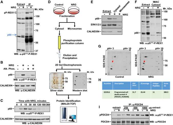
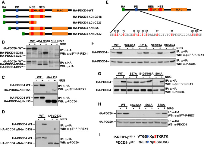
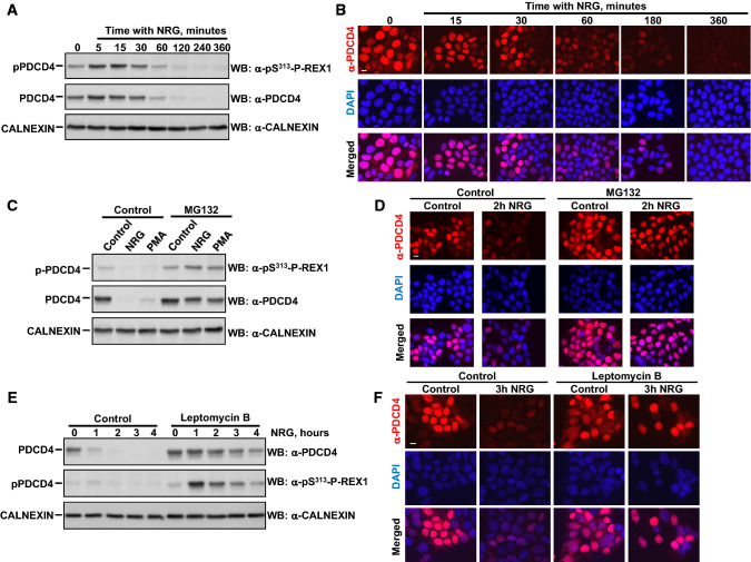
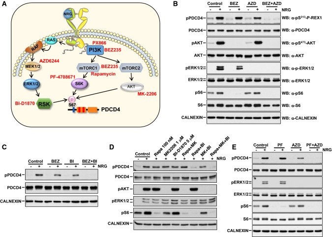
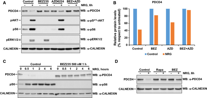
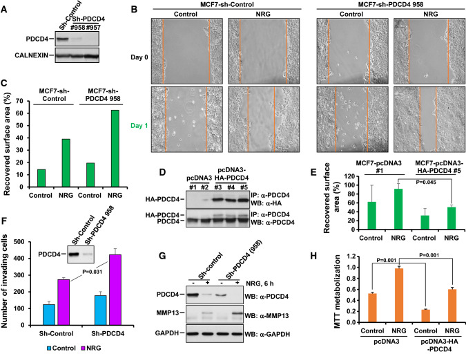
The neuregulins and their ErbB/HER receptors play essential roles in mammalian development and tissue homeostasis. In addition, deregulation of their function has been linked to the pathogenesis of diseases such as cancer or schizophrenia. These circumstances have stimulated research into the biology of this ligand-receptor system. Here we show the identification of programmed cell death protein-4 (PDCD4) as a novel neuregulin-ErbB signaling mediator. Phosphoproteomic analyses identified PDCD4 as protein whose phosphorylation increased in cells treated with neuregulin. Mutagenesis experiments defined serine 67 of PDCD4 as a site whose phosphorylation increased upon activation of neuregulin receptors. Phosphorylation of that site promoted degradation of PDCD4 by the proteasome, which depended on exit of PDCD4 from the nucleus to the cytosol. Mechanistic studies defined mTORC1 and ERK1/2 as routes implicated in neuregulin-induced serine 67 phosphorylation and PDCD4 degradation. Functionally, PDCD4 regulated several important biological functions of neuregulin, such as proliferation, migration, or invasion.

Keywords: Breast cancer; ErbB receptors; Neuregulin; PDCD4.


Conflict of interest statement

The authors declare that they have no conflict of interest.

Figures

Fig. 1
Fig. 1
Identification of a novel NRG-ErbB intermediate. a MCF7 cells stimulated with or without NRG (10 nM) for 15 min were lysed and cell extracts or anti-P-REX1 immunoprecipitates analyzed by Western blot with an anti-pS313-P-REX1 antibody. The position of the Mr markers is shown at the right. b p60 is phosphorylated. Extracts from MCF7 cells stimulated with or without NRG were treated with alkaline phosphatase and the samples were analyzed by Western with the anti-pS313-P-REX1 antibody. Calnexin was used as a loading control. c Kinetics of phospho-p60 in response to NRG. Lysates from MCF7 cells treated for different times with NRG were analyzed by Western with the anti-pS313-P-REX1 antibody. d Schematic representation of the steps used to identify p60. e p60 is a cytosolic protein. MCF7 cells treated or not with NRG were homogenized and the cytosolic and microsomal fractions analyzed by Western blot with the anti-pS313-P-REX1 antibody. ERK1/2 and calnexin were used as controls of cytosolic or microsomal fractions, respectively. f Analysis of p60 in eluates from phosphopeptide purification IMAC columns. g After preparation of cytosolic fractions from control and NRG-treated MCF7 cells, eluted phosphoproteins were precipitated and resolved in 2D gels that were then transferred to PVDF membranes. p60 was detected in these blots with the anti-pS313-P-REX1 antibody. Red arrows indicate p60. h The table shows the main protein identified in the proteomic analysis of the region corresponding to p60. i p60 is PDCD4. Cell extracts from MCF7 cells treated with or without NRG were inmunoprecipitated with anti-PDCD4 antibodies
Fig. 2
Fig. 2
The anti-pS313-P-REX1 antibody cross-reacts with pSer67 in PDCD4. a Schematic representation of different domains of HA-tagged wild type PDCD4 and different deletion mutants. HA: hemagglutinin, PD: phosphodegron (aa 70–76), the MA-3 domains are in orange (aa 163–284 and aa 326–449), red boxes are the potential nuclear export signals (NES), aa 241–251 and aa 182–192. bd MCF7 cells transfected with HA-tagged wild type PDCD4 or HA-PDCD4 deletion mutants in: ∆C-t G318, ∆C-t C227 (b), ∆N-t I55 (c) or ∆N-t D132 (d) were treated without or with NRG (10 nM). Cell extracts were immunoprecipitated with the anti-HA antibody and the blots were probed with the indicated antibodies. e Serines present in the region spanning amino acids 55 and 132. fh MCF7 cells transfected with wild type HA-PDCD4 or different mutants in which serine residues were replaced by alanine were treated with or without NRG. Cell extracts were immunoprecipitated with anti-HA and the blots were probed with anti-pS313-P-REX1. These blots were reprobed with the anti-PDCD4 antibody. i Sequence of P-REX1 used to generate the anti-pS313-P-REX1 antibody, aligned to the region surrounding S67
Fig. 3
Fig. 3
Role of the proteasome and nucleocytoplasmic circulation in the regulation of PDCD4 levels by NRG receptors. a MCF7 cells were treated with NRG for the indicated times. Lysates were analyzed by Western blot with anti-pS313-P-REX1 and anti-PDCD4 antibodies. b Immunofluorescence analysis of levels of PDCD4 in MCF7 cells treated with NRG. Cells were stained for PDCD4 (red) and DNA (blue). Scale bar = 10 μm. c, d NRG induces the degradation of PDCD4 via proteasome. MCF7 cells were pre-treated with or without MG132 (10 μM) and then stimulated with NRG or PMA (1 μM) during 6 h (c), or with NRG during 2 h (d). e, f Degradation of PDCD4 requires exit to the cytosol. MCF7 cells were pre-treated with leptomycin B (20 μg/ml) and then stimulated with NRG at different times. Analyses of total and pS67-PDCD4 were performed by Western blot (e), and localization and levels of PDCD4 were analyzed by immunofluorescence (f)
Fig. 4
Fig. 4
Phosphorylation of PDCD4 in serine 67 is required for NRG-induced degradation. a Phosphorylation of PDCD4 in serine 67 is not required for the cytosol to nucleus transit of PDCD4 induced by NRG. MCF7 cells transfected with wild type HA-PDCD4 or HA-PDCD4-S67A were seeded on coverslips and treated with NRG (10 nM, 15 min). Location of exogenous HA-PDCD4 and endogenous PDCD4 were performed by immunofluorescence. b MCF7 cell transfected as above were treated with NRG for times indicated. Levels of HA-PDCD4, HA-PDCD4-S67A and endogenous PDCD4 were analyzed by Western. GAPDH was used as a loading control. c Quantitative analysis of the relative protein levels of HA-PDCD4-WT and HA-PDCD4-S67A of the experiment shown in b. The numbers indicate hours of treatment with NRG
Fig. 5
Fig. 5
NRG controls phosphorylation of serine 67 through several signaling routes. a Schematic representation of pathways activated by neuregulin-ErbB receptors and analyzed using drugs that block them (shown in red). be MCF7 cells were preincubated with BEZ235 (1 μM), AZD6224 (5 μM), combination of BEZ235 plus AZD6224, BI-D1870 (5 μM), combination of BEZ235 plus BI-D1870, rapamycin (100 nM), MK2206 (1 μM), BI-D1870 (5 μM), rapamycin plus MK2206, rapamycin plus BI-D1870, MK2206 plus BI-D1870, triple combination of rapamycin, MK2206 plus BI-D1870, PF-4708671 (20 μM), AZD6224 (5 μM), and combination of PF-4708671 plus AZD6224. All preincubations with the drugs were for 3 h and treatments with NRG (10 nM) for 15 min
Fig. 6
Fig. 6
NRG controls PDCD4 stability through several signaling routes. a MCF7 cells were preincubated with BEZ235 (1 μM), AZD6224 (5 μM) or a combination of BEZ235 plus AZD6224 for 3 h, and then treated without or  with NRG (10 nM) during 6 h. Levels of PDCD4, pAKTS473, pS6, pERK1/2 and calnexin were analyzed by Western blotting. b Quantitative representation of the relative protein levels of PDCD4 respect to cells not treated with NRG in each condition of the experiment shown in a. c MCF7 cells were preincubated with BEZ235 (500 nM) and then treated without or with NRG for the times indicated. d Role of the mTORC1 pathway in the control of PDCD4 stability. MCF7 cells were preincubated with BEZ235 (1 μM) or rapamycin (100 nM) and then treated with or without NRG for 6 h
Fig. 7
Fig. 7
Control of NRG-induced migration, invasion and proliferation by PDCD4. a Knockdown of PDCD4 in MCF7 cells. Cells were infected with lentivirus containing the shRNA control (Sh-Control) or two shRNA sequences targeting PDCD4. Levels of PDCD4 were analyzed by Western blotting, using calnexin as a loading control. b Representative images from the wound healing assay showing the effect of PDCD4 knockdown on NRG-induced migration of MCF7 cells. The colored lines delimit the wounded region area. c Bar graph representing the recovered surface area with respect to the initial wounded area in each condition of the experiment performed in b. d Overexpression of PDCD4 in MCF7 cells. Clones of MCF7 cells transfected with HA-PDCD4 or empty vector were lysed and expression of the exogenous HA-PDCD4 or endogenous PDCD4 detected by Western. e Increased expression of PDCD4 prevented NRG-induced migration. The bar graph represents the recovered surface area with respect to the initial area in each condition. f Bar graph showing the number of MCF7-sh-Control or MCF7-sh-PDCD4 invading cells 48 h after NRG stimulation. Invading cells (those able to pass through the Matrigel layer) were fixed, stained with crystal violet, and counted. The Western image above the graph shows the level of PDCD4 in MCF7-sh-Control or MCF7-sh-PDCD4 cells. g Western blotting analysis of the effect of PDCD4 knockdown on the regulation of MMP13 after NRG stimulation for 6 h in MCF7 cells. h Increased expression of PDCD4 decreased resting and NRG-induced proliferation of MCF7 cells. Cell proliferation was determined by MTT metabolization 3 days after NRG (10 nM) stimulation

References

    1. Arteaga CL, Engelman JA. ERBB receptors: from oncogene discovery to basic science to mechanism-based cancer therapeutics. Cancer Cell. 2014;25(3):282–303. doi: 10.1016/j.ccr.2014.02.025. - DOI - PMC - PubMed
    1. Massague J, Pandiella A. Membrane-anchored growth factors. Annu Rev Biochem. 1993;62:515–541. doi: 10.1146/annurev.bi.62.070193.002503. - DOI - PubMed
    1. Montero JC, Rodriguez-Barrueco R, Ocana A, Diaz-Rodriguez E, Esparis-Ogando A, Pandiella A. Neuregulins and cancer. Clin Cancer Res. 2008;14(11):3237–3241. doi: 10.1158/1078-0432.CCR-07-5133. - DOI - PubMed
    1. Meyer D, Birchmeier C. Multiple essential functions of neuregulin in development. Nature. 1995;378(6555):386–390. doi: 10.1038/378386a0. - DOI - PubMed
    1. Britsch S, Li L, Kirchhoff S, Theuring F, Brinkmann V, Birchmeier C, Riethmacher D. The ErbB2 and ErbB3 receptors and their ligand, neuregulin-1, are essential for development of the sympathetic nervous system. Genes Dev. 1998;12(12):1825–1836. doi: 10.1101/gad.12.12.1825. - DOI - PMC - PubMed

LinkOut - more resources