Three-Dimensional Printed Molds for Image-Guided Surgical Biopsies: An Open Source Computational Platform
- PMID: 32804543
- PMCID: PMC7469624
- DOI: 10.1200/CCI.20.00026
Three-Dimensional Printed Molds for Image-Guided Surgical Biopsies: An Open Source Computational Platform
Abstract
Purpose: Spatial heterogeneity of tumors is a major challenge in precision oncology. The relationship between molecular and imaging heterogeneity is still poorly understood because it relies on the accurate coregistration of medical images and tissue biopsies. Tumor molds can guide the localization of biopsies, but their creation is time consuming, technologically challenging, and difficult to interface with routine clinical practice. These hurdles have so far hindered the progress in the area of multiscale integration of tumor heterogeneity data.
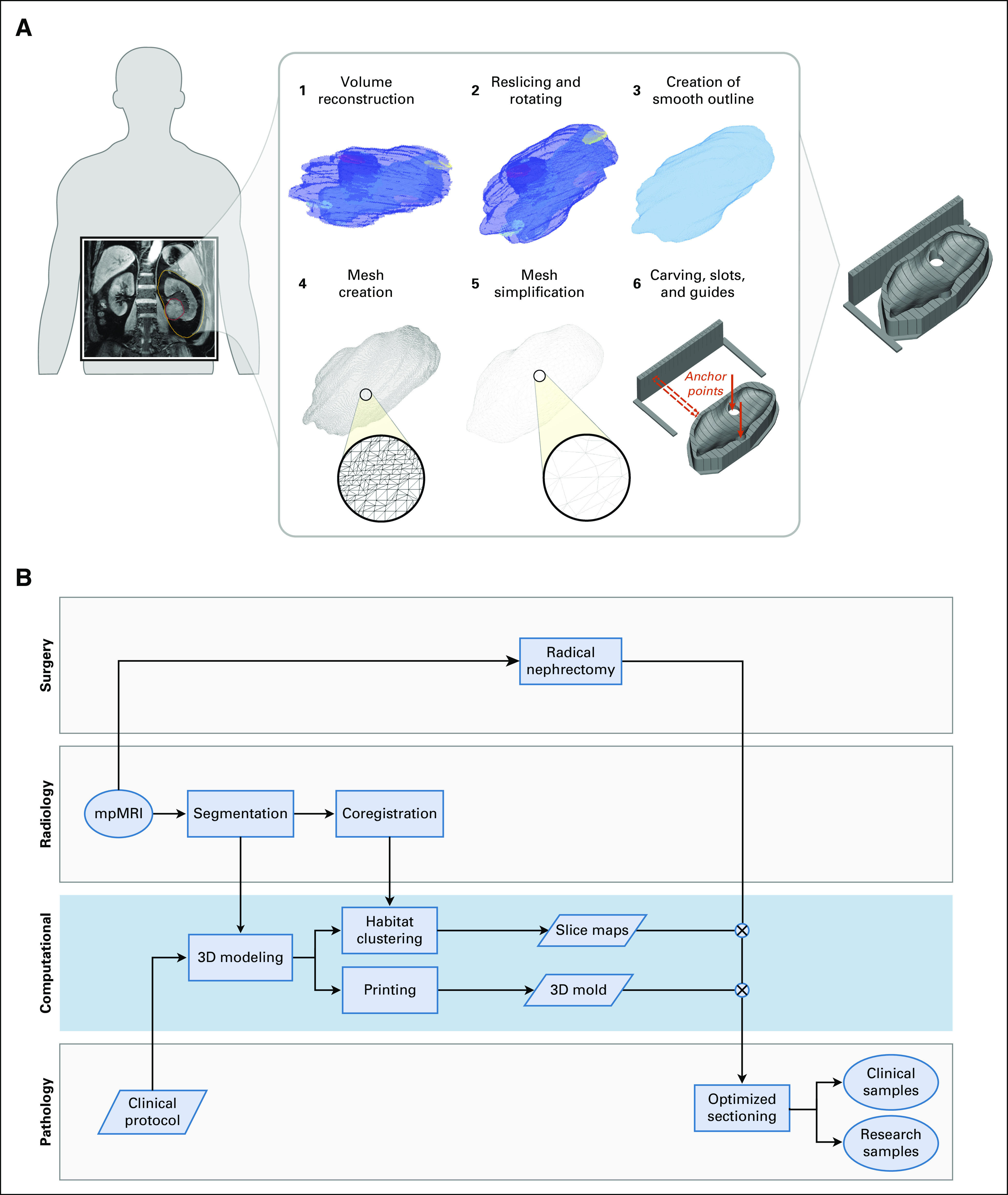
Methods: We have developed an open-source computational framework to automatically produce patient-specific 3-dimensional-printed molds that can be used in the clinical setting. Our approach achieves accurate coregistration of sampling location between tissue and imaging, and integrates seamlessly with clinical, imaging, and pathology workflows.
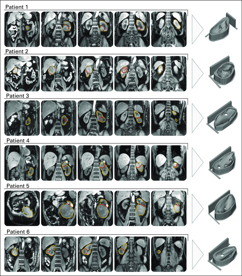
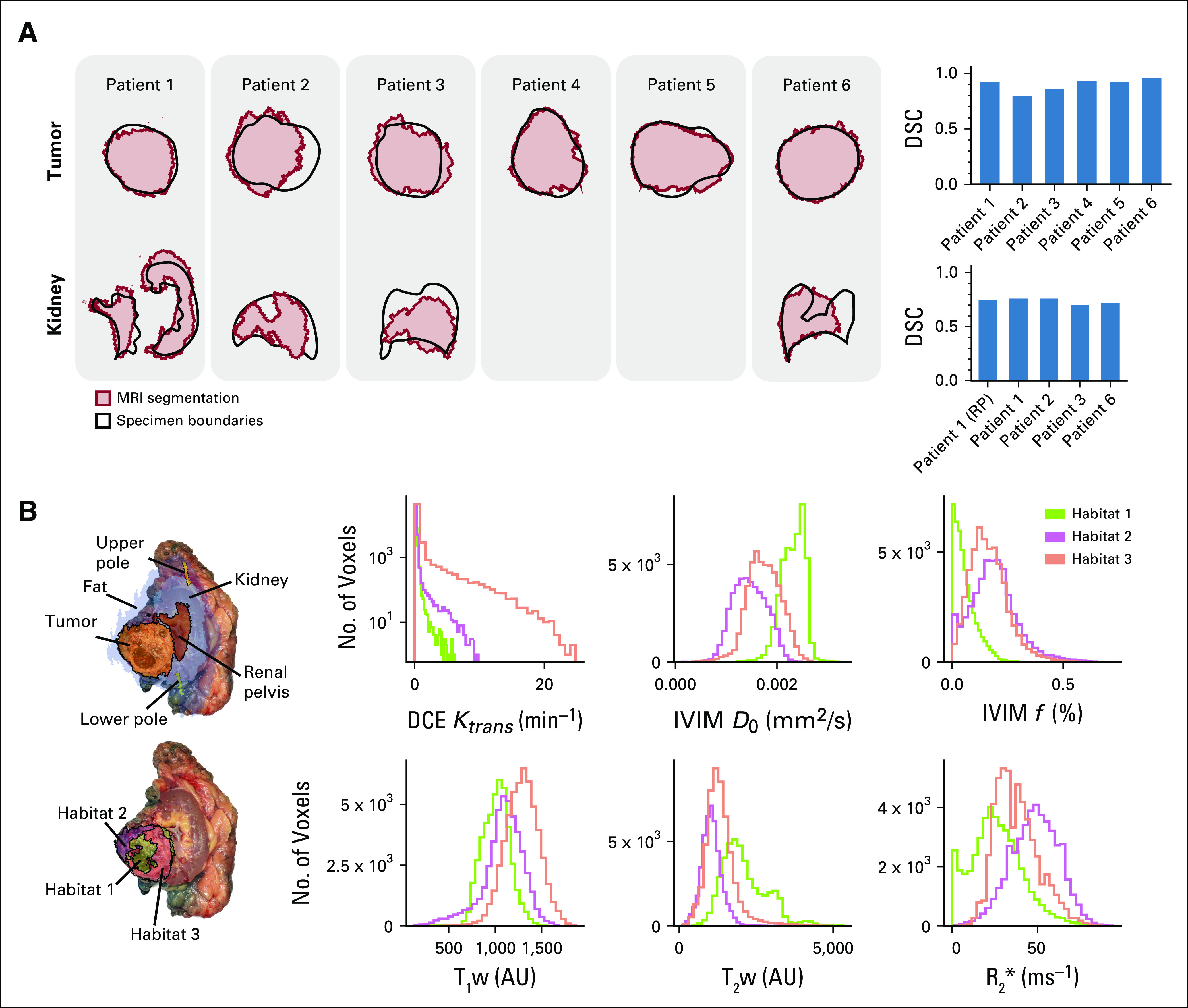
Results: We applied our framework to patients with renal cancer undergoing radical nephrectomy. We created personalized molds for 6 patients, obtaining Dice similarity coefficients between imaging and tissue sections ranging from 0.86 to 0.96 for tumor regions and between 0.70 and 0.76 for healthy kidneys. The framework required minimal manual intervention, producing the final mold design in just minutes, while automatically taking into account clinical considerations such as a preference for specific cutting planes.
Conclusion: Our work provides a robust and automated interface between imaging and tissue samples, enabling the development of clinical studies to probe tumor heterogeneity on multiple spatial scales.
Conflict of interest statement
Mireia Crispin-Ortuzar
Marcel Gehrung
Ferdia A. Gallagher
Andrew N. Priest
Anne Y. Warren
Grant D. Stewart
Evis Sala
Florian Markowetz
No other potential conflicts of interest were reported.
Figures
References
Publication types
MeSH terms
Grants and funding
- C19212/A911376/CRUK_/Cancer Research UK/United Kingdom
- 28290/CRUK_/Cancer Research UK/United Kingdom
- 19274/CRUK_/Cancer Research UK/United Kingdom
- C197/A16465/CRUK_/Cancer Research UK/United Kingdom
- 27176/CRUK_/Cancer Research UK/United Kingdom
- C9685/A25117/CRUK_/Cancer Research UK/United Kingdom
- WT_/Wellcome Trust/United Kingdom
- C19212/A27150/CRUK_/Cancer Research UK/United Kingdom
- 16628/CRUK_/Cancer Research UK/United Kingdom
- C8742/A18097/CRUK_/Cancer Research UK/United Kingdom
- C19212/A16628/CRUK_/Cancer Research UK/United Kingdom
- C14303/A17197/CRUK_/Cancer Research UK/United Kingdom
- 095962/WT_/Wellcome Trust/United Kingdom
- C14303/A19274/CRUK_/Cancer Research UK/United Kingdom
LinkOut - more resources
Full Text Sources
Medical