

Generation of Loa loa infective larvae by experimental infection of the vector, Chrysops silacea
- PMID: 32804951
- PMCID: PMC7470323
- DOI: 10.1371/journal.pntd.0008415
Generation of Loa loa infective larvae by experimental infection of the vector, Chrysops silacea
Abstract
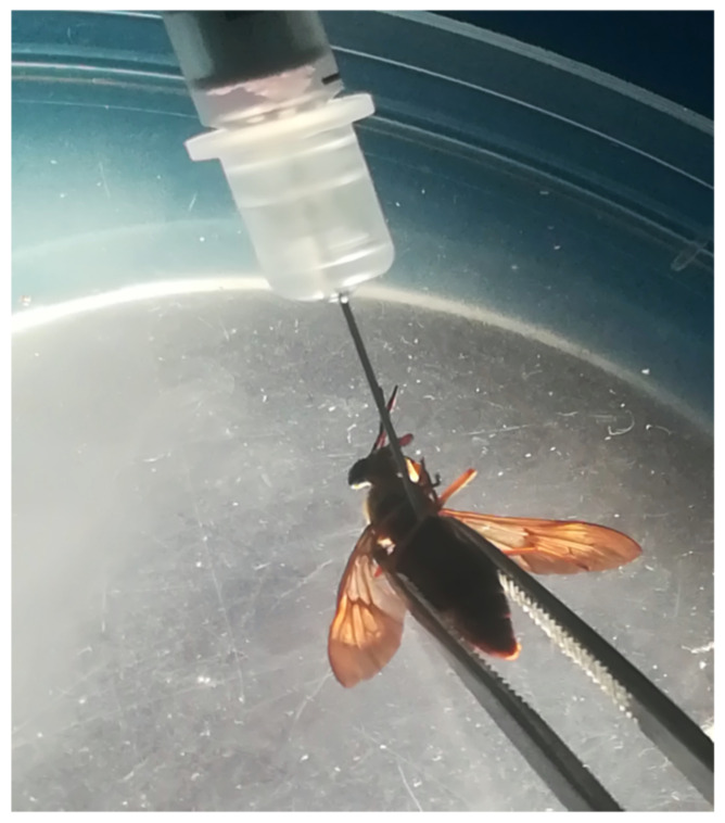
Basic and translational research on loiasis, a filarial nematode infection of medical importance, is impeded by a lack of suitable Loa loa infection models and techniques of obtaining and culturing life cycle stages. We describe the development of a new method for routine production of infective third-stage larvae (L3) of L. loa from the natural intermediate arthropod vector host, Chrysops silacea, following experimental infection with purified microfilariae. At 14-days post-infection of C. silacea, the fly survival rate was 43%. Survival was significantly higher in flies injected with 50 mf (55.2%) than those that received 100 mf (31.0%). However, yield per surviving fly and total yield of L3 was markedly higher in the group of flies inoculated with 100 mf (3474 vs 2462 L3 produced). The abdominal segment hosted the highest percentage recovery of L3 (47.7%) followed by head (34.5%) and thorax (17.9%). L. loa larval survival was higher than 90% after 30 days of in vitro culture. The in vitro moulting success rate to the L4 larval stage was 59.1%. After experimental infection of RAG2-/-IL-2γc-/-mice, the average L. loa juvenile adult worm recovery rate was 10.5% at 62 dpi. More than 87% of the worms were recovered from the muscles and subcutaneous tissues. Worms recovered measured an average 24.3 mm and 11.4 mm in length for females (n = 5) and males (n = 5), respectively. In conclusion, L. loa mf injected into C. silacea intrathoracically develop into infective larvae that remain viable and infective comparable to L3 obtained through natural feeding on the human host. This technique further advances the development of a full laboratory life cycle of L. loa where mf derived from experimentally-infected animals may be utilized to passage life cycle generations via intrathoracic injections of wild-caught vector hosts.
Conflict of interest statement
The authors have declared that no competing interests exist.
Figures
Similar articles
-
Comparison of immune responses to Loa loa stage-specific antigen extracts in Loa loa-exposed BALB/c mice upon clearance of infection.Parasit Vectors. 2020 Feb 7;13(1):51. doi: 10.1186/s13071-020-3921-x. Parasit Vectors. 2020. PMID: 32033624 Free PMC article.
-
Chrysops silacea biting densities and transmission potential in an endemic area of human loiasis in south-west Cameroon.Trop Med Int Health. 2002 Apr;7(4):371-7. doi: 10.1046/j.1365-3156.2002.00845.x. Trop Med Int Health. 2002. PMID: 11952954
-
Large scale collection of viable infective larvae of Loa loa.Trop Med Parasitol. 1995 Sep;46(3):203-4. Trop Med Parasitol. 1995. PMID: 8533026
-
Loa loa vectors Chrysops spp.: perspectives on research, distribution, bionomics, and implications for elimination of lymphatic filariasis and onchocerciasis.Parasit Vectors. 2017 Apr 5;10(1):172. doi: 10.1186/s13071-017-2103-y. Parasit Vectors. 2017. PMID: 28381279 Free PMC article. Review.
-
Loiasis: African eye worm.Trans R Soc Trop Med Hyg. 2008 Oct;102(10):983-9. doi: 10.1016/j.trstmh.2008.03.022. Epub 2008 May 7. Trans R Soc Trop Med Hyg. 2008. PMID: 18466939 Review.
Cited by
-
Biotransformation of anthelmintics in nematodes in relation to drug resistance.Int J Parasitol Drugs Drug Resist. 2025 Apr;27:100579. doi: 10.1016/j.ijpddr.2025.100579. Epub 2025 Jan 7. Int J Parasitol Drugs Drug Resist. 2025. PMID: 39827513 Free PMC article. Review.
-
Comparative development of human filariae Loa loa, Onchocerca volvulus and Mansonella perstans in immunocompromised mouse strains.Front Trop Dis. 2024 Apr 15;5:1293632. doi: 10.3389/fitd.2024.1293632. Front Trop Dis. 2024. PMID: 38655273 Free PMC article.
-
Advances in preclinical platforms of Loa loa for filarial neglected tropical disease drug and diagnostics research.Front Trop Dis. 2021 Nov 8;2:778724. doi: 10.3389/fitd.2021.778724. Front Trop Dis. 2021. PMID: 38654889 Free PMC article.
-
Direct Observation of Feeding Behavior of Adult Tabanidae (Diptera) on Beef Cattle from Khon Kaen Province in Thailand.Insects. 2024 Aug 10;15(8):602. doi: 10.3390/insects15080602. Insects. 2024. PMID: 39194807 Free PMC article.
-
Large-scale production of Mansonella perstans infective larvae from engorged Culicoides milnei.Front Trop Dis. 2024 Dec 10;5:fitd.2024.1391823. doi: 10.3389/fitd.2024.1391823. Front Trop Dis. 2024. PMID: 39811392 Free PMC article.
References
-
- Wanji S, Tendongfor N, Esum M, Enyong P. Chrysops silacea biting densities and transmission potential in an endemic area of human loiasis in South-West Cameroon. Trop Med Int Health. 2001;7:371–377. - PubMed
Publication types
MeSH terms
LinkOut - more resources
Full Text Sources
