. 2020 Aug 17;15(8):e0237704.
doi: 10.1371/journal.pone.0237704. eCollection 2020.

Bacterial biofilms colonizing plastics in estuarine waters, with an emphasis on Vibrio spp. and their antibacterial resistance

Affiliations

Bacterial biofilms colonizing plastics in estuarine waters, with an emphasis on Vibrio spp. and their antibacterial resistance

Amanda L Laverty et al. PLoS One. .

Abstract

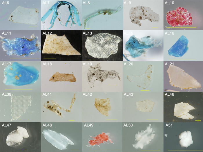
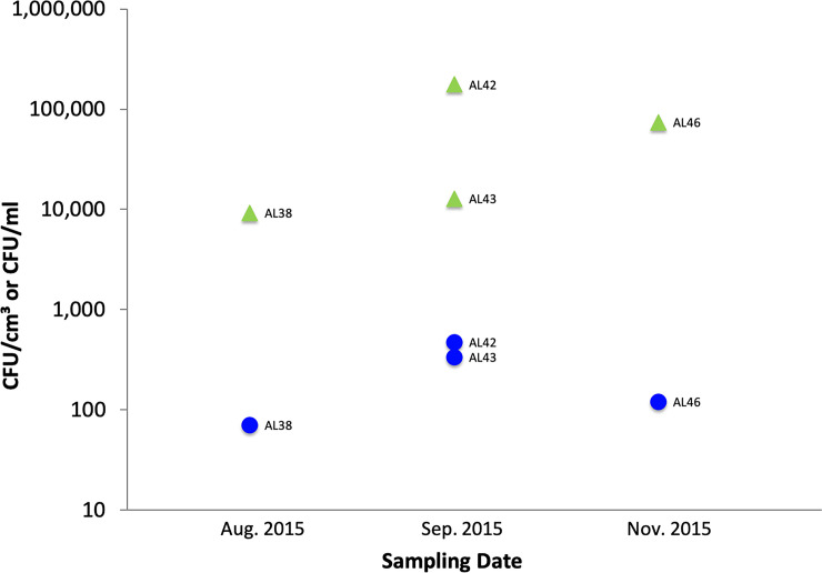
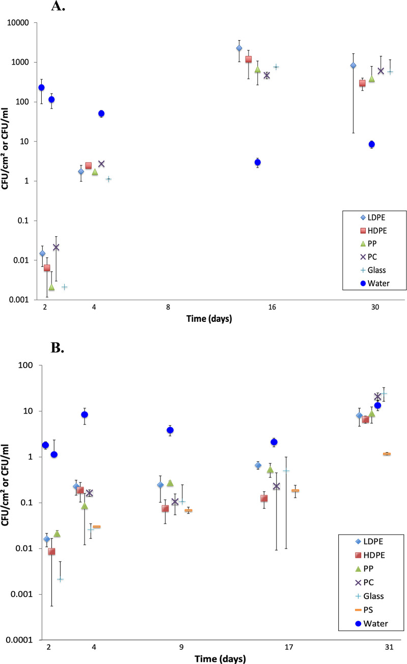
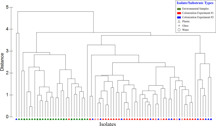
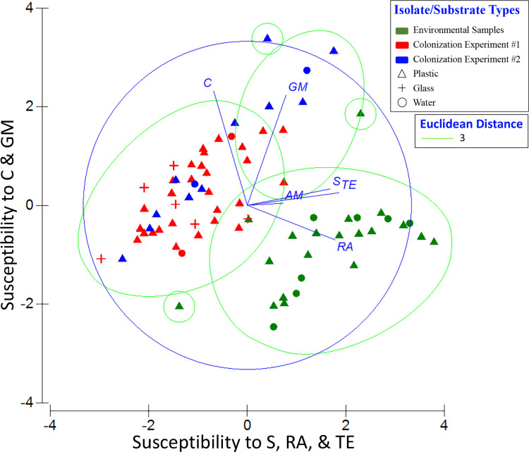
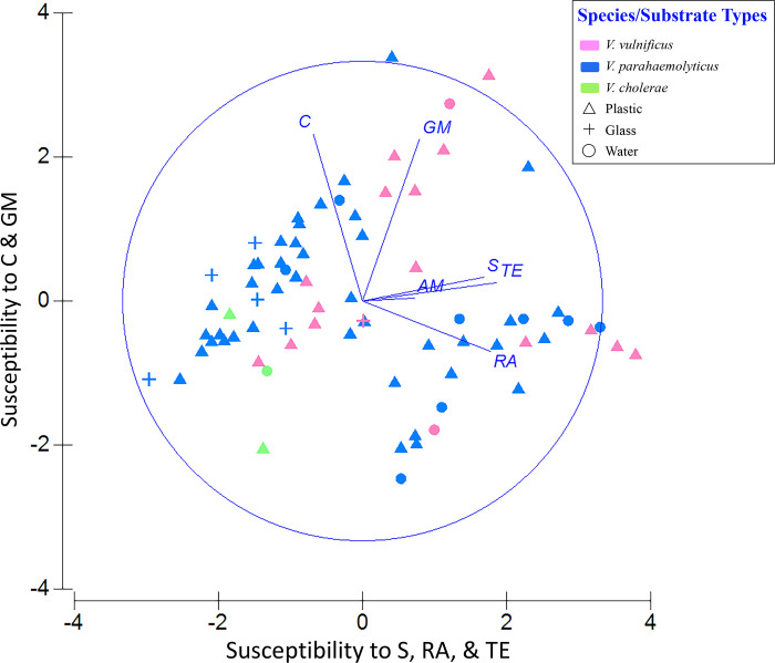
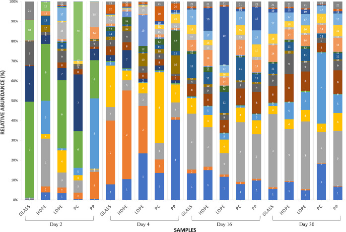
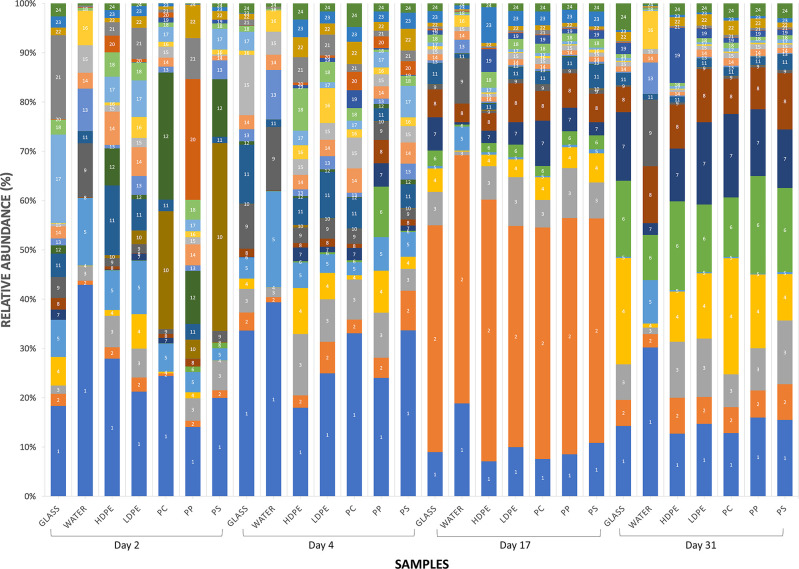
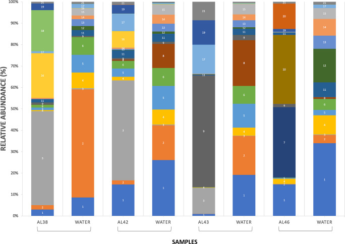
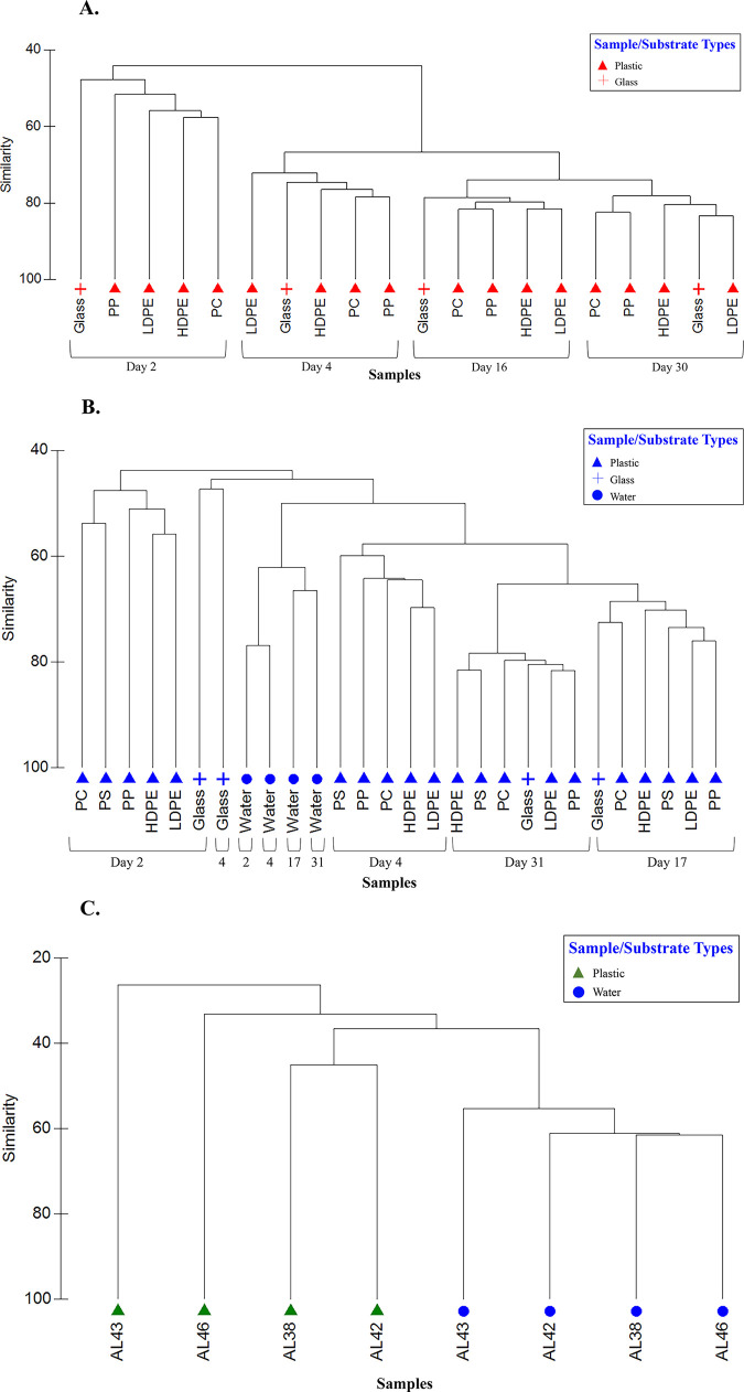
Since plastics degrade very slowly, they remain in the environment on much longer timescales than most natural organic substrates and provide a novel habitat for colonization by bacterial communities. The spectrum of relationships between plastics and bacteria, however, is little understood. The first objective of this study was to examine plastics as substrates for communities of Bacteria in estuarine surface waters. We used next-generation sequencing of the 16S rRNA gene to characterize communities from plastics collected in the field, and over the course of two colonization experiments, from biofilms that developed on plastic (low-density polyethylene, high-density polyethylene, polypropylene, polycarbonate, polystyrene) and glass substrates placed in the environment. Both field sampling and colonization experiments were conducted in estuarine tributaries of the lower Chesapeake Bay. As a second objective, we concomitantly analyzed biofilms on plastic substrates to ascertain the presence and abundance of Vibrio spp. bacteria, then isolated three human pathogens, V. cholerae, V. parahaemolyticus, and V. vulnificus, and determined their antibiotic-resistant profiles. In both components of this study, we compared our results with analyses conducted on paired samples of estuarine water. This research adds to a nascent literature that suggests environmental factors govern the development of bacterial communities on plastics, more so than the characteristics of the plastic substrates themselves. In addition, this study is the first to culture three pathogenic vibrios from plastics in estuaries, reinforcing and expanding upon earlier reports of plastic pollution as a habitat for Vibrio species. The antibiotic resistance detected among the isolates, coupled with the longevity of plastics in the aqueous environment, suggests biofilms on plastics have potential to persist and serve as focal points of potential pathogens and horizontal gene transfer.


Conflict of interest statement

The authors have declared that no competing interests exist.

Figures

Fig 1
Fig 1. Representative putative microplastics from environmental samples photographed with a stereo-zoom microscope/camera after their collection from surface waters of the Elizabeth River at Old Dominion University’s Sailing Center.
Samples were collected from June through November 2015. Most pieces were distinctly biofouled. Bars represent one millimeter.
Fig 2
Fig 2. Concentrations of putative Vibrio spp. on PE (environmental samples; green triangles) and in paired water samples (filled blue circles).
Fig 3
Fig 3
A. Colonization Experiment #1 (12 Oct– 10 Nov 2015): Mean (n = 3, ± 1 sd) concentrations of putative Vibrio spp. from biofilms on plastics and glass, and from paired water samples. Water temperature ranged from 15–22°C. For clarity, points are jittered about the day of sampling. Values for day 1 all were zero and are not displayed. Substrate types: low-density polyethylene (LDPE), high-density polyethylene (HDPE), polypropylene (PP), polycarbonate (PC), and glass (Glass). B. Colonization Experiment #2 (12 Jan– 10 Feb 2016): Mean (+ 1 sd) concentrations of putative Vibrio spp. from biofilms on plastics and glass, and from paired water samples. Water temperature ranged from 2.1–9.5°C. Note the decreased range of the Y-axis relative to Fig 3A. For clarity, points are jittered about the day of sampling. Values for day 1 all were zero and are not displayed. Polystyrene (PS) was added to the list of substrates referenced in Fig 3A.
Fig 4
Fig 4. Hierarchical clustering dendrogram showing isolates’ relationship based on their antibiotic susceptibility profiles.
ZOI data for each isolate was compared with that of all other isolates using Euclidean distance similarity, then clustered using a group average algorithm. Red symbols, Colonization Experiment #1; blue symbols, Colonization Experiment #2; green symbols, environmental samples. Plastic substrates (all types), triangles; glass substrate; +; water, filled circles.
Fig 5
Fig 5. Principal components analysis (PCA) of antibiotic profiles of Vibrio spp. (n = 97) by sample type.
PC1 represents increasing susceptibility to streptomycin (S), rifampin (RA), and tetracycline (TE), and PC2 represents increasing susceptibility to chloramphenicol (C) and gentamicin (GM). Eigenvectors for each antibiotic are shown as lines adjacent to the corresponding labels. Symbols as in Fig 4. The PCA is overlain with Euclidean distance (value of 3) from the cluster analysis (Fig 4).
Fig 6
Fig 6. Principal components analysis (PCA) of antibiotic profiles identifying the 97 Vibrio isolates.
PC1 and PC2 as in Fig 5. Eigenvectors for each antibiotic are shown as lines. Isolates are color coded: magenta, V. vulnificus; blue, V. parahaemolyticus; green, V. cholerae.
Fig 7
Fig 7. Relative abundances of sequenced Bacteria in Colonization Experiment #1.
Substrate types: low-density polyethylene (LDPE), high-density polyethylene (HDPE), polypropylene (PP), polycarbonate (PC), and glass (G). Paired water samples were not collected. The most abundant OTUs are listed as follows: (1) Bacteria, Bacteria Unclassified; (2) Bacteria, Proteobacteria, Gammaproteobacteria, Pseudomonadales, Pseudomonadaceae, Pseudomonas; (3) Bacteria, Proteobacteria, Gammaproteobacteria, Oceanospirillales, Oceanospirillaceae; (4) Bacteria, Bacteroidetes, Flavobacteria, Flavobacteriales, Flavobacteriaceae, Tenacibaculum; (5) Bacteria, Proteobacteria, Gammaproteobacteria, Pseudomonadales, Pseudomonadaceae, Pseudomonas; (6) Bacteria, Bacteroidetes, Flavobacteria, Flavobacteriales, Flavobacteriaceae, Maribacter; (7) Bacteria, Verrucomicrobia, Verrucomicrobiae, Verrucomicrobiales, Rubritaleaceae, Rubritalea; (8) Bacteria, Bacteroidetes, Flavobacteria, Flavobacteriales, Flavobacteriaceae, Flavobacterium; (9) Bacteria, Proteobacteria, Gammaproteobacteria, Alteromonadales, Pseudoalteromonadaceae, Pseudoalteromonas; (10) Bacteria, Proteobacteria, Gammaproteobacteria, Oceanospirillales, Oceanospirillaceae, Oleibacter; (11) Bacteria, Actinobacteria, Actinobacteria, Actinomycetales, Corynebacteriaceae, Corynebacterium; (12) Bacteria, Proteobacteria, Alphaproteobacteria, Rhodobacterales, Rhodobacteraceae; (13) Bacteria, Bacteroidetes, Flavobacteria, Flavobacteriales, Flavobacteriaceae; (14) Bacteria, Proteobacteria, Gammaproteobacteria, Alteromonadales, Alteromonadaceae, Glaciecola; (15) Bacteria, Bacteria Unclassified; (16) Bacteria, Proteobacteria, Gammaproteobacteria, Vibrionales, Vibrionaceae, Vibrio; (17) Bacteria, Proteobacteria, Alphaproteobacteria, Sphingomonadales, Sphingomonadaceae, Sphingomonas; (18) Bacteria, Bacteroidetes, Flavobacteria, Flavobacteriales, Flavobacteriaceae, Polaribacter; (19) Bacteria, Proteobacteria, Alphaproteobacteria, Sphingomonadales, Erythrobacteraceae, Erythrobacter; (20) Bacteria, Proteobacteria, Alphaproteobacteria, Rhodobacterales, Rhodobacteraceae; (21) Bacteria, Proteobacteria, Alphaproteobacteria, Rhodobacterales, Rhodobacteraceae.
Fig 8
Fig 8. Relative abundances of sequenced Bacteria in Colonization Experiment #2.
As in Fig 7, with the addition of polystyrene (PS) as a substrate and paired water samples. The most abundant OTUs are listed as follows: (1) Bacteria, Proteobacteria, Gammaproteobacteria, Oceanospirillales, Oceanospirillaceae; (2) Bacteria, Bacteria Unclassified; (3) Bacteria, Proteobacteria, Alphaproteobacteria, Sphingomonadales, Sphingomonadaceae, Sphingomonas; (4) Bacteria, Bacteria Unclassified; (5) Bacteria,Verrucomicrobia, Verrucomicrobiae, Verrucomicrobiales, Verrucomicrobiaceae, Luteolibacter; (6) Bacteria, Bacteroidetes, Flavobacteria, Flavobacteriales, Flavobacteriaceae, Flavobacterium, (7) Bacteria, Bacteroidetes, Flavobacteria, Flavobacteriales, Flavobacteriaceae, (8) Bacteria, Proteobacteria, Betaproteobacteria, Burkholderiales, Comamonadaceae, Delflia, (9) Bacteria, Proteobacteria, Alphaproteobacteria, Rhodobacterales, Rhodobacteraceae, Loktanella; (10) Bacteria, Proteobacteria, Gammaproteobacteria, Oceanospirillales, Oceanospirillaceae; (11) Bacteria, Bacteroidetes, Flavobacteria, Flavobacteriales, Flavobacteriaceae, Tenacibaculum, (12) Bacteria, Bacteroidetes, Flavobacteria, Flavobacteriales, Flavobacteriaceae, Polaribacter; (13) Bacteria, Proteobacteria, Betaproteobacteria, Burkholderiales, Comamonadaceae; (14) Bacteria, Bacteroidetes, Flavobacteria, Flavobacteriales, Flavobacteriaceae, Flavobacterium; (15) Bacteria, Proteobacteria, Gammaproteobacteria, Alteromonadales, Pseudoalteromonadaceae, Pseudoalteromonas, (16) Bacteria, Proteobacteria, Gammaproteobacteria, Pseudomonadales, Pseudomonadaceae, Pseudomonas; (17) Bacteria, Bacteroidetes, Flavobacteria, Flavobacteriales, Flavobacteriaceae, Maribacter; (18) Bacteria, Bacteria Unclassified; 19) Bacteria, Proteobacteria, Alphaproteobacteria, Rhodobacterales, Rhodobacteraceae; (20) Bacteria, Proteobacteria, Gammaproteobacteria, Vibrionales, Vibrionaceae, Vibrio; (21) Bacteria, Proteobacteria, Gammaproteobacteria, Alteromonadales, Alteromonadaceae, Glaciecola; (22) Bacteria, Actinobacteria, Actinobacteria, Actinomycetales, Corynebacteriaceae, Corynebacterium, (23) Bacteria, Bacteroidetes, Flavobacteria, Flavobacteriales, Flavobacteriaceae, Maribacter; (24) Bacteria, Proteobacteria, Alphaproteobacteria, Rhodobacterales, Rhodobacteraceae.
Fig 9
Fig 9. Relative abundances of sequenced Bacteria on polyethylene microplastics (ATR-FTIR confirmed) and paired water samples.
Sample number is indicated as AL##. The most abundant OTUs are listed as follows: (1) Bacteria, Proteobacteria, Gammaproteobacteria, Oceanospirillales, Oceanospirillaceae; (2) Bacteria, Proteobacteria, Gammaproteobacteria, Alteromonadales, Alteromonadaceae, Glaciecola; (3) Bacteria, Proteobacteria, Gammaproteobacteria, Pseudomonadales, Pseudomonadaceae, Pseudomonas; (4) Bacteria, Proteobacteria, Gammaproteobacteria, Alteromonadales, Pseudoalteromonadaceae, Pseudoalteromonas; (5) Bacteria, Bacteroidetes, Flavobacteria, Flavobacteriales, Cryomorphaceae, Lishizhenia; (6) Bacteria, Proteobacteria, Gammaproteobacteria, Alteromonadales, Alteromonadaceae, Glaciecola; (7) Bacteria, Proteobacteria, Alphaproteobacteria, Rhodobacterales, Rhodobacteraceae, Pseudoruegeria; (8) Bacteria, Proteobacteria, Gammaproteobacteria, Vibrionales, Vibrionaceae, Vibrio; (9) Bacteria, Bacteroidetes, Flavobacteria, Flavobacteriales, Flavobacteriaceae, Maribacter; (10) Bacteria, Verrucomicrobia, Verrucomicrobiae, Verrucomicrobiales, Rubritaleaceae, Rubritalea; (11) Bacteria, Bacteroidetes, Flavobacteria, Flavobacteriales, Flavobacteriaceae, Flavobacterium; (12) Bacteria, Proteobacteria, Gammaproteobacteria, Pseudomonadales, Pseudomonadaceae, Pseudomonas; (13) Bacteria, Proteobacteria, Betaproteobacteria, Betaproteobacteria Unclassified; (14) Bacteria, Proteobacteria, Betaproteobacteria, Burkholderiales, Comamonadaceae; (15) Bacteria, Verrucomicrobia, Verrucomicrobiae, Verrucomicrobiales, Verrucomicrobiaceae, Luteolibacter; (16) Bacteria, Proteobacteria, Alphaproteobacteria, Rhodobacterales, Rhodobacteraceae; (17) Bacteria, Bacteroidetes, Flavobacteria, Flavobacteriales, Flavobacteriaceae, Tenacibaculum; (18) Bacteria, Bacteroidetes, Flavobacteria, Flavobacteriales, Flavobacteriaceae, Krokinobacter; (19) Bacteria, Bacteria Unclassified; (20) Bacteria, Actinobacteria, Actinobacteria, Acidimicrobiales, Acidimicrobiaceae, Ilumatobacter; (21) Bacteria, Proteobacteria, Alphaproteobacteria, Rhodobacterales, Rhodobacteraceae.
Fig 10
Fig 10. Hierarchical clustering dendrogram of bacterial communities showing sample relationship based on sequence data.
Abundance data (Log (x+1)) was compared between samples using Bray-Curtis similarity, then clustered using a group-average algorithm. A) Colonization Experiment #1; B) Colonization Experiment #2; C) environmental samples (polyethylene microplastics and paired water samples). Substrate types: low-density polyethylene (LDPE), high-density polyethylene (HDPE), polypropylene (PP), polycarbonate (PC), glass (Glass), and polystyrene (PS).

References

    1. Derraik JG. The pollution of the marine environment by plastic debris: a review. Marine pollution bulletin. 2002. September 1;44(9):842–52. 10.1016/s0025-326x(02)00220-5 - DOI - PubMed
    1. Lavers JL, Bond AL. Exceptional and rapid accumulation of anthropogenic debris on one of the world’s most remote and pristine islands. Proceedings of the National Academy of Sciences. 2017. June 6;114(23):6052–5. - PMC - PubMed
    1. Eriksen M, Lebreton LC, Carson HS, Thiel M, Moore CJ, Borerro JC, et al. Plastic pollution in the world's oceans: more than 5 trillion plastic pieces weighing over 250,000 tons afloat at sea. PloS one. 2014. December 10;9(12):e111913 10.1371/journal.pone.0111913 - DOI - PMC - PubMed
    1. Obbard RW, Sadri S, Wong YQ, Khitun AA, Baker I, Thompson RC. Global warming releases microplastic legacy frozen in Arctic Sea ice. Earth’s Future 2: 315–320.
    1. Kelly A, Lannuzel D, Rodemann T, Meiners KM, Auman HJ. Microplastic contamination in east Antarctic sea ice. Marine Pollution Bulletin. 2020. May 1;154:111130 10.1016/j.marpolbul.2020.111130 - DOI - PubMed

Publication types