. 2020 Sep;12(3):e84.
doi: 10.1002/cpch.84.

In Vitro Activity Assays to Quantitatively Assess the Endogenous Reversible Oxidation State of Protein Tyrosine Phosphatases in Cells

Affiliations

In Vitro Activity Assays to Quantitatively Assess the Endogenous Reversible Oxidation State of Protein Tyrosine Phosphatases in Cells

Avinash D Londhe et al. Curr Protoc Chem Biol. 2020 Sep.

Abstract

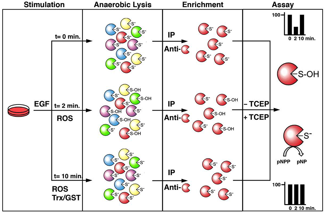
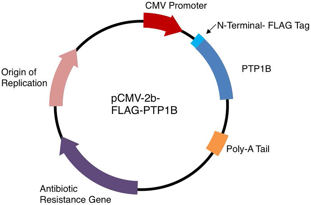
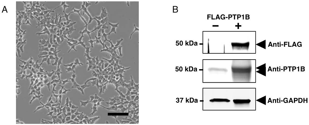
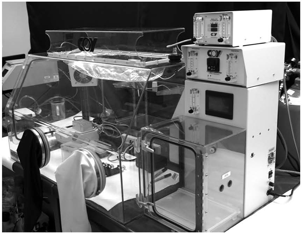
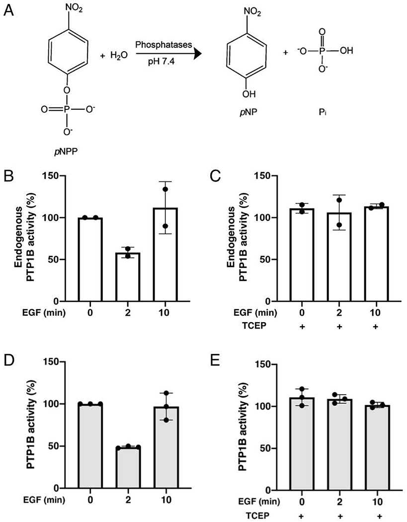
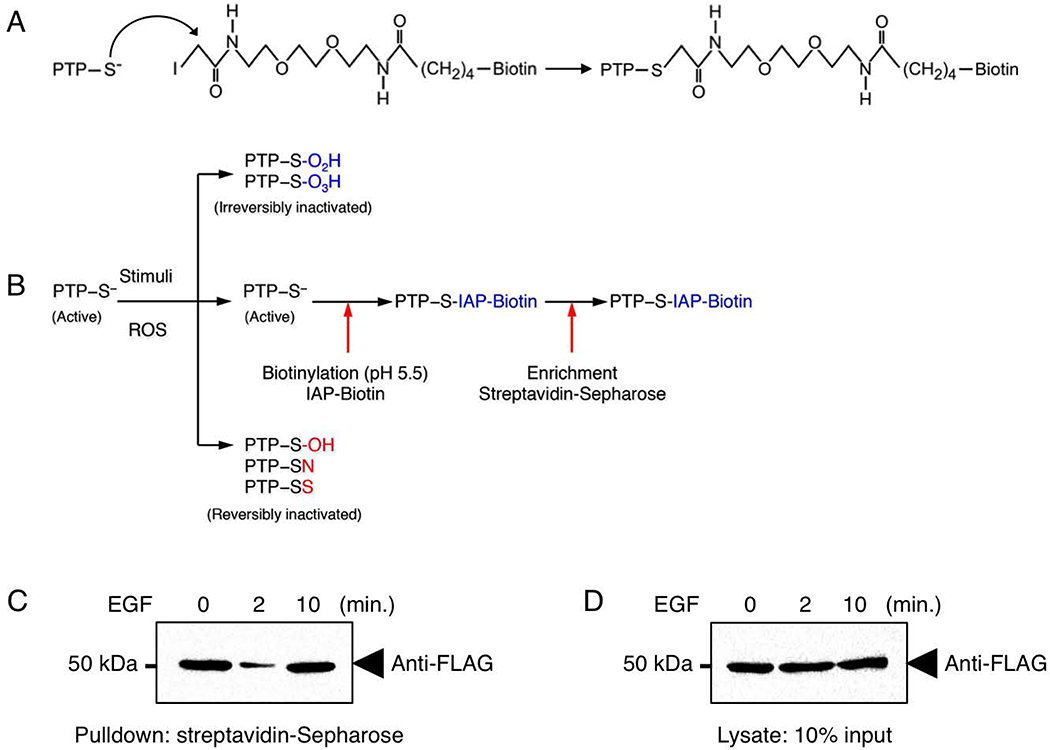
The reversible oxidation of protein tyrosine phosphatases (PTPs) impairs their ability to dephosphorylate substrates in vivo. This transient inactivation of PTPs occurs as their conserved catalytic cysteine residue reacts with cellular oxidants thereby abolishing the ability of this reactive cysteine to attack the phosphate of the target substrate. Hence, in vivo, the inhibition of specific PTPs in response to regulated and localized rises in cellular oxidants enables phospho-dependent signaling. We present assays that measure the endogenous activity of specific PTPs that become transiently inactivated in cells exposed to growth factors. Here, we describe the methods and highlight the pitfalls to avoid post-lysis oxidation of PTPs in order to assess the inactivation and the reactivation of PTPs targeted by cellular oxidants in signal transduction. © 2020 Wiley Periodicals LLC. Basic Protocol 1: Cell transfection (optional) Support Protocol: Preparation of degassed lysis buffers Basic Protocol 2: Cellular extraction in anaerobic conditions Basic Protocol 3: Enrichment and activity assay of specific PTPs Alternate Protocol: Measurement of active PTPs via direct cysteinyl labeling.

Keywords: activity assay; biotin labeling; pNPP; protein tyrosine phosphatases; redox signaling.


Figures

Figure 1.
Figure 1.. Schematic outline of the endogenous PTP-activity assay.
In resting cells, active PTPs are reduced and their invariable catalytic cysteinyl residue exists as a thiolate ion. This results from the unique structure and conserved residues of the PTP fold at the active site of this family of enzymes, which lowers the pKa of this specific cysteinyl residue. The nucleophilic nature of this thiolate ion makes it reactive towards phospho-amino acid residues and reactive oxygen species (ROS). Following cell exposure to a ROS-promoting stimulus such as epidermal growth factor (EGF) in the first step, specific PTPs become inactivated by reversible oxidation. In the second step of the assay, cellular lysis is performed with a degassed buffer in a hypoxic glove-box equilibrated with argon gas. This lysis in an anaerobic environment in absence of reducing agents minimizes post-lysis oxidation and maintains the reduced or oxidized state of PTPs. The third step consists of the enrichment of a PTP of interest by immunoprecipitation using degassed buffers that maintain the samples in a non-oxidizing environment. The final step entails an immune-complex activity assay of the purified PTPs using a synthetic chromogenic substrate, para-nitrophenylphosphate (pNPP). Conveniently, the dephosphorylated product of pNPP, para-nytrophenol (pNP), is a yellow compound that can be quantified using a spectrophotometer. As a positive control, one set of immunoprecipitated samples are reduced with TCEP to reactivate reversibly oxidized PTPs in order to measure the total PTP activity of the immune-complex. By comparing the endogenous activity of PTPs with control experimental conditions that activate all PTPs, this approach allows one to assess the reactivation of specific PTPs that become reversibly oxidized in cells exposed to growth factors.
Figure 2.
Figure 2.. Plasmid used in these protocols for expression of FLAG-tag PTP1B.
pCMV-2B is a mammalian expression vector for tagging proteins with an N-terminal FLAG epitope. A CMV promoter allows for elevated protein expression in HEK293T cells. The pCMV-2B-FLAG-PTP1B vector was a generous gift from Dr. Tonks (Cold Spring Harbor Laboratory).
Figure 3.
Figure 3.
A) Phase-contrast image of HEK293T cells. Subconfluent (~50%) HEK293T cells 24 hours following trypsinization and passage (1:5). Bar: 100 μm B) Detection of FLAG-PTP1B expression in HEK293T cells. 50% confluent HEK293T cells in Growth Medium were transfected with 4 μg of pCMV-2B-FLAG-PTP1B and incubated for ~ 48 hours in a cell incubator. Cells were then lysed with lysis buffer 1, and 10 μg lysate was separated by SDS-PAGE and electrophoretically transferred onto nitrocellulose membranes. Membranes were blotted using FLAG-HRP, PTP1B (FG6) or GAPDH antibodies and the proteins were detected with HRP-conjugated secondary antibodies and visualized by ECL. Overexpression of FLAG-PTP1B was observed following transfection of HEK293T cells. The overexpressed protein was visible as a sharp immunoreactive band above the endogenous PTP1B protein at 50 kDa. GAPDH immunodetection is indicative of equal loading.
Figure 4.
Figure 4.
The hypoxic glove-box used to generate cell lysate in an anaerobic environment.
Figure 5.
Figure 5.. Quantitative analysis of the endogenous activity of PTP1B from immune-complexes.
A) Dephosphorylation of para-nitrophenylphosphate (pNPP) into para-nitrophenol (A405) and inorganic phosphate. B) PTP1B inactivation was studied using the endogenous PTP-activity assay. Serum-deprived HEK293T cells stimulated with EGF (100 ng/ml) for the indicated times were lysed using degassed Lysis Buffer 2 in anaerobic conditions. Endogenous PTP1B was immunoprecipitated from lysates using 3 μg FG6 anti-PTP1B antibody coupled to Protein A/G beads. Endogenous activity of PTP1B was measured in absence of reducing agents, using pNPP as substrate. The depshophorylated product, pNP, was measured after 40 minutes at A405 using a spectrophotometer. C) In a duplicate experiment, endogenous PTP1B was immunoprecipitated from lysates from A and subjected to a pNPP activity assay in the presence of 5 mM TCEP to measure total PTP1B activity in these samples. The average readout value of technical replicates is represented by the dot-plot bar graph. Each experiment was performed two independent times. D) Serum-deprived HEK293T cells stimulated with EGF (100 ng/ml) for the indicated times were lysed using degassed Lysis Buffer 2 in anaerobic conditions. FLAG-PTP1B were purified from lysates using anti-FLAG magnetic beads. Endogenous activity of PTP1B was measured in absence of reducing agents, using pNPP as substrate. The depshophorylated product, pNP, was measured after 20 minutes at A405 using a spectrophotometer. E) In a duplicate experiment, FLAG-PTP1B was purified from lysates from A and subjected to a pNPP activity assay in the presence of 5 mM TCEP to measure total PTP1B activity in these samples. The average readout value of technical replicates is represented by the dot-plot bar graph. Each experiment was performed three independent times.
Figure 6.
Figure 6.. Direct Cysteinyl Labeling of active PTP1B in HEK293T cell lysates.
A) Mechanism of PTP labeling by the IAP probe. B) Schematic outline of the direct cysteinyl labeling assay. In resting cells, active PTPs are reduced and their invariable catalytic cysteinyl residue exists as a thiolate ion. The nucleophilic nature of this thiolate ion makes it reactive towards phospho-amino acid residues, reactive oxygen species (ROS) or certain sulhydryl-reactive compounds. Following cell exposure to a ROS-promoting stimulus in the first step, specific PTPs become inactivated by reversible oxidation. Following a ROS-promoting stimulus, specific PTPs are inactivated by oxidation. Reversible oxidation [sulfenic acids (SOH), cyclic sulfenamides (SN) and intramolecular disulfides (SS)] are depicted in red, whereas irreversible oxidation [sulfenic acid (SO2H) and sulfinic acid (SO3H)] are depicted in blue. In the second step of the direct cysteinyl labeling assay, cellular lysis is performed with a degassed buffer at pH 5.5 containing a biotinylated sulfhydryl-reactive probe [iodoacetyl-polyethylene oxide (IAP)]. This step of the assay is performed in a hypoxic glove-box equilibrated with argon gas. This lysis in an anaerobic environment in absence of reducing agents minimizes post-lysis oxidation and maintains the endogenous redox state of PTPs. PTPs that remained in their active state following cell stimulation react and become biotinylated by IAP, whereas oxidized PTPs are protected from this biotinylation step. In the last step of the assay, biotinylated PTPs are purified by streptavidin pulldown and visualized by immunoblot. As a positive control, lysates are used to measure the total level of PTPs. By comparing the decreased biotinylation of PTPs with the total level of PTPs present in the lysate, this approach allows one to assess the reactivation of specific PTPs that become reversibly oxidized in cells exposed to growth factors. C) PTP1B inactivation was studied using a direct cysteinyl labeling approach. Serum-deprived HEK293T cells stimulated with EGF (100 ng/ml) for the indicated times were lysed at pH 5.5 in the presence of IAP-biotin. Biotin-labeled proteins were purified from lysates using streptavidin-Sepharose beads. Proteins were separated by SDS-PAGE, transferred onto nitrocellulose membranes and probed for FLAG-PTP1B. D) Lysates from C were probed for FLAG-PTP1B to control for protein expression.

References

    1. Barrett WC, DeGnore JP, Konig S, Fales HM, Keng YF, Zhang ZY, … Chock PB (1999). Regulation of PTP1B via glutathionylation of the active site cysteine 215. Biochemistry, 38(20), 6699–6705. - PubMed
    1. Boivin B, Chaudhary F, Dickinson BC, Haque A, Pero SC, Chang CJ, & Tonks NK (2013). Receptor Protein-tyrosine Phosphatase α Regulates Focal Adhesion Kinase Phosphorylation and ErbB2 Oncoprotein-mediated Mammary Epithelial Cell Motility. J Biol Chem, 288(52), 36926–36935. - PMC - PubMed
    1. Boivin B, & Tonks NK (2010). Analysis of the redox regulation of protein tyrosine phosphatase superfamily members utilizing a cysteinyl-labeling assay. Methods Enzymol, 474, 35–50. - PubMed
    1. Boivin B, Yang M, & Tonks NK (2010). Targeting the reversibly oxidized protein tyrosine phosphatase superfamily. Sci Signal, 3(137), pl2. - PMC - PubMed
    1. Boivin B, Zhang S, Arbiser JL, Zhang ZY, & Tonks NK (2008). A modified cysteinyl-labeling assay reveals reversible oxidation of protein tyrosine phosphatases in angiomyolipoma cells. Proc Natl Acad Sci U S A , 105(29), 9959–9964. - PMC - PubMed

Publication types

MeSH terms

Substances

LinkOut - more resources