Comparative Study
. 2020 Aug 12;12(8):882.
doi: 10.3390/v12080882.

Modelling West Nile Virus and Usutu Virus Pathogenicity in Human Neural Stem Cells

Affiliations
Comparative Study

Modelling West Nile Virus and Usutu Virus Pathogenicity in Human Neural Stem Cells

Silvia Riccetti et al. Viruses. .

Abstract

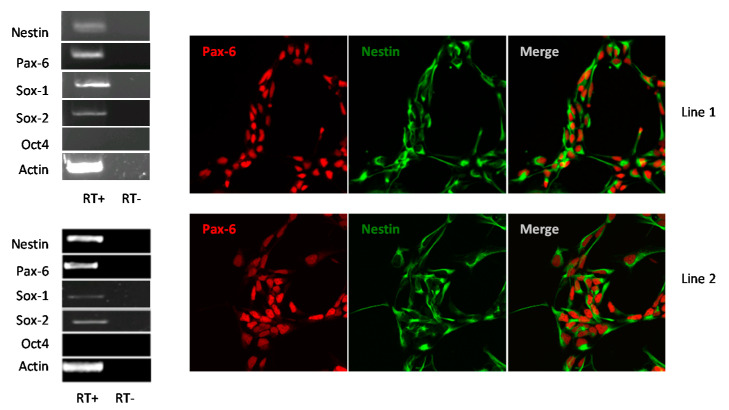
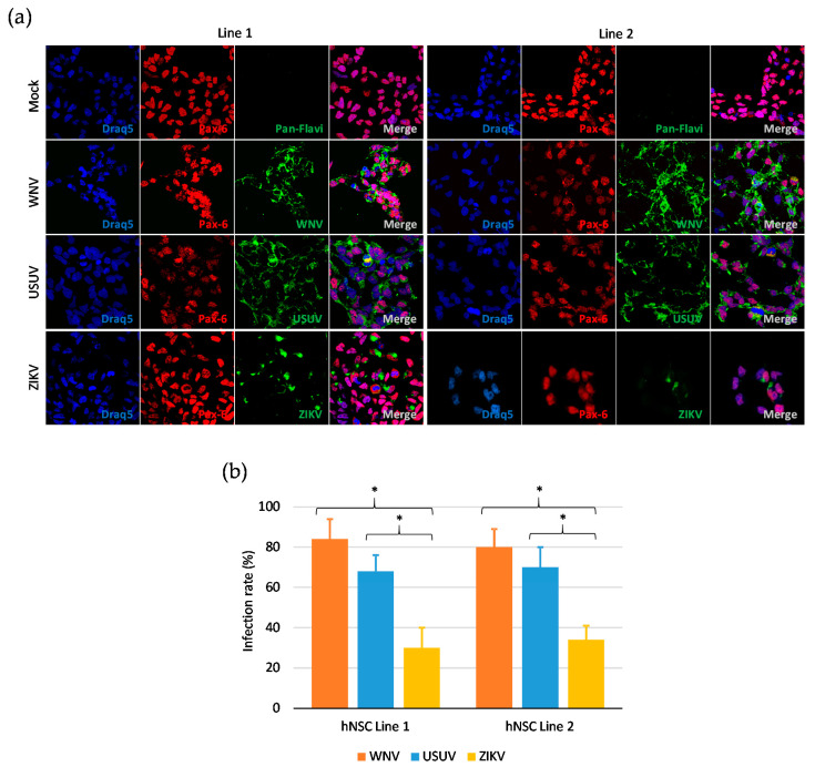
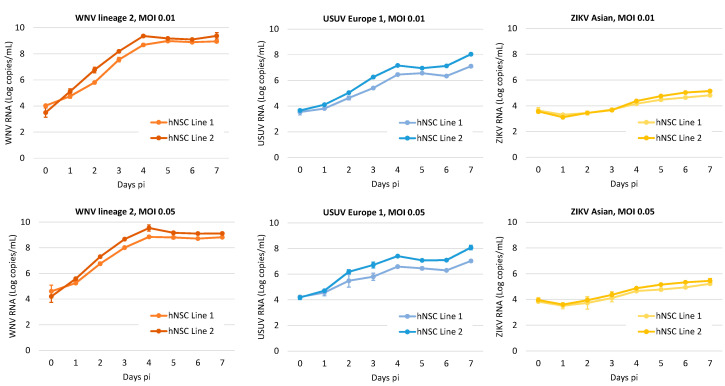
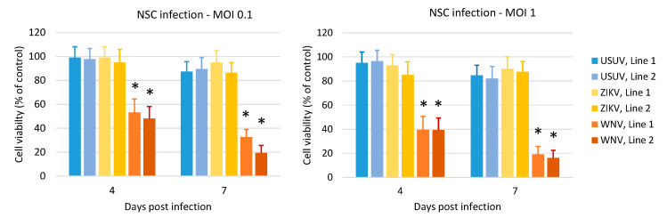
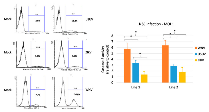
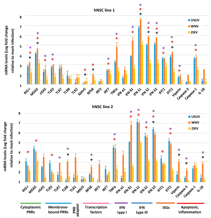
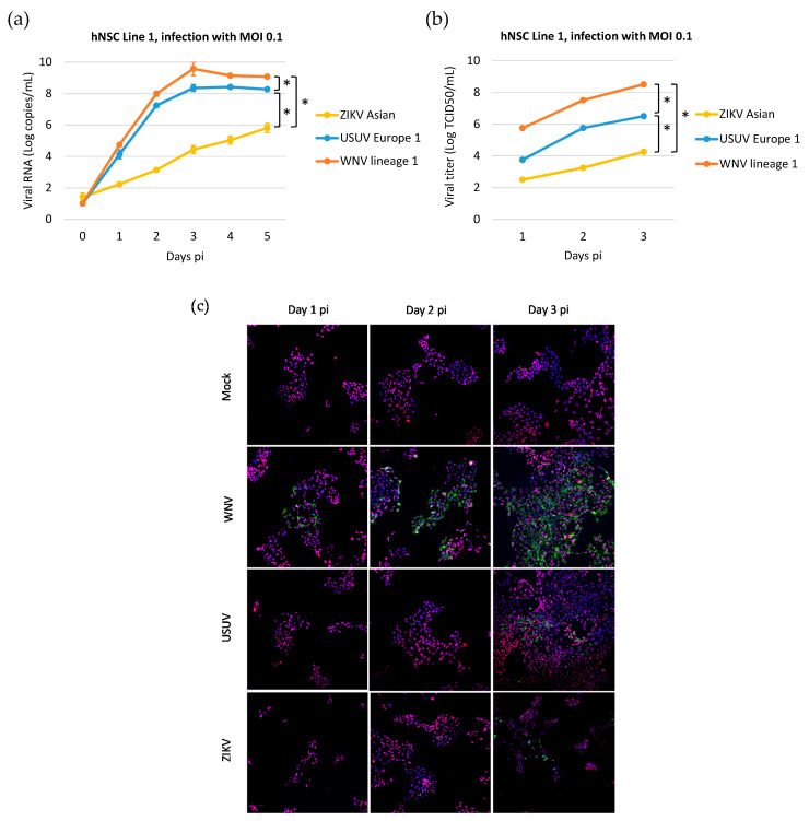
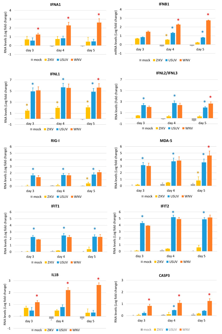
West Nile virus (WNV) and Usutu virus (USUV) are genetically related neurotropic mosquito-borne flaviviruses, which frequently co-circulate in nature. Despite USUV seeming to be less pathogenic for humans than WNV, the clinical manifestations induced by these two viruses often overlap and may evolve to produce severe neurological complications. The aim of this study was to investigate the effects of WNV and USUV infection on human induced pluripotent stem cell-derived neural stem cells (hNSCs), as a model of the neural progenitor cells in the developing fetal brain and in adult brain. Zika virus (ZIKV), a flavivirus with known tropism for NSCs, was used as the positive control. Infection of hNSCs and viral production, effects on cell viability, apoptosis, and innate antiviral responses were compared among viruses. WNV displayed the highest replication efficiency and cytopathic effects in hNSCs, followed by USUV and then ZIKV. In these cells, both WNV and USUV induced the overexpression of innate antiviral response genes at significantly higher levels than ZIKV. Expression of interferon type I, interleukin-1β and caspase-3 was significantly more elevated in WNV- than USUV-infected hNSCs, in agreement with the higher neuropathogenicity of WNV and the ability to inhibit the interferon response pathway.

Keywords: Usutu virus; West Nile virus; Zika virus; apoptosis; flavivirus; human induced pluripotent stem cells; inflammasome; innate antiviral response; neural stem cells; virus replication.


Conflict of interest statement

The authors declare no conflict of interest. The funders had no role in the design of the study; in the collection, analyses, or interpretation of data; in the writing of the manuscript, or in the decision to publish the results.

Figures

Figure 1
Figure 1
Characterization of induced pluripotent stem cell (hiPSC)s-derived neural stem cells (NSCs) lines 1 and 2: RT-PCR analysis of NSCs markers PAX6, NESTIN, SOX1, SOX2, the negative control OCT4 and the housekeeping gene ACTIN; confocal imaging of NSCs immunolabeled for the neural progenitor markers Pax6 and Nestin. NSC Lines 1 and 2 are shown at 60× magnification. RT: Reverse transcriptase.
Figure 2
Figure 2
Flavivirus infection of human NSCs. (a) Confocal microscopy of NSCs of line 1 and line 2 at 48 hpi with West Nile virus (WNV) lineage 2, Usutu virus (USUV) Europe 1 and Zika virus (ZIKV) Asian lineage at multiplicity of infection (MOI) 1. NSCs were immunolabeled with a pan-flavivirus antibody targeting flavivirus E glycoprotein and with an antibody targeting the NSC marker Pax-6. Nuclei were stained with DRAQ5. 60× magnification zoomed two times. (b) Mean ± SD of the percentage of infected NSCs of line 1 and line 2 at 48 hpi with WNV, USUV and ZIKV at MOI 1. The percentage of infected cells was estimated by counting the number of immunolabeled cells to the total number of cells in 5 fields of view containing at least 100 cells, in triplicate experiments. * p < 0.05, Student’s t-test.
Figure 3
Figure 3
WNV, USUV and ZIKV production kinetics on hiPSC-derived NSCs. Cells were infected with WNV lineage 2, USUV Europe 1 and ZIKV Asian lineage at MOI 0.01, and 0.05 and viral RNA load was measured in cell culture supernatant by qRT-PCR daily, from day 0 to day 7 post infection (pi). Cell growth medium was replaced daily. Viral RNA load is reported as Log copies/mL. Analyses were performed in duplicate and repeated in three independent experiments. Data represent mean values ± SD of all experiments.
Figure 4
Figure 4
Effect of WNV, USUV and ZIKV infection on NSC viability. Cell viability was measured by MTT assay at 4 and 7 days post infection with WNV lineage 2, USUV Europe 1 and ZIKV Asian lineage at MOI 0.1 and MOI 1. Analysis was performed in eight replicates and repeated in three independent experiments. Data represent mean values ± SD of all experiments. WNV vs. USUV and vs. ZIKV, * p < 0.05, Student’s t-test.
Figure 5
Figure 5
Effect of WNV, USUV and ZIKV infection on NSC apoptosis. Activated-caspase-3 was measured by flow cytometry in cells collected at 4 days post infection with WNV lineage 2, USUV Europe 1 and ZIKV Asian lineage at MOI 1. Flow cytometry data show representative experiments (infected cells are shown in the right panel and the respective mock infection control in the left panel). The experiments with the three viruses were performed independently at different times and a mock infection control was included in each experiment. Mean ± SD of caspase activity at 4 dpi of duplicate samples in triplicate experiments is reported in the graph. * p < 0.05, Student’s t-test.
Figure 6
Figure 6
Antiviral innate immune response to WNV, USUV and ZIKV infection. qRT-PCR analysis of innate antiviral immune response gene expression in hNSC lines 1 and 2 at 4 dpi with WNV lineage 2, USUV Europe 1 and ZIKV Asian MOI 1. mRNA levels are represented as geometric mean ± SD of Log fold regulation (compared to mock control). Analyses were performed in triplicates and repeated in three independent experiments. * USUV vs. WNV; * USUV vs. ZIKV; * WNV vs. ZIKV; p < 0.05; Student’s t-test.
Figure 7
Figure 7
WNV, USUV and ZIKV production kinetics on hiPSC-derived human neural stem cell (hNSC) line 1. (a) Cells were infected with WNV lineage 1, USUV Europe 1 and ZIKV Asian at MOI 0.1 and viral RNA load was measured in cell culture supernatant by qRT-PCR daily, from day 0 to day 5 p.i.; results are reported as Log viral RNA copies/mL. Analyses were performed in triplicate and repeated in two independent experiments. (b) Viral titer in supernatant of NSCs infected with viruses at MOI 0.1 was measured by TCID50 assay on Vero cells on days 1, 2 and 3 p.i.; results are reported as Log TCID50/mL. Analyses were performed in triplicate and repeated in two independent experiments. Data represent mean values ± SD of all experiments. * WNV vs. USUV and vs. ZIKV, p < 0.05 by Student’s t-test. (c) Representative confocal microscopy images of hNSC line 1 at different days p.i. with WNV lineage 1, USUV Europe 1 and ZIKV Asian lineage at MOI 0.1 or mock infection. Cells were immunolabeled with a pan-flavivirus antibody targeting flavivirus E glycoprotein (green) and with an antibody targeting the NSC marker Pax-6 (red); nuclei were stained with DRAQ5 fluorescent probe solution (blue). Merged images are shown at 20× magnification.
Figure 8
Figure 8
Antiviral innate immune response to WNV, USUV and ZIKV infection. qRT-PCR analysis of innate antiviral immune response gene expression in hNSC line 1 at 3, 4 and 5 dpi with WNV lineage 1, USUV Europe 1 and ZIKV Asian lineage at MOI 0.1. RNA levels are represented as geometric mean ± SD of Log fold change (compared to mock infection at day 3). Analyses were performed in quadruplicated experiments. * WNV vs. USUV; * USUV vs. ZIKV; * ZIKV vs. mock; p < 0.05; Student’s t-test.

References

    1. Barzon L. Ongoing and emerging arbovirus threats in Europe. J. Clin. Virol. 2018;107:38–47. doi: 10.1016/j.jcv.2018.08.007. - DOI - PubMed
    1. Woodall J.P. The viruses isolated from arthropods at the East African Virus Research Institute in the 26 years ending December 1963. Proc. E Afr. Acad. 1964;2:141–146.
    1. Weissenböck H., Bakonyi T., Rossi G., Mani P., Nowotny N. Usutu virus, Italy, 1996. Emerg. Infect. Dis. 2013;19:274–277. doi: 10.3201/eid1902.121191. - DOI - PMC - PubMed
    1. Weissenböck H., Kolodziejek J., Url A., Lussy H., Rebel-Bauder B., Nowotny N. Emergence of Usutu virus, an African mosquito-borne flavivirus of the Japanese encephalitis virus group, central Europe. Emerg. Infect. Dis. 2002;8:652–656. doi: 10.3201/eid0807.020094. - DOI - PMC - PubMed
    1. Zannoli S., Sambri V. West Nile virus and Usutu virus co-circulation in Europe: Epidemiology and implications. Microorganisms. 2019;7:184. doi: 10.3390/microorganisms7070184. - DOI - PMC - PubMed

Publication types

Supplementary concepts