. 2020 Aug 12;21(16):5789.
doi: 10.3390/ijms21165789.

Dermal Fibroblasts Internalize Phosphatidylserine-Exposed Secretory Melanosome Clusters and Apoptotic Melanocytes

Affiliations

Dermal Fibroblasts Internalize Phosphatidylserine-Exposed Secretory Melanosome Clusters and Apoptotic Melanocytes

Hideya Ando et al. Int J Mol Sci. .

Abstract

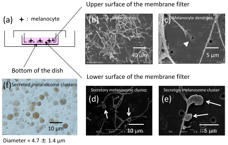
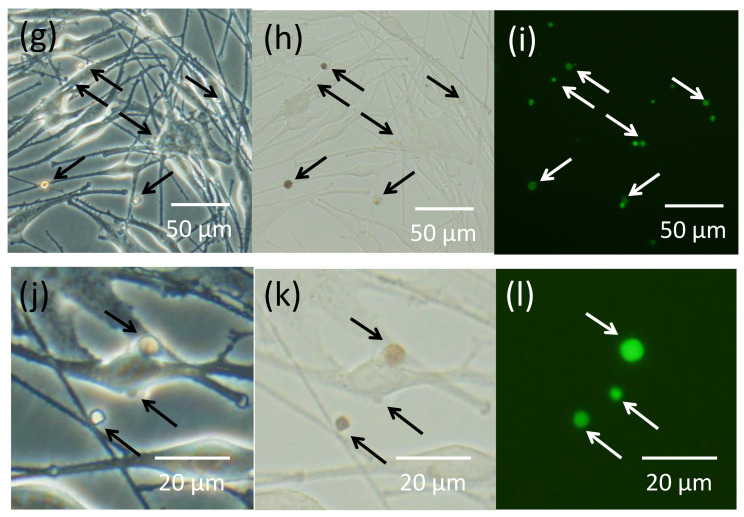
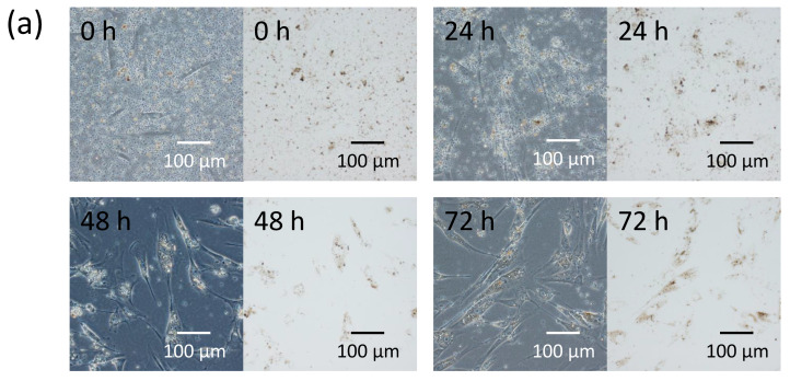
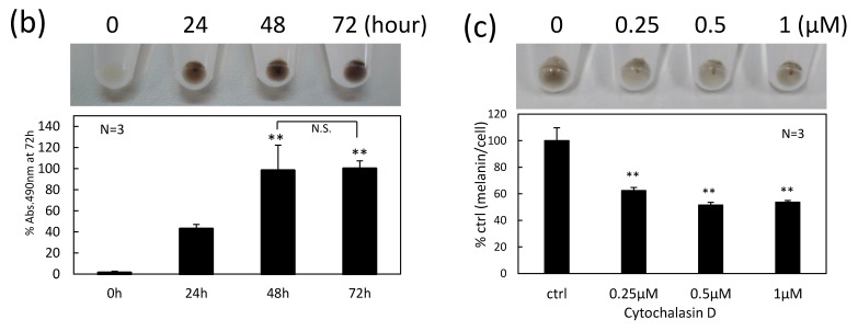
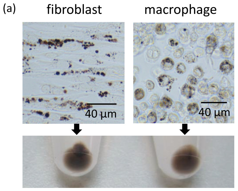
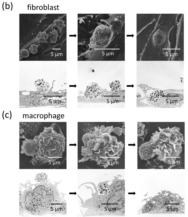
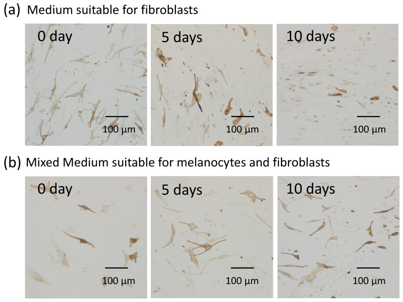
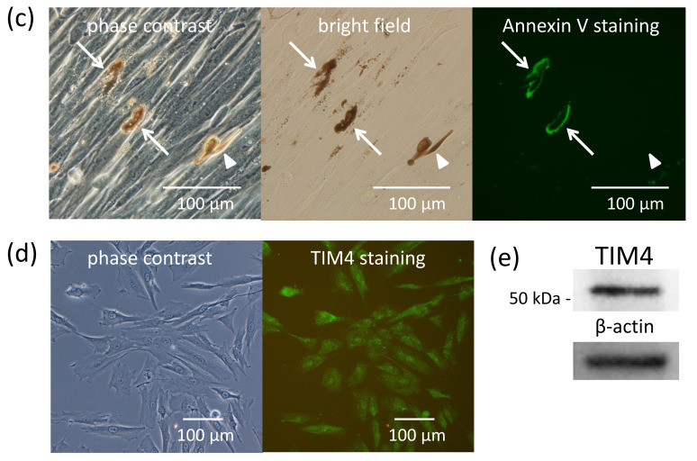
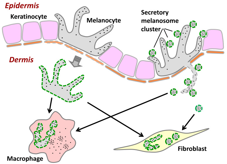
Pigmentation in the dermis is known to be caused by melanophages, defined as melanosome-laden macrophages. In this study, we show that dermal fibroblasts also have an ability to uptake melanosomes and apoptotic melanocytes. We have previously demonstrated that normal human melanocytes constantly secrete melanosome clusters from various sites of their dendrites. After adding secreted melanosome clusters collected from the culture medium of melanocytes, time-lapse imaging showed that fibroblasts actively attached to the secreted melanosome clusters and incorporated them. Annexin V staining revealed that phosphatidylserine (PtdSer), which is known as an 'eat-me' signal that triggers the internalization of apoptotic cells by macrophages, is exposed on the surface of secreted melanosome clusters. Dermal fibroblasts were able to uptake secreted melanosome clusters as did macrophages, and those fibroblasts express TIM4, a receptor for PtdSer-mediated endocytosis. Further, co-cultures of fibroblasts and melanocytes demonstrated that dermal fibroblasts internalize PtdSer-exposed apoptotic melanocytes. These results suggest that not only macrophages, but also dermal fibroblasts contribute to the collection of potentially toxic substances in the dermis, such as secreted melanosome clusters and apoptotic melanocytes, that have been occasionally observed to drop down into the dermis from the epidermis.

Keywords: apoptosis; dermis; epidermis; fibroblast; macrophage; melanin; melanocyte; melanosome; phosphatidylserine; pigmentation.


Conflict of interest statement

The authors declare no conflict of interest.

Figures

Figure 1
Figure 1
Normal human melanocytes produce secretory melanosome clusters along their dendrites with exposed PtdSer. (a) Normal human melanocytes were cultured for 6 days on microporous (1 µm-pore) membrane filters. Scanning electron microscopic (SEM) images of (b,c) the upper surface and (d,e) the lower surface of a membrane filter are shown. The arrow-head indicates a melanocyte dendrite inserted into a pore and the arrows indicate secretory melanosome clusters. Light microscopic bright field image of secreted melanosome clusters that had dropped down to the bottom of the culture dish released from melanocyte dendrites that had penetrated through (f) the microporous membrane filter. The same field of melanocytes (gi) at lower magnification and (jl) at higher magnification is shown with (g,j) phase contrast, (h,k) bright field and (i,l) immunofluorescence staining of PtdSer by Annexin V. Arrows indicate secretory melanosome clusters.
Figure 1
Figure 1
Normal human melanocytes produce secretory melanosome clusters along their dendrites with exposed PtdSer. (a) Normal human melanocytes were cultured for 6 days on microporous (1 µm-pore) membrane filters. Scanning electron microscopic (SEM) images of (b,c) the upper surface and (d,e) the lower surface of a membrane filter are shown. The arrow-head indicates a melanocyte dendrite inserted into a pore and the arrows indicate secretory melanosome clusters. Light microscopic bright field image of secreted melanosome clusters that had dropped down to the bottom of the culture dish released from melanocyte dendrites that had penetrated through (f) the microporous membrane filter. The same field of melanocytes (gi) at lower magnification and (jl) at higher magnification is shown with (g,j) phase contrast, (h,k) bright field and (i,l) immunofluorescence staining of PtdSer by Annexin V. Arrows indicate secretory melanosome clusters.
Figure 2
Figure 2
Normal human fibroblasts internalize secreted melanosome clusters in an actin-dependent manner. (a) Time-dependent photos from time-lapse imaging (Supplementary Materials Video V1) and cell pellets and (b) melanin content per cell after the addition of secreted melanosome clusters to the culture medium of normal human fibroblasts. Cell pellets of fibroblasts pretreated for 30 min with or without an actin polymerization inhibitor, cytochalasin D (CytoD) at 0.25, 0.5 or 1 µM were produced after equal amounts of secreted melanosome clusters were added to the culture medium and were incubated for 2 days; (c) melanin content per cell incorporated in fibroblasts was measured spectrophotometrically. Data are expressed as a percentage of the value at 72 h (in b) or the control (ctrl) (in c) and are mean values ± SD. Statistical analysis was performed using the Dunnett II test (** p < 0.01 versus 24 h (in b) or the control (in c), N.S.: Not significant).
Figure 2
Figure 2
Normal human fibroblasts internalize secreted melanosome clusters in an actin-dependent manner. (a) Time-dependent photos from time-lapse imaging (Supplementary Materials Video V1) and cell pellets and (b) melanin content per cell after the addition of secreted melanosome clusters to the culture medium of normal human fibroblasts. Cell pellets of fibroblasts pretreated for 30 min with or without an actin polymerization inhibitor, cytochalasin D (CytoD) at 0.25, 0.5 or 1 µM were produced after equal amounts of secreted melanosome clusters were added to the culture medium and were incubated for 2 days; (c) melanin content per cell incorporated in fibroblasts was measured spectrophotometrically. Data are expressed as a percentage of the value at 72 h (in b) or the control (ctrl) (in c) and are mean values ± SD. Statistical analysis was performed using the Dunnett II test (** p < 0.01 versus 24 h (in b) or the control (in c), N.S.: Not significant).
Figure 3
Figure 3
Normal human fibroblasts uptake secreted melanosome clusters as do macrophages. The same amounts of secreted melanosome clusters were added to the culture medium of fibroblasts or macrophages and were incubated for 2 days. (a) The upper two light microscopic images show the incorporation of secreted melanosome clusters in fibroblasts (left) and in macrophages (right). The lower photo shows the cell pellets of fibroblasts (left) and macrophages (right) collected from those cells. Sequential SEM images (upper panels) and TEM images (lower panels) of (b) fibroblasts and (c) macrophages that internalized secreted melanosome clusters are shown.
Figure 3
Figure 3
Normal human fibroblasts uptake secreted melanosome clusters as do macrophages. The same amounts of secreted melanosome clusters were added to the culture medium of fibroblasts or macrophages and were incubated for 2 days. (a) The upper two light microscopic images show the incorporation of secreted melanosome clusters in fibroblasts (left) and in macrophages (right). The lower photo shows the cell pellets of fibroblasts (left) and macrophages (right) collected from those cells. Sequential SEM images (upper panels) and TEM images (lower panels) of (b) fibroblasts and (c) macrophages that internalized secreted melanosome clusters are shown.
Figure 4
Figure 4
Normal human fibroblasts internalize apoptotic melanocytes and express TIM4. Time-dependent observations of melanocytes and fibroblasts co-cultured in medium suitable only for (a) fibroblasts or (b) in medium suitable for melanocytes and fibroblasts. The same fields with (left) phase contrast, (middle) bright field and (right) immunofluorescence staining of PtdSer by Annexin V showing (c) the manner of internalization of apoptotic melanocytes into fibroblasts. Arrows indicate apoptotic melanocytes with exposed PtdSer. Arrow-heads indicate a clear-shaped and distinctive melanocyte with no staining of Annexin V. (d) Immunofluorescence staining and (e) Western blotting of TIM4, a receptor for PtdSer, in fibroblasts is shown.
Figure 4
Figure 4
Normal human fibroblasts internalize apoptotic melanocytes and express TIM4. Time-dependent observations of melanocytes and fibroblasts co-cultured in medium suitable only for (a) fibroblasts or (b) in medium suitable for melanocytes and fibroblasts. The same fields with (left) phase contrast, (middle) bright field and (right) immunofluorescence staining of PtdSer by Annexin V showing (c) the manner of internalization of apoptotic melanocytes into fibroblasts. Arrows indicate apoptotic melanocytes with exposed PtdSer. Arrow-heads indicate a clear-shaped and distinctive melanocyte with no staining of Annexin V. (d) Immunofluorescence staining and (e) Western blotting of TIM4, a receptor for PtdSer, in fibroblasts is shown.
Figure 5
Figure 5
Hypothetical scheme of the internalization of secreted melanosome clusters and apoptotic melanocytes by macrophages and fibroblasts. When melanosomes or melanocytes drop down into the dermis in cases when the basement membrane is disrupted or disappears in abnormal conditions such as excessive inflammation, the secreted melanosome clusters and apoptotic melanocytes might be incorporated not only in macrophages, but also in fibroblasts. Green dashed lines indicate exposed PtdSer.

References

    1. Wu X., Hammer J.A. Melanosome transfer: It is best to give and receive. Curr. Opin. Cell Biol. 2014;29:1–7. doi: 10.1016/j.ceb.2014.02.003. - DOI - PMC - PubMed
    1. Braun-Falco O., Schoefinius H.H. Lentigo senilis. Hautarzt. 1971;22:277–283. - PubMed
    1. Hashmi S., Marinkovich M.P. Molecular organization of the basement membrane zone. Clin. Dermatol. 2011;29:398–411. doi: 10.1016/j.clindermatol.2011.01.009. - DOI - PubMed
    1. Nagao S., Iijima S. Light and electron microscopic study of Riehl’s melanosis. Possible mode of its pigmentary incontinence. J. Cutan. Pathol. 1974;1:165–175. doi: 10.1111/j.1600-0560.1974.tb00622.x. - DOI - PubMed
    1. Masu S., Seiji M. Pigmentary incontinence in fixed drug eruptions. Histologic and electron microscopic findings. J. Am. Acad. Dermatol. 1983;8:525–532. doi: 10.1016/S0190-9622(83)70060-5. - DOI - PubMed

MeSH terms

LinkOut - more resources