. 2020 Oct 27;142(17):1667-1683.
doi: 10.1161/CIRCULATIONAHA.120.045470. Epub 2020 Aug 18.

4HNE Impairs Myocardial Bioenergetics in Congenital Heart Disease-Induced Right Ventricular Failure

Affiliations

4HNE Impairs Myocardial Bioenergetics in Congenital Heart Disease-Induced Right Ventricular Failure

HyunTae V Hwang et al. Circulation. .

Abstract

Background: In patients with complex congenital heart disease, such as those with tetralogy of Fallot, the right ventricle (RV) is subject to pressure overload stress, leading to RV hypertrophy and eventually RV failure. The role of lipid peroxidation, a potent form of oxidative stress, in mediating RV hypertrophy and failure in congenital heart disease is unknown.

Methods: Lipid peroxidation and mitochondrial function and structure were assessed in right ventricle (RV) myocardium collected from patients with RV hypertrophy with normal RV systolic function (RV fractional area change, 47.3±3.8%) and in patients with RV failure showing decreased RV systolic function (RV fractional area change, 26.6±3.1%). The mechanism of the effect of lipid peroxidation, mediated by 4-hydroxynonenal ([4HNE] a byproduct of lipid peroxidation) on mitochondrial function and structure was assessed in HL1 murine cardiomyocytes and human induced pluripotent stem cell-derived cardiomyocytes.

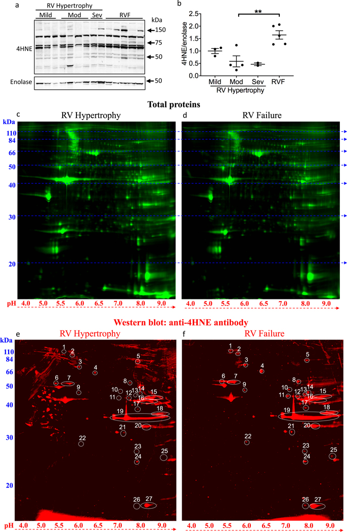
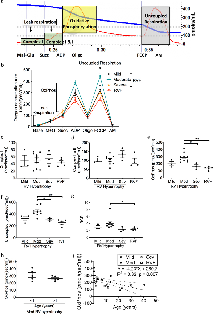
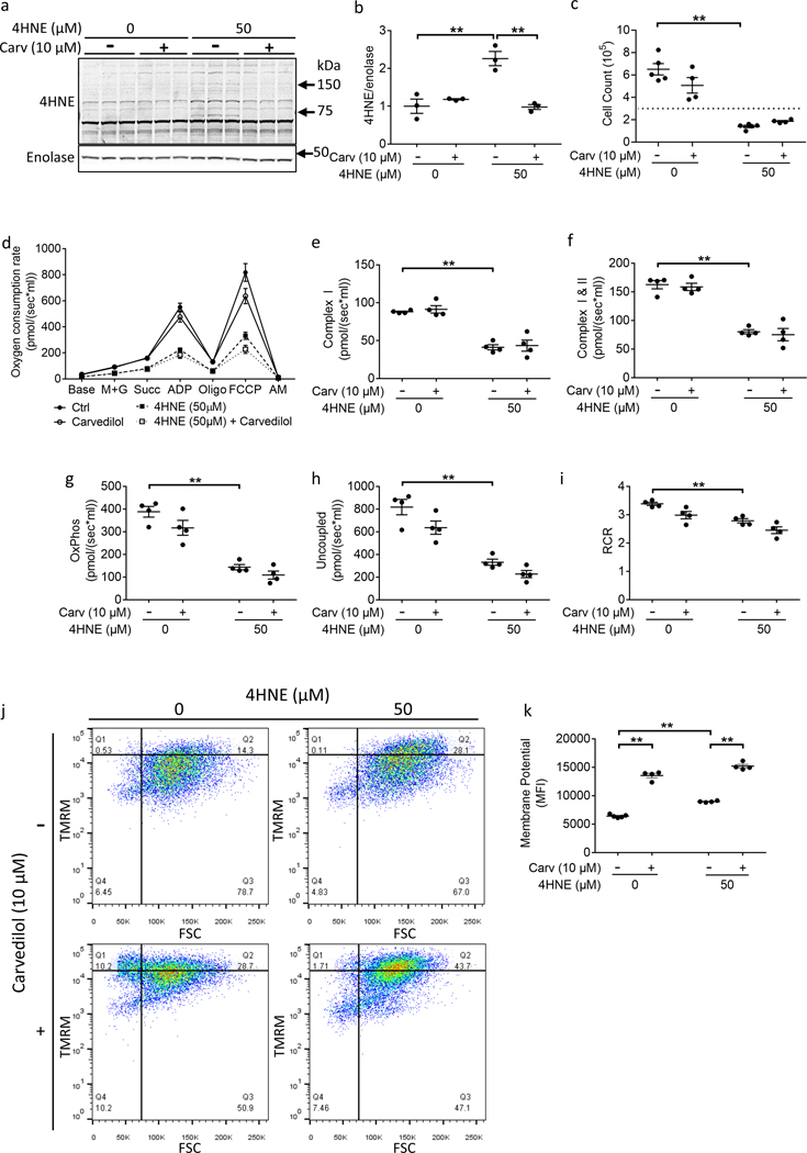
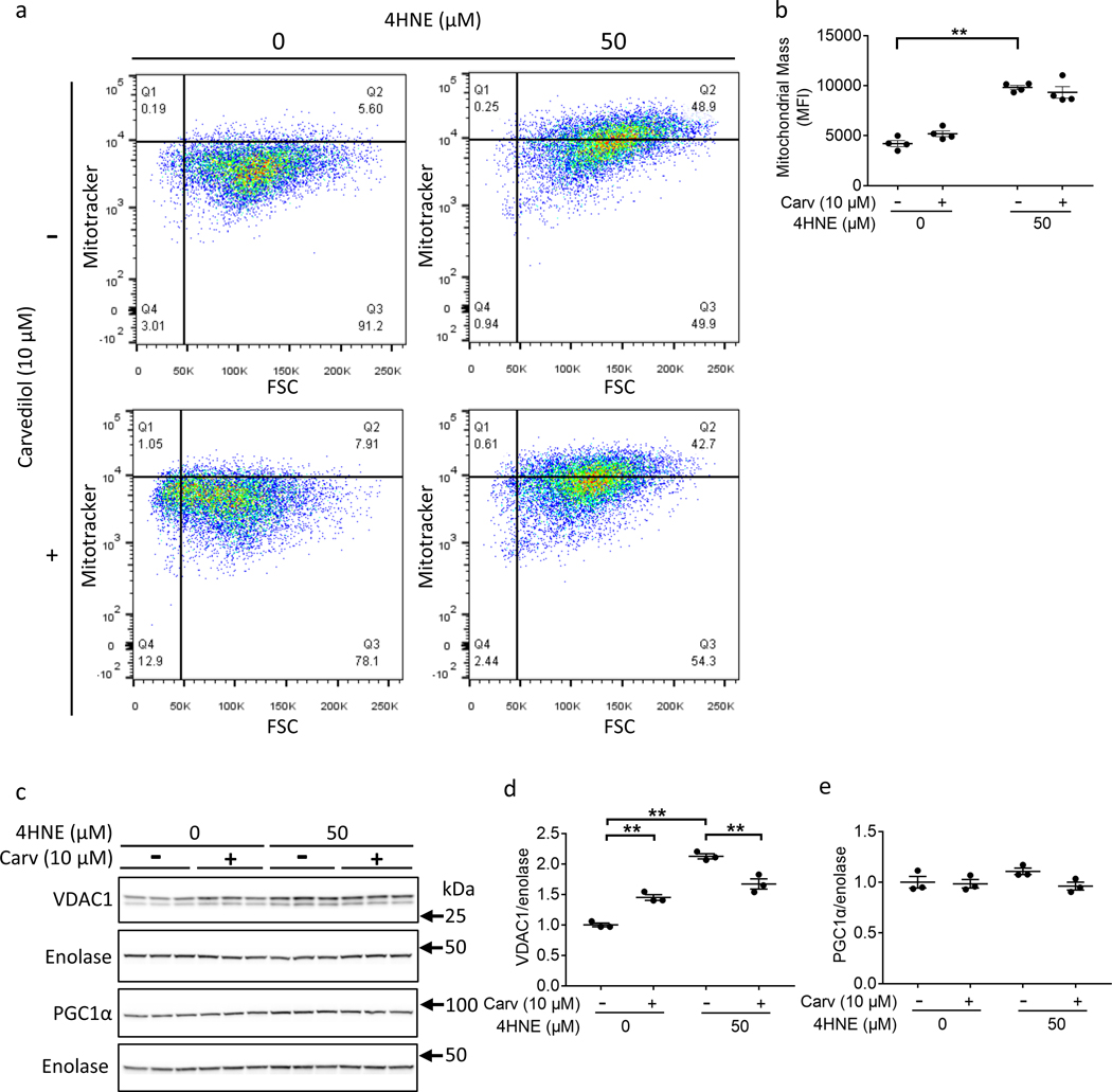
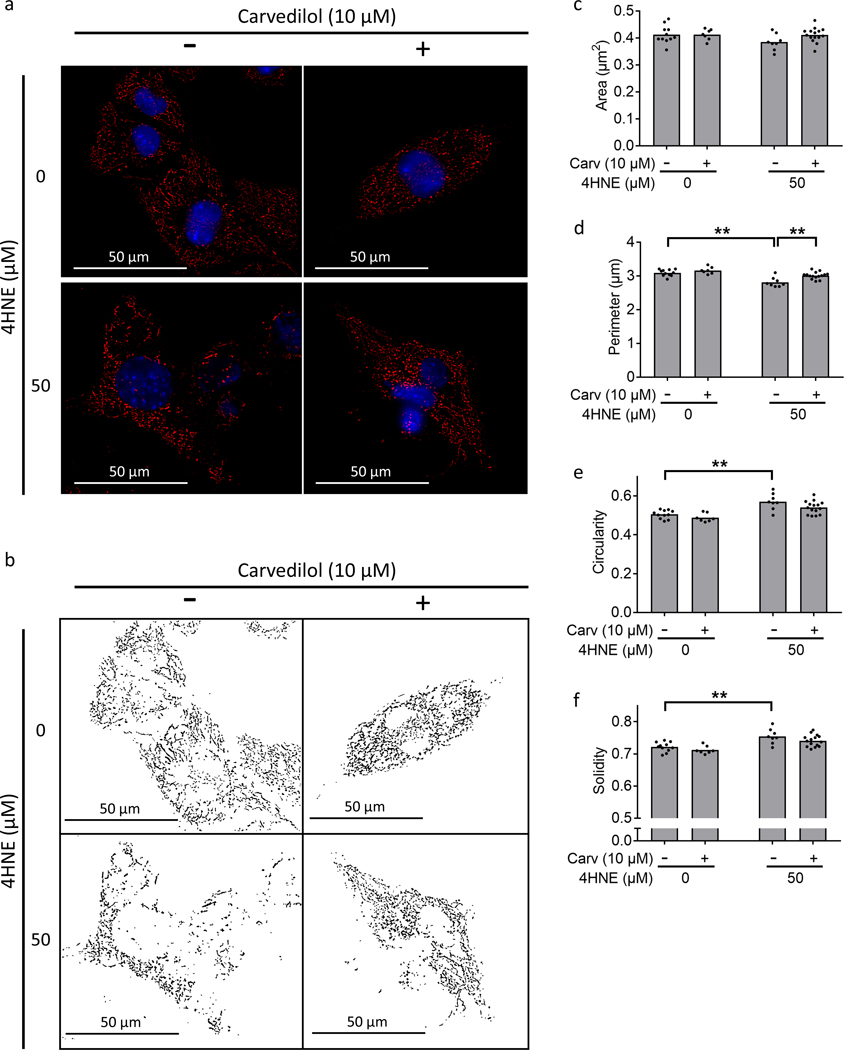
Results: RV failure was characterized by an increase in 4HNE adduction of metabolic and mitochondrial proteins (16 of 27 identified proteins), in particular electron transport chain proteins. Sarcomeric (myosin) and cytoskeletal proteins (desmin, tubulin) also underwent 4HNE adduction. RV failure showed lower oxidative phosphorylation (moderate RV hypertrophy, 287.6±19.75 versus RV failure, 137.8±11.57 pmol/[sec×mL]; P=0.0004), and mitochondrial structural damage. Using a cell model, we show that 4HNE decreases cell number and oxidative phosphorylation (control, 388.1±23.54 versus 4HNE, 143.7±11.64 pmol/[sec×mL]; P<0.0001). Carvedilol, a known antioxidant did not decrease 4HNE adduction of metabolic and mitochondrial proteins and did not improve oxidative phosphorylation.

Conclusions: Metabolic, mitochondrial, sarcomeric, and cytoskeletal proteins are susceptible to 4HNE-adduction in patients with RV failure. 4HNE decreases mitochondrial oxygen consumption by inhibiting electron transport chain complexes. Carvedilol did not improve the 4HNE-mediated decrease in oxygen consumption. Strategies to decrease lipid peroxidation could improve mitochondrial energy generation and cardiomyocyte survival and improve RV failure in patients with congenital heart disease.

Keywords: heart failure; heart ventricles; hypertrophy; lipid peroxidation; mitochondria.


Conflict of interest statement

Disclosures: The authors have declared that no conflict of interest exists.

Figures

Figure 1.
Figure 1.
Lipid peroxidation is increased in RV failure. (a, b) 4HNE adducts increased with RV failure vs moderate RV hypertrophy, N=2–5/group. (c, d) 2D-gel electrophoresis and western blot were used to detect total protein in representative RV hypertrophy (N=1) and RV failure (N=1) samples. (e, f) 2D-gel electrophoresis and western blot were used to detect the proteins with increased 4HNE-adducts. 27 protein spots where 4HNE signals were higher in RV failure vs. RV hypertrophy were numbered for identification. Mod – moderate; Sev – severe; RV – right ventricle; RVF – right ventricular failure; 4HNE - 4-hydroxynonenal. Data are presented as mean±SEM. **p<0.01.
Figure 2.
Figure 2.
Patients with severe RV hypertrophy and RV failure demonstrate decreased oxidative phosphorylation. (a) A representative oxygen consumption tracing is shown for myocardial tissue (red curve) along with the available oxygen in the assay chamber (blue curve). We evaluated leak respiration (green shaded zones), oxidative phosphorylation (yellow shaded zone), and uncoupled respiration (gray shaded zone). (b) Summary of oxygen consumption in right ventricular myocardial tissue (N=3–10/group). (c, d) Complex I and Complex II mediated oxygen consumption was unchanged across all groups. (e, f) Oxidative phosphorylation and uncoupled respiration trended toward an increase from mild to moderate RV hypertrophy, decreased from moderate RV hypertrophy to severe hypertrophy, and decreased with RV failure. Oxidative phosphorylation remained significantly decreased after adjusting for age (p=0.0004) in RV failure. (g) Similarly, respiratory control ratio trended toward an increase in moderate hypertrophy and subsequently significantly decreased from moderate RV hypertrophy to RV failure. (h) Oxidative phosphorylation did not differ in patients with moderate RV hypertrophy between those <1 year of age versus >1 year of age (N=5/group). (i) Linear regression analysis demonstrates that age plays a role in the changes seen in oxidative phosphorylation but the actual change in oxidative phosphorylation per year (Slope) is small at −4.23pmol/sec*ml (N=21). RV – right ventricle; RVF – right ventricular failure; M+G – malate and glutamate; Succ – succinate; Oligo – oligomycin; FCCP - carbonyl cyanide-p-trifluoromethoxyphenylhydrazone; AM – antimycin A; Mod – moderate; Sev – severe; OxPhos - oxidative phosphorylation; RCR – respiratory control ratio. Data are presented as mean±SEM. *p<0.05, **p<0.01.
Figure 3.
Figure 3.
Patients with RV failure demonstrate mitochondrial structural changes. (a-c) Both VDAC1 (mitochondrial protein) and PGC1α (mitochondrial biogenesis factor) trended toward an increase in RV failure (N=2–5/group). (d) Mitochondrial DNA copy number was not different between mild and moderate RV hypertrophy groups, but was significantly increased from moderate RV hypertrophy to RV failure (N=3–5/group). (e) Histogram of mitochondrial size distribution for each group by transmission electron microscopy demonstrates no difference in mitochondrial size. Each bin represents increments in mitochondrial area of 0.1 μm2 from left to right. Range is 0 to 2 μm2. (f) No significant difference was observed in median mitochondrial area. (g) Representative transmission electron microscopy images. Mitochondria become more rounded in moderate RV hypertrophy and more irregular in shape in RV failure, with a loss of defined cristae structure. Scale bars are 2 μm. N=3/group. Mod – moderate; Sev – severe; mtDNA – mitochondrial DNA; RV – right ventricle; RVF – right ventricular failure. Data are presented as mean±SEM. *p<0.05.
Figure 4.
Figure 4.
4HNE treatment decreases ATP synthesis but increases mitochondrial membrane potential. HL1 Cardiomyocytes were treated with 50 μM 4HNE for 24 hours and rescue was assessed with 10 μM carvedilol. (a, b) 4HNE adducts increased following 4HNE treatment. Carvedilol decreased 4HNE adducts(N=3/group). (c) 4HNE decreased cell count - the horizontal dotted line denotes the original number of plated cells (N=4–5/group). (d-i) Oxygen consumption was assessed in response to 4HNE using high resolution respirometry. (d) Summary of the oxygen consumption data. 4HNE decreased (e, f) leak respiration, (g) oxidative phosphorylation, (h) uncoupled respiration, and (i) respiratory control ratio (N=4/group). (j, k) However, 4HNE increased mitochondrial membrane potential, which further increased with carvedilol treatment (N=4–5/group). Carv - carvedilol; 4HNE - 4-hydroxynonenal; M+G - malate and glutamate; Succ - succinate; Oligo-oligomycin; FCCP - carbonyl cyanide 4-(trifluoromethoxy) phenylhydrazone; AM - antimycin; OxPhos - oxidative phosphorylation; RCR – respiratory control ratio; TMRM - tetramethylrhodamine, methyl ester; FSC – forward scatter; MFI – median fluorescence intensity. Data are presented as mean±SEM. **p<0.01.
Figure 5.
Figure 5.
4HNE treatment increases mitochondrial mass. HL1 Cardiomyocytes were treated with 50 μM 4HNE and/or 10 μM carvedilol. (a, b) 4HNE increased flow cytometric signal of Mitotracker Red CMXRos, a measure of mitochondrial mass (N=4/group). (c, d) 4HNE increased VDAC1 expression, another marker of mitochondrial mass, but this was inhibited by carvedilol. (e) 4HNE did not decrease PGC1α expression, a marker of mitochondrial biogenesis (N=3/group). 4HNE - 4-hydroxynonenal; Carv – carvedilol; FSC – forward scatter. Data are presented as mean±SEM. **p<0.01.
Figure 6.
Figure 6.
4HNE decreases mitochondrial network connectivity. Cardiomyocytes were stained with Mitotracker Red CMXRos (red) and Hoechst 33342 (blue) for mitochondrial and nuclear staining, respectively. 50 μM 4HNE treatment for 24 hours (a, b) disrupted mitochondrial network connectivity and Carvedilol improved network connectivity, (c) did not demonstrate a change in mitochondrial area, (d) decreased perimeter, (e, f) increased circularity and solidity. Scale bars are 50 μm. 4HNE - 4-hydroxynonenal. Data are presented as mean±SEM. **p<0.01.

Similar articles

Cited by

References

    1. Altmann K, Printz BF, Solowiejczyk DE, Gersony WM, Quaegebeur J and Apfel HD. Two-dimensional echocardiographic assessment of right ventricular function as a predictor of outcome in hypoplastic left heart syndrome. Am J Cardiol. 86:964–968. - PubMed
    1. Bogaard HJ, Abe K, Vonk Noordegraaf A and Voelkel NF. The right ventricle under pressure: cellular and molecular mechanisms of right-heart failure in pulmonary hypertension. Chest. 2009;135:794–804. - PubMed
    1. Fine NM, Chen L, Bastiansen PM, Frantz RP, Pellikka PA, Oh JK and Kane GC. Outcome prediction by quantitative right ventricular function assessment in 575 subjects evaluated for pulmonary hypertension. Circ Cardiovasc Imaging. 2013;6:711–721. - PubMed
    1. Haddad F, Hunt SA, Rosenthal DN and Murphy DJ. Right ventricular function in cardiovascular disease, part I: Anatomy, Physiology, Aging, and Functional Assessment of the Right Ventricle. Circulation. 2008;117:1436–1448. - PubMed
    1. Reddy S, Bernstein D and Newburger JW. Renin-Angiotensin-Aldosterone System Inhibitors for Right Ventricular Dysfunction in Tetralogy of Fallot: Quo Vadis? Circulation. 2018;137:1472–1474. - PMC - PubMed

Publication types