

Median nerve conduction studies in rabbits
- PMID: 32807101
- PMCID: PMC7433080
- DOI: 10.1186/s12868-020-00584-2
Median nerve conduction studies in rabbits
Abstract
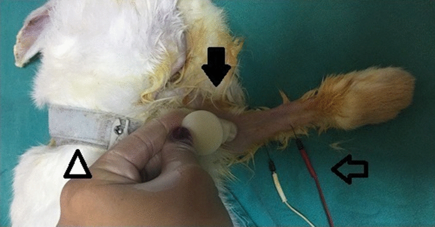
Background: When planning nerve conduction studies using animal models, the sciatic nerve is often used and the upper extremity nerves are not preferred due to the size of laboratory animals. This study aimed to present the method and mean values of median nerve conduction studies in laboratory rabbits. Fifty-five six-month-old male New Zealand white rabbits weighing 2 to 2.5 kg were included in nerve conduction studies performed under anesthesia. The compound muscle action potential amplitude and distal latency values were recorded for the median motor nerve with the electrodes placed on the flexor digitorum superficialis muscle and tendon.
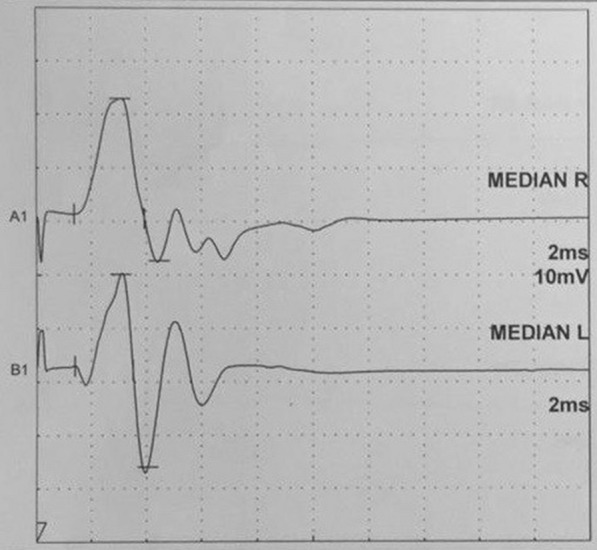
Results: A total of 110 median nerves were evaluated. The mean amplitude of the median nerve was 30.6 ± 6.8, mV the median nerve distal latency was 1.3 ± 0.2 ms, and the mean intensity of stimulation inducing a response was 2.5 ± 1 mA.
Conclusions: The mean values obtained by the median motor nerve conduction method in this study can act as a guide for future nerve interventions undertaken in the upper extremities.
Keywords: Compound muscle action potential amplitude; Distal latency; Median nerve; Nerve conduction studies; Rabbit.
Conflict of interest statement
The authors declare that they have no competing interests.
Figures
References
-
- Kouyoumdjian JA. Peripheral nerve injuries: a retrospective survey of 456 cases. Muscle Nerve. 2006;34(6):785–788. - PubMed
-
- Oh SJ. Anatomical and physiological basis for electromyography studies. In: Oh SJ, editor. Clinical Electromyography Nerve Conduction Studies. Baltimore: Wilkins and Wilkins; 1993. pp. 3–14.
MeSH terms
LinkOut - more resources
Full Text Sources
