. 2020 Aug 17;16(1):296.
doi: 10.1186/s12917-020-02521-1.

Exogenous Jaagsiekte Sheep Retrovirus type 2 (exJSRV2) related to ovine pulmonary adenocarcinoma (OPA) in Romania: prevalence, anatomical forms, pathological description, immunophenotyping and virus identification

Affiliations

Exogenous Jaagsiekte Sheep Retrovirus type 2 (exJSRV2) related to ovine pulmonary adenocarcinoma (OPA) in Romania: prevalence, anatomical forms, pathological description, immunophenotyping and virus identification

Corina Toma et al. BMC Vet Res. .

Abstract

Background: Ovine pulmonary adenocarcinoma (OPA) is a neoplastic disease caused by exogenous Jaagsiekte Sheep Retrovirus (exJSRV). The prevalence of JSRV-related OPA in Eastern European countries, including Romania is unknown. We aimed to investigate: the prevalence and morphological features of OPA (classical and atypical forms) in the Transylvania region (Romania), the immunophenotype of the pulmonary tumors and their relationships with exJSRV infection. A total of 2693 adult ewes slaughtered between 2017 and 2019 in two private slaughterhouses from Transylvania region (Romania) was evaluated. Lung tumors were subsequently assessed by cytology, histology, immunocytochemistry, immunohistochemistry, electron microscopy and DNA testing.

Results: Out of 2693 examined sheep, 34 had OPA (1.26% prevalence). The diaphragmatic lobes were the most affected. Grossly, the classical OPA was identified in 88.24% of investigated cases and the atypical OPA in 11.76% that included solitary myxomatous nodules. Histopathology results confirmed the presence of OPA in all suspected cases, which were classified into acinar and papillary types. Myxoid growths (MGs) were diagnosed in 6 classical OPA cases and in 2 cases of atypical form. Lung adenocarcinoma was positive for MCK and TTF-1, and MGs showed immunoreaction for Vimentin, Desmin and SMA; Ki67 expression of classical OPA was higher than atypical OPA and MGs. JSRV-MA was identified by IHC (94.11%) in both epithelial and mesenchymal cells of OPA. Immunocytochemistry and electron microscopy also confirmed the JSRV within the neoplastic cells. ExJSRV was identified by PCR in 97.05% of analyzed samples. Phylogenetic analysis revealed the presence of the exJSRV type 2 (MT809678.1) in Romanian sheep affected by lung cancer and showed a high similarity with the UK strain (AF105220.1).

Conclusions: In this study, we confirmed for the first time in Romania the presence of exJSRV in naturally occurring OPA in sheep. Additionally, we described the first report of atypical OPA in Romania, and to the best of our knowledge, in Eastern Europe. Finally, we showed that MGs have a myofibroblastic origin.

Keywords: Atypical OPA; Epidemiology; Jaagsiekte sheep retrovirus type 2; Myxoid growths; Ovine pulmonary adenocarcinoma.


Conflict of interest statement

The authors declare no conflicts of interests.

Figures

Fig. 1
Fig. 1
Gross features of spontaneous OPA in sheep. a Distribution of pulmonary lesions; b Classical form of OPA, involving the diaphragmatic lobe (arrow); c Classical form of OPA, affecting multiple pulmonary lobes (arrows); d Classical form of OPA on cut surface showing the junction (interrupted line) between the tumor and normal tissue; e and e1 Atypical form of OPA showing a subpleural white-grey nodule (arrow). On cut surface, the tumor is pearly white and dried (arrow); f Gross aspects of pulmonary myxoma-like nodule (arrow); g the myxomatous mass shows a multilobular, white-gray, gelatinous feature on cut surface
Fig. 2
Fig. 2
Microscopical findings of OPA. a Cytological exam of pulmonary tumors showing multiple nest of cubical to polygonal epithelial cells, interpreted as type II alveolar cells, DQ stain; b Histological appearance of OPA-classical form: tubular type and c papillary type, H&E stain; d and e The microphotographs of classical OPA showing myxoid growths (white arrow) interspersed with neoplastic epithelial component (blue arrow), H&E stain; f Histological exam of atypical OPA showing a well-delimited neoplastic nodule, surrounded by fibrous tissue and infilammatory infiltrates (arrow), H&E stain; g Myxoma-like tumor without neoplastic epithelial component. The mass is multinodular (white arrow) and composed of spindle to stellates cells, and abundant extracellular matrix (the inset), H&E stain; h and i AB-PAS stained sections from myxoma-like nodules showing abundant, pale blue extracellular myxomatous matrix; the Goblet cells of bronchial lining epithelium are used as positive control for AB-PAS stain
Fig. 3
Fig. 3
Immunophenotypical characterization of OPA. a The neoplastic epithelial cells of classical form are diffusely and intensely immunopositive for MCK; b TTF1-positive nuclei of the epithelial cells are present within the neoplastic masses. Inset: the bronchial epithelium was used as positive control for TTF1. c All epithelial cells lining neoplastic acini are negative for vimentin (blue arrow) in contrast to immunopositive stromal cells (white arrow); d Myxoid growths showing an intense immunopositive reaction for Vimentin. Inset: detail of diffuse cytoplasmic labeling for Vimentin; e Mesenchymal cells of myxoid growths have diffuse and strong cytoplasmic labeling for Desmin. Inset: The smooth muscle cells of pulmonary arteries and bronchioles are strongly positive for desmin and served as internal positive control; f The cells of myxoid growths have selective and moderate cytoplasmic labeling for alpha-SMA. Inset: The bronchiolar smooth muscle cells are positive for SMA, internal positive control; g The myxoid component of OPA showing a negative immunoreaction of MCK (blue arrow) compared to the neoplastic epithelial cells which are strongly immunopositive; h Neoplastic cells of myxoma-like nodules showing a negative reaction for S100 protein. Inset: the bronchial cartilage is diffusely positive for S100 protein, internal positive control; i The proliferative index, characterized by ki67 immunopositive nuclei, was higher in the epithelial neoplasia compared to the MGs and myxoma-like nodules (blue arrows) (j). Inset: Bronchial-associated lymphoid tissue (BALT) hyperplasia was used as internal positive control for ki67. DAB and hematoxylin counterstain
Fig. 4
Fig. 4
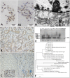
JSRV detection from neoplastic tissues. a and a1 Immunocytochemistry showing intracytoplasmic JSRV-MA expression within the neoplastic epithelial cells. DAB and hematoxylin counterstain; B Diffuse expression of the JSRV-MA marker by neoplastic epithelial cells forming tubular structures; c Expression of the JSRV-MA marker by MGs and epithelial tumor. Left inset: Cell cytoplasm of MGs (white arrow) is strongly labelled with JSRV-MA antibody. Right inset: Neoplastic epithelial cells have diffuse cytoplasmic labelling for JSRV-MA (bue arrow). DAB and hematoxylin counterstain; d JSRV-infected type 2 pneumocytes. Note the presence of intracytoplasmic lamellar body (white arrow) and aggregates and solitary intracytoplasmic viral particles (grey arrow). TEM. Bar, 1 μm. Inset: Detail of a viral replication site showing specific virions compatible with JSRV (white arrow). TEM. Bar, 100 nm; e Electrophoresis profile of the amplified DNA fragments (128 bp) evidencing the presence of proviral Jaagsiekte DNA in ovine pulmonary adenocarcinoma (OPA). L - DNA ladder; PCR positive (+) samples; PCR negative (−) samples; M1 and M2 control samples amplified from healthy ovine lung tissue; NTC - no template control (negative control); f Phylogenetic analysis of different JSRV strains and other retroviruses based on the LTR region. The Romanian strains of exJSRV (JSRV-RO) (GenBank MT809678.1) showed homology at nucleotide level of 97% with UK strain (GenBank AF105220.1), and 83% with the South African strain (GenBank M80216)

Similar articles

Cited by

References

    1. Palmarini M, Sharp JM, De las Heras M, Fan H. Jaagsiekte sheep retrovirus is necessary and sufficient to induce a contagious lung cancer in sheep. J Virol. 1999;73:6964–6972. - PMC - PubMed
    1. York D, Querat G. A history of ovine pulmonary adenocarcinoma (Jaagsiekte) and experiments leading to the deduction of the JSRV nucleotide sequence. In: Fan H, editor. Jaagsiekte sheep retrovirus and lung Cancer, Curr top in Microbiol Immunol. Berlin: Springer; 2003. pp. 1–25. - PubMed
    1. Bai J, Zhu RY, Stedman K, Cousens C, Carlson J, Sharp JM, DeMartini JC. Unique long terminal repeat U3 sequences distinguish exogenous jaagsiekte sheep retroviruses associated with ovine pulmonary carcinoma from endogenous loci in the sheep genome. J Virol. 1996;70:3159–3168. - PMC - PubMed
    1. García-Goti M, González L, Cousens C, Cortabarría N, Extramiana AB, Minguijón E, Ortín A, De las Heras M, Sharp JM. Sheep pulmonary adenomatosis: characterization of two pathological forms associated with jaagsiekte retrovirus. J Comp Pathol. 2000;122:55–65. - PubMed
    1. Palmarini M, Cousens C, Dalziel RG, Bai J, Stedman K, DeMartini JC, Sharp JM. The exogenous form of jaagsiekte retrovirus is specifically associated with a contagious lung cancer of sheep. J Virol. 1996;70:1618–1623. - PMC - PubMed

MeSH terms