

Bilateral posterior cerebral artery territory infarction in a SARS-Cov-2 infected patient: discussion about an unusual case
- PMID: 32807489
- PMCID: PMC7321046
- DOI: 10.1016/j.jstrokecerebrovasdis.2020.105095
Bilateral posterior cerebral artery territory infarction in a SARS-Cov-2 infected patient: discussion about an unusual case
Abstract
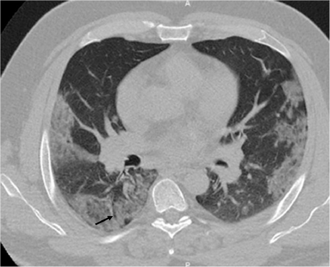
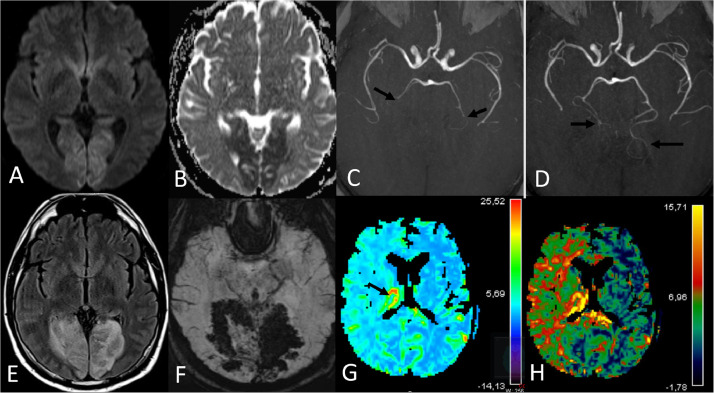
In time of SARS-Cov2 pandemic, neurologists need to be vigilant for cerebrovascular complications of Covid-19. We present a case of bilateral occipito-temporal infarction revealed by a sudden cortical blindness with haemorrhagic transformation after intravenous thrombolysis in a diabetic patient infected by Covid-19. Differential diagnoses are discussed in front of this unusual presentation and evolution.
Keywords: COVID-19; Infarction; MR perfusion; SARS-Cov2; Visual loss.
Copyright © 2020 Elsevier Inc. All rights reserved.
Conflict of interest statement
Declaration of Competing Interest The authors report no disclosures. Informed consent for publication has been signed by the wife of the patient.
Figures
References
-
- Kidwell CS, Saver JL, Mattiello J. Diffusion-perfusion MRI characterization of post-recanalization hyperperfusion in humans. Neurology. 2001;57:2015–2021. - PubMed
-
- Yamada H, Kikuchi R, Nakamura A, Miyazaki H. Severe reversible cerebral vasoconstriction syndrome with large posterior cerebral infarction. J Stroke Cerebrovasc Dis. 2018;27:3043–3045. - PubMed
Publication types
MeSH terms
Substances
LinkOut - more resources
Full Text Sources
Miscellaneous
