

Lung Ultrasound May Support Diagnosis and Monitoring of COVID-19 Pneumonia
- PMID: 32807570
- PMCID: PMC7369598
- DOI: 10.1016/j.ultrasmedbio.2020.07.018
Lung Ultrasound May Support Diagnosis and Monitoring of COVID-19 Pneumonia
Abstract
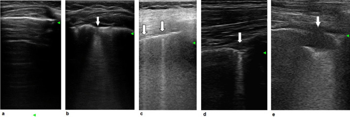
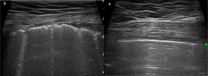
Severe acute respiratory syndrome coronavirus 2 (SARS-CoV-2) disease (COVID-19) is characterized by severe pneumonia and/or acute respiratory distress syndrome in about 20% of infected patients. Computed tomography (CT) is the routine imaging technique for diagnosis and monitoring of COVID-19 pneumonia. Chest CT has high sensitivity for diagnosis of COVID-19, but is not universally available, requires an infected or unstable patient to be moved to the radiology unit with potential exposure of several people, necessitates proper sanification of the CT room after use and is underutilized in children and pregnant women because of concerns over radiation exposure. The increasing frequency of confirmed COVID-19 cases is striking, and new sensitive diagnostic tools are needed to guide clinical practice. Lung ultrasound (LUS) is an emerging non-invasive bedside technique that is used to diagnose interstitial lung syndrome through evaluation and quantitation of the number of B-lines, pleural irregularities and nodules or consolidations. In patients with COVID-19 pneumonia, LUS reveals a typical pattern of diffuse interstitial lung syndrome, characterized by multiple or confluent bilateral B-lines with spared areas, thickening of the pleural line with pleural line irregularity and peripheral consolidations. LUS has been found to be a promising tool for the diagnosis of COVID-19 pneumonia, and LUS findings correlate fairly with those of chest CT scan. Compared with CT, LUS has several other advantages, such as lack of exposure to radiation, bedside repeatability during follow-up, low cost and easier application in low-resource settings. Consequently, LUS may decrease utilization of conventional diagnostic imaging resources (CT scan and chest X-ray). LUS may help in early diagnosis, therapeutic decisions and follow-up monitoring of COVID-19 pneumonia, particularly in the critical care setting and in pregnant women, children and patients in areas with high rates of community transmission.
Keywords: Acute respiratory disease syndrome; B-Lines; COVID-19; Interstitial syndrome; Lung ultrasound; Pneumonia.
Copyright © 2020 World Federation for Ultrasound in Medicine & Biology. Published by Elsevier Inc. All rights reserved.
Conflict of interest statement
Conflict of interest disclosure Dr. Allinovi, Dr. Parise, Dr. Giacalone, Prof. Amerio, Dr. Delsante, Prof. Odone, Dr. Gigliotti, Dr. Franci, Dr. Amadasi, Prof. Delmonte, Dr. Parri, and Dr. Mangia report no conflicts of interest.
Figures
References
-
- Alberici F., Delbarba E., Manenti C., Econimo L., Valerio F., Pola A., Maffei C., Possenti S., Piva S., Latronico N., Focà E., Castelli F., Gaggia P., Movilli E., Bove S., Malberti F., Farina M., Bracchi M., Costantino E.M., Bossini N., Gaggiotti M., Scolari F., Brescia Renal COVID Task Force Management of patients on dialysis and with kidney transplant during SARS-COV-2 (COVID-19) pandemic in Brescia, Italy. Kidney Int Rep. 2020;5:580–585. - PMC - PubMed
-
- Asano M., Watanabe H., Sato K., Okuda Y., Sakamoto S., Hasegawa Y., Sudo K., Takeda M., Sano M., Kibira S., Ito H. Validity of ultrasound lung comets for assessment of the severity of interstitial pneumonia. J Ultrasound Med. 2018;37:1523–1531. - PubMed
-
- Bai H.X., Hsieh B., Xiong Z., Halsey K., Choi J.W., Tran T.M.L., Pan I., Shi L.B., Wang D.C., Mei J., Jiang X.L., Zeng Q.H., Egglin T.K., Hu P.F., Agarwal S., Xie F., Li S., Healey T., Atalay M.K., Liao W.H. Performance of radiologists in differentiating COVID-19 from viral pneumonia on chest CT. Radiology. 2020;10 - PMC - PubMed
Publication types
MeSH terms
LinkOut - more resources
Full Text Sources
Other Literature Sources
Medical
Miscellaneous
