Review
. 2020 Sep 30;43(9):774-783.
doi: 10.14348/molcells.2020.0059.

WNT Signaling in Lung Repair and Regeneration

Affiliations
Review

WNT Signaling in Lung Repair and Regeneration

Ahmed A Raslan et al. Mol Cells. .

Abstract

The lung has a vital function in gas exchange between the blood and the external atmosphere. It also has a critical role in the immune defense against external pathogens and environmental factors. While the lung is classified as a relatively quiescent organ with little homeostatic turnover, it shows robust regenerative capacity in response to injury, mediated by the resident stem/progenitor cells. During regeneration, regionally distinct epithelial cell populations with specific functions are generated from several different types of stem/progenitor cells localized within four histologically distinguished regions: trachea, bronchi, bronchioles, and alveoli. WNT signaling is one of the key signaling pathways involved in regulating many types of stem/progenitor cells in various organs. In addition to its developmental role in the embryonic and fetal lung, WNT signaling is critical for lung homeostasis and regeneration. In this minireview, we summarize and discuss recent advances in the understanding of the role of WNT signaling in lung regeneration with an emphasis on stem/progenitor cells.

Keywords: WNT signaling; lung homeostasis; lung regeneration; lung stem/progenitor cells; β-catenin.


Conflict of interest statement

CONFLICT OF INTEREST

The authors have no potential conflicts of interest to disclose.

Figures
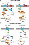
Fig. 1
Fig. 1. WNT signaling pathway.
Canonical (A) and non-canonical (B) WNT signaling pathways are shown. Both WNT and RSPO ligands synergistically act to generate a maximum activation in canonical WNT signaling. WNT, wingless-type MMTV integration site family; LRP5/6, low density lipoprotein receptor-related protein 5/6; FZD, frizzled; RNF43, ring finger protein 43; ZNRF3, zinc and ring finger 3; LGR, leucine-rich repeat containing G protein-coupled receptor; AXIN, axis inhibition protein; CK1, casein kinase 1; APC, adenomatous polyposis coli; DVL, dishevelled; GSK-3β, glycogen synthase kinase 3 beta; TCF, T-cell factor; RSPO, R-spondin; ROR, retinoic acid-related orphan receptors; RYK, receptor tyrosine kinases; PLC, phospholipase C; PKC, protein kinase C; CDC42, cell division cycle 42; CaN, calcineurin; NFAT, nuclear factor of activated T cells; CaMKII, calcium/calmodulin-dependent protein kinase II; TAK1, TGF-beta-activated kinase 1; NLK, Nemo-like kinase; RAC1, Rac family small GTPase 1; JNK, c-Jun N-terminal kinase; RhoA, ras homolog family member A; ROCK, Rho-associated coiled-coil containing protein kinase.
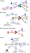
Fig. 2
Fig. 2. Heterogeneity and distribution of epithelial cells in different regions of the mouse lung.
Cell distribution within four different regions—Trachea, Bronchi, Bronchioles, and Alveoli—are shown along the proximal-distal axis of mouse lung. BASC, bronchioalveolar stem cell; LNEP, lineage-negative epithelial stem/progenitor cells.
Fig. 3
Fig. 3. Major stem cell populations in the lung.
(a, a’, a’’) Hematoxylin and eosin staining of the trachea, bronchioles and alveoli area in the adult lung. Scale bars = 50 μm. (b-d) Immunofluorescence staining of basal cells marker, cytokeratin 5 (KRT5) in the trachea. Scale bar = 20 μm. (b’-d’) Immunofluorescence staining of club cells marker, secretoglobin 1A1 (SCGB1A1) in bronchioles. Scale bar = 20 μm. (b’’-d’’) Immunofluorescence staining of AT2 cells marker, surfactant protein C (SFTPC) in alveoli. Scale bar = 20 μm.
Fig. 4
Fig. 4. WNT signaling in lung repair and regeneration.
WNT signaling acting positively and negatively are shown in blue and red colors, respectively. Dotted lines with an arrow indicate the presumed interaction among different cell types. BC, basal cell; MEC, myoepithelial cell; ASMC, airway smooth muscle cell; BASC, bronchioalveolar stem cell; LNEP, lineage-negative epithelial stem/progenitor cells; AT2, alveolar type 2 cell; AT1, alveolar type 1 cell.

Similar articles

Cited by

References

    1. Andersson-Sjoland A., Karlsson J.C., Rydell-Tormanen K. ROS-induced endothelial stress contributes to pulmonary fibrosis through pericytes and Wnt signaling. Lab. Invest. 2016;96:206–217. doi: 10.1038/labinvest.2015.100. - DOI - PubMed
    1. Ardhanareeswaran K., Mirotsou M. Lung stem and progenitor cells. Respiration. 2013;85:89–95. doi: 10.1159/000346500. - DOI - PMC - PubMed
    1. Barkauskas C.E., Cronce M.J., Rackley C.R., Bowie E.J., Keene D.R., Stripp B.R., Randell S.H., Noble P.W., Hogan B.L. Type 2 alveolar cells are stem cells in adult lung. J. Clin. Invest. 2013;123:3025–3036. doi: 10.1172/JCI68782. - DOI - PMC - PubMed
    1. Barker N., Tan S., Clevers H. Lgr proteins in epithelial stem cell biology. Development. 2013;140:2484–2494. doi: 10.1242/dev.083113. - DOI - PubMed
    1. Basil M.C., Katzen J., Engler A.E., Guo M., Herriges M.J., Kathiriya J.J., Windmueller R., Ysasi A.B., Zacharias W.J., Chapman H.A., et al. The cellular and physiological basis for lung repair and regeneration: past, present, and future. Cell Stem Cell. 2020;26:482–502. doi: 10.1016/j.stem.2020.03.009. - DOI - PMC - PubMed

LinkOut - more resources