. 2020 Aug 17;3(1):448.
doi: 10.1038/s42003-020-01161-3.

FAM46C/TENT5C functions as a tumor suppressor through inhibition of Plk4 activity

Affiliations

FAM46C/TENT5C functions as a tumor suppressor through inhibition of Plk4 activity

Karineh Kazazian et al. Commun Biol. .

Abstract

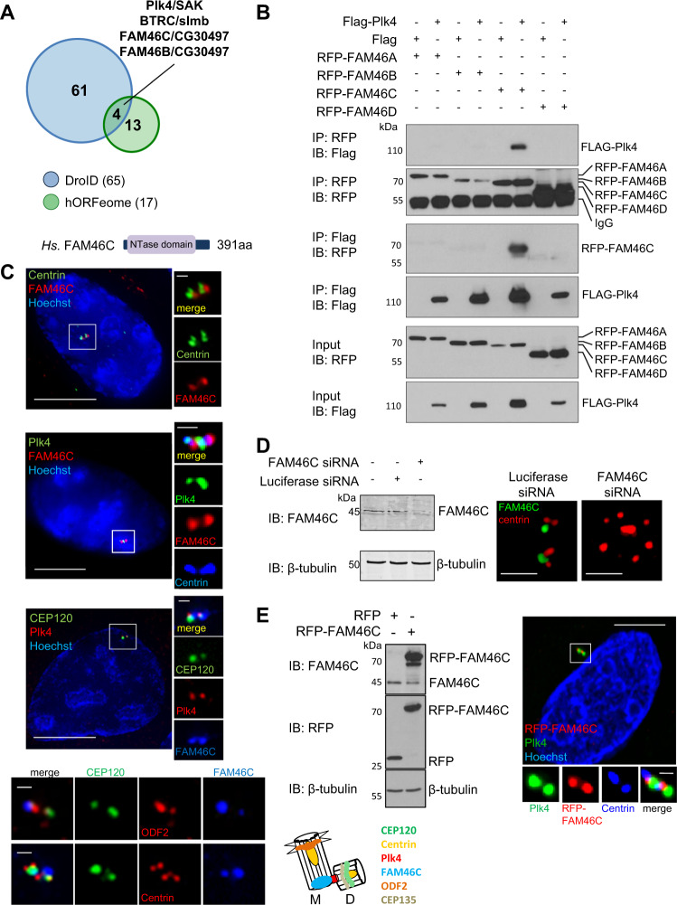
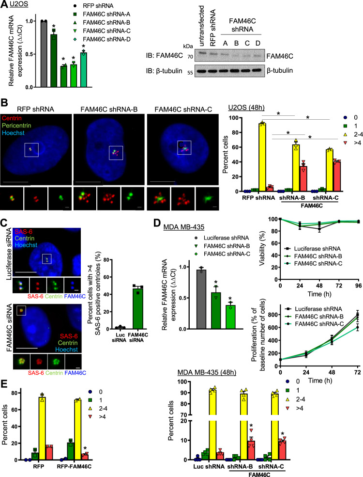
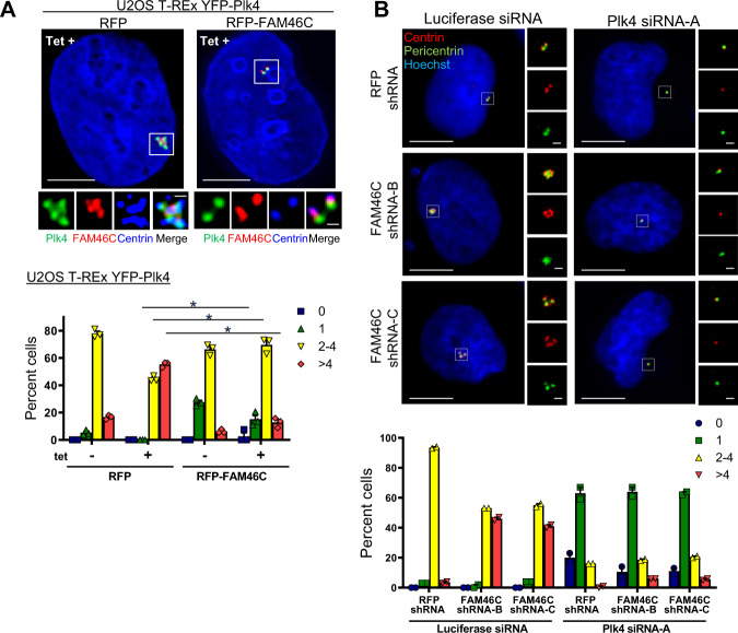
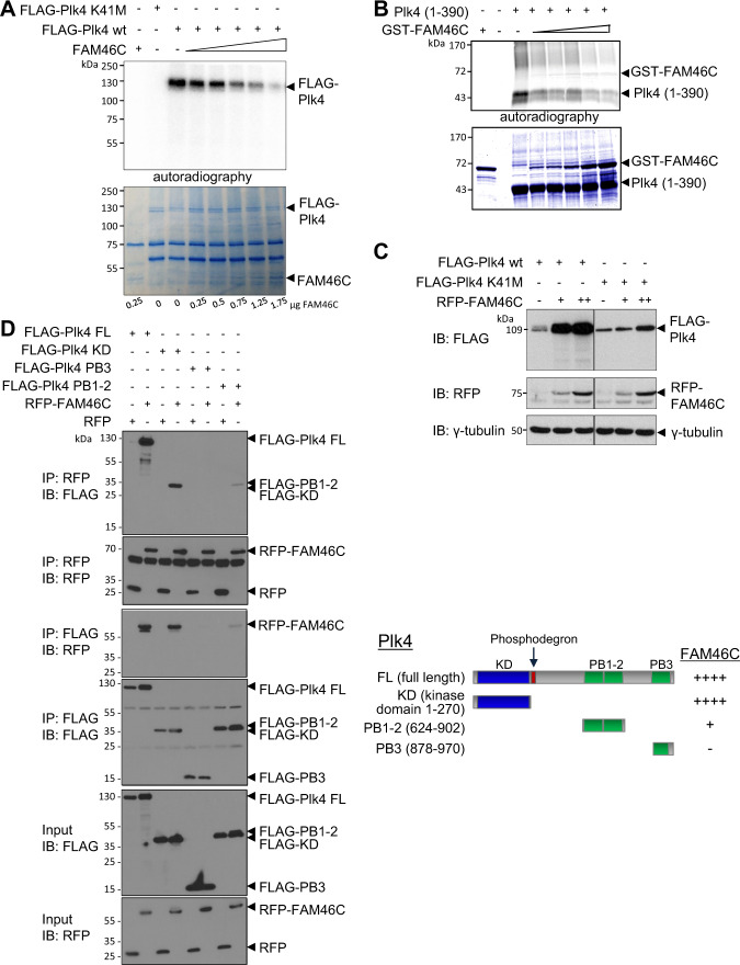
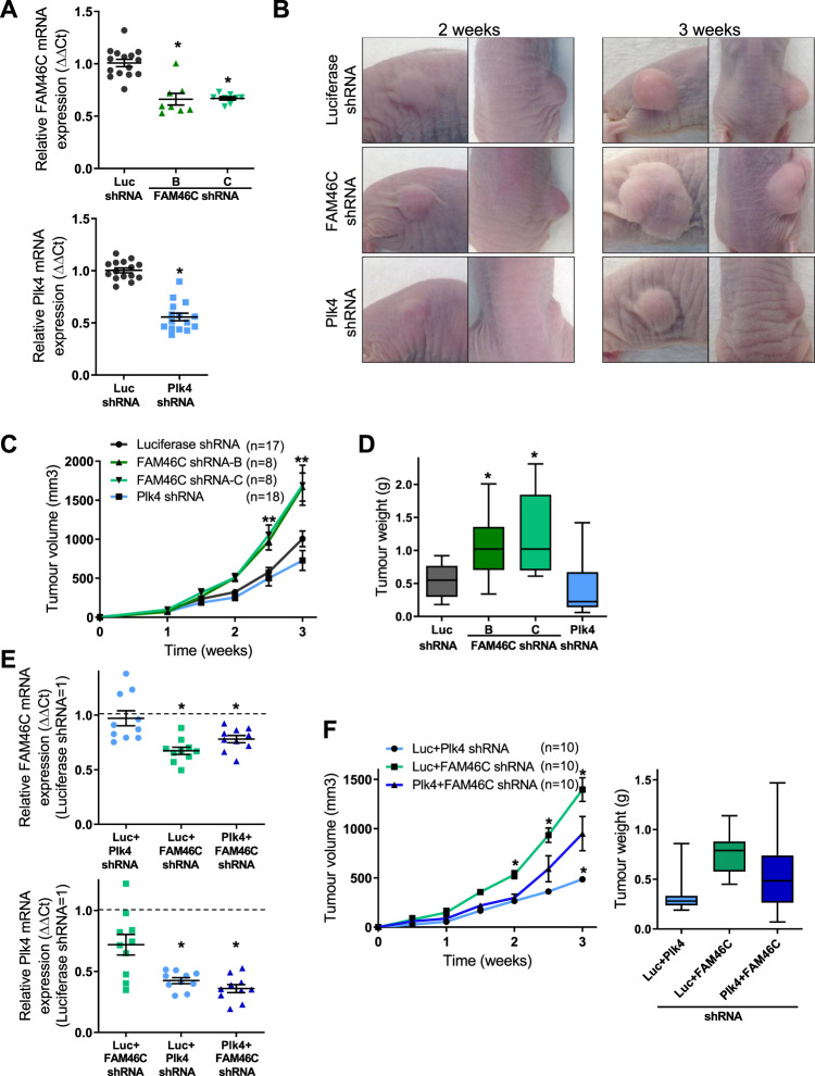
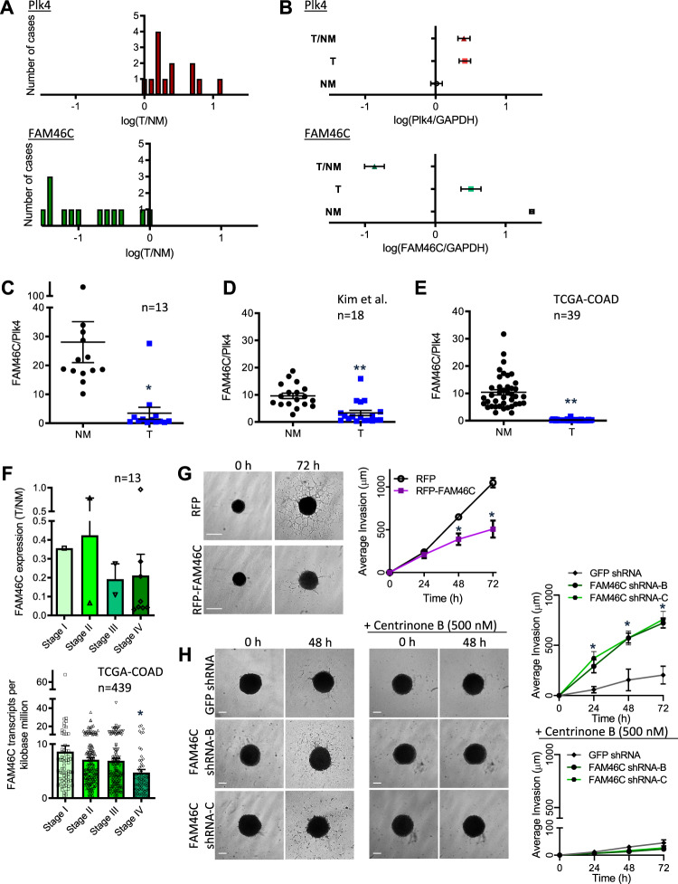
Polo like kinase 4 (Plk4) is a tightly regulated serine threonine kinase that governs centriole duplication. Increased Plk4 expression, which is a feature of many common human cancers, causes centriole overduplication, mitotic irregularities, and chromosomal instability. Plk4 can also promote cancer invasion and metastasis through regulation of the actin cytoskeleton. Herein we demonstrate physical interaction of Plk4 with FAM46C/TENT5C, a conserved protein of unknown function until recently. FAM46C localizes to centrioles, inhibits Plk4 kinase activity, and suppresses Plk4-induced centriole duplication. Interference with Plk4 function by FAM46C was independent of the latter's nucleotidyl transferase activity. In addition, FAM46C restrained cancer cell invasion and suppressed MDA MB-435 cancer growth in a xenograft model, opposing the effect of Plk4. We demonstrate loss of FAM46C in patient-derived colorectal cancer tumor tissue that becomes more profound with advanced clinical stage. These results implicate FAM46C as a tumor suppressor that acts by inhibiting Plk4 activity.


Conflict of interest statement

The authors declare no competing interests.

Figures

Fig. 1
Fig. 1. FAM46C is a conserved protein that interacts with Plk4.
a Diagram summarizing results of yeast two-hybrid screens for Plk4/SAK interactors from the HuRI (the human reference protein interactome mapping project, http://interactome.baderlab.org) and DroID (Drosophila interactions database, http://www.droidb.org) databases, showing overlap of four interactors, including H. Sapiens FAM46C/D. Melanogaster CG30497. Hs.FAM46C is a protein of 391 amino acids (aa) that contains a nucleotidyltransferase (NTase) domain, that has been recently renamed TENT5C. b Immunoblots of Flag-Plk4 and four individual Hs.RFP-FAM46 proteins after coexpression in HEK293T cells, showing reciprocal co-immunoprecipitation of Plk4 only with FAM46C. Uncropped blots for this and subsequent figures can be found in Supplementary Fig. 10. c Localization of FAM46C to centrioles, identified by staining with centriole markers including centrin, CEP120 (daughter centriole marker) and ODF2 (mother centriole marker), and overlap with Plk4. Representative immunofluorescence images of U2OS cells labeled with antibodies to: top panel, centrin (green) and FAM46C (red); second panel, Plk4 (green), FAM46C (red), centrin (blue); third panel, CEP120 (green, daughter), Plk4 (red), FAM46C (blue), and with Hoechst (blue). The right panels/inserts show magnified centrosomes (boxed in white). Bottom panels show magnified centrosomes labeled with antibodies to CEP120 (green, preferential labeling of daughter), ODF2 (red, top row, preferential labeling of mother), centrin (red, bottom row) and blue (FAM46C). FAM46C localizes predominantly to the mother centriole in unsynchronized cells (see Supplementary Fig. 4b for localization in synchronized cells). Cartoon summarizes putative FAM46C localization in relation to centriolar markers probed in this study, showing localization at the proximal mother centriole (M mother, D daughter; Bar 1 µm). d Left panel, representative immunoblot of Luciferase or FAM46C siRNA transfected U2OS cell extracts probed using anti-FAM46C antibody, with β-tubulin as loading control, demonstrating 87% depletion of FAM46C. Right panels, representative immunofluorescence images of Luciferase or FAM46C siRNA-transfected U2OS cell centrosomes labeled with antibodies to centrin (red) and FAM46C (green). FAM46C depletion was confirmed and resulted in a rosette-like centriolar phenotype. Bar: 1 µm. e Left panel, immunoblot showing expression of RFP and RFP-FAM46C in U2OS cells transfected with RFP or RFP-FAM46C, respectively, using anti-FAM46C and anti-RFP antibodies with β-tubulin as a loading control, representative of n = 3 independent experiments. Right panel, localization of exogenous FAM46C to centrioles shown in representative immunofluorescence images of U2OS cells transfected with RFP-FAM46C (red) for 42 h and labeled with antibodies to Plk4 (green) and centrin (blue, bottom panels/inserts) and with Hoechst (blue, top panel). The bottom panels/inserts show a magnified centrosome (boxed in white). Bars: 10 µm, inserts 1 µm.
Fig. 2
Fig. 2. FAM46C regulates centriole duplication.
a Depletion of FAM46C in U2OS cells using four individual FAM46C shRNAs, confirmed by reduction of FAM46C mRNA levels (left panel) and reduction of FAM46C protein levels (right panel) relative to RFP shRNA cell lines, n = 2 independent experiments, *p < 0.001 vs. RFP shRNA. b Centriolar overduplication phenotype in U2OS cells depleted of FAM46C. Left panels show representative immunofluorescence images of U2OS cells labeled with antibodies to centrin (red) and pericentrin (green), and with Hoechst (blue). The bottom panels/inserts show magnified centrosomes (boxed in white) for each condition. Right panel, bar graph showing proportion of cells with indicated number of centrioles per cell, quantified by scoring centrin-positive foci. n = 3 independent experiments with >50 cells measured in each, *p < 0.001 vs. RFP shRNA. c Downstream marker of Plk-4 driven centriolar duplication SAS-6 is amplified by FAM46C depletion, as shown in representative immunofluorescence images (left panels) of Luciferase- or FAM46C- siRNA transfected U2OS cells labeled with antibodies to SAS-6 (red), centrin (green), FAM46C (blue, bottom panels/inserts) and with Hoechst (blue, top panels). Right panel: Bar graph showing proportion of cells with more than four SAS-6 positive centrioles, n = 3. Bars: 10 µm; insert 1 µm. d Depletion of FAM46C in MDA MB-435 cells using two individual FAM46C shRNAs. Left panel, confirmation of reduction of FAM46C mRNA levels relative to Luciferase shRNA cell lines, n = 2 independent experiments, *p = 0.016 vs. Luciferase shRNA. Right panels, viability and proliferation of MDA MB-435 cells, treated as indicated. Cells were labeled with Hoechst and Propidium Iodide, then imaged using the Celigo Cell Imaging Cytometer at the indicated times. Dead cells were distinguished from the live cells based on the mean intensity of the Propidium Iodide signal. Number of independent experiments was 3 for Viability, 3 for Proliferation. p = NS vs. control, using ANOVA with Bonferroni correction. Bottom panel, bar graph showing proportion of cells with indicated number of centrioles per cell, quantified by scoring centrin-positive foci. n = 4 independent experiments with >60 cells measured in each, *p < 0.015 vs. Luciferase shRNA. e Reduction in centriole number in response to RFP-FAM46C expression for 42 h in U2OS cells. Bar graph shows proportion of cells with indicated number of centrioles per cell, quantified by scoring centrin-positive foci. n = 2 independent experiments with >50 cells measured in each, *p = 0.0098 vs. RFP. Bars: 10 µm, inset 1 µm. Data are means ± SEM.
Fig. 3
Fig. 3. FAM46C interacts functionally with Plk4 to regulate centriole duplication.
a Suppression of Plk4 overduplication phenotype by FAM46C shown in representative immunofluorescence images of U2OS T-REx YFP-Plk4 (green) cells with Plk4 expression induced by tetracycline (Tet+) and transfected with RFP or RFP-FAM46C X40h (top panels), with quantification of centriole numbers (bottom panel). Cells were labeled with antibodies to FAM46C (red, stains endogenous FAM46C in left panel, and both endogenous and transfected FAM46C in right panel), and centrin (blue, inserts), and with Hoechst (blue, top panels). The inserts show magnified centrosomes (boxed in white) for each condition. Bar graph shows proportion of cells with indicated number of centrioles per cell, quantified by scoring centrin-positive foci. n = 3 independent experiments with 80 cells measured in each, *p < 0.034 vs. RFP + Tet. b The FAM46C shRNA centriolar overduplication phenotype is dependent on Plk4 expression, as illustrated by representative immunofluorescence images of U2OS RFP shRNA or FAM46C shRNA cells transfected with Luciferase siRNA or Plk4 siRNA-A X48h (top panels), and labeled with antibodies to centrin (red) and pericentrin (green), and with Hoechst (blue). Inserts to the right show magnified centrosomes (boxed in white) for each condition. Bottom panel: Bar graph showing proportion of cells with indicated number of centrioles per cell, quantified by scoring centrin-positive foci. n = 2 independent experiments with >50 cells measured in each. Data are means ± SEM.
Fig. 4
Fig. 4. FAM46C regulates Plk4 kinase activity.
a In vitro kinase assay showing dose-dependent reduction in wild-type (wt) Plk4 autophosphorylation by FAM46C. Increasing amounts of FAM46C are indicated by wedge, specified for each lane under the colloidal blue-stained gel of input proteins. Autoradiographs show incorporation of γ-33P during incubation, reflecting active phosphorylation. The kinase-dead construct FLAG-Plk4 K41M lacks autophosphorylation. b In vitro kinase assay using bacterially expressed purified proteins, showing dose-dependent reduction in Plk4 kinase domain (1–390) autophosphorylation by GST-FAM46C. Increasing amounts of GST-FAM46C are indicated by wedge. c Increase in Plk4 protein level in response to forced expression of FAM46C, shown in representative immunoblot of HEK293T cells transfected with FLAG-Plk4 wt or FLAG-Plk4 K41M and increasing amounts of RFP-FAM46C, as indicated, using anti-FLAG and anti-RFP antibodies, with γ-tubulin as a loading control. The effect on kinase-dead Plk4 protein level was much less pronounced. d Immunoblots of RFP-FAM46C and the indicated wild-type Plk4 fragment, after coexpression in HEK293T cells in a reciprocal coimmunoprecipitation assay. Right panel, summary of domain-dependent interactions between Plk4 fragments coexpressed with RFP-FAM46C as in left panel, showing interaction of RFP-FAM46C with the Plk4 kinase domain and PB1-2.
Fig. 5
Fig. 5. FAM46C suppresses MDA MB-435 xenograft tumor growth.
ad Tumors were generated by injecting MDA MB-435 cells transduced with shRNAs as indicated, and suspended in Matrigel, subcutaneously into the right flank of nude mice, and followed over the ensuing 3 weeks. a Quantification of FAM46C and Plk4 mRNA levels in tumors at 3 weeks, showing persistence of FAM46C or Plk4 depletion, as appropriate, relative to control, *p < 0.001 vs. Luciferase (Luc) shRNA. Quantification is relative to the housekeeping gene RPII. b Representative images of flank tumors in nude mice at the indicated times after injection. c Tumor volume of xenografts at indicated times after injection, showing larger tumor size of FAM46C shRNA compared to Luciferase shRNA xenografts at 2.5 and 3 weeks, **p < 0.012 vs. Luc shRNA, and showing smaller size of Plk4 shRNA compared to Luciferase shRNA xenografts at 3 weeks. d Corresponding tumor weights at time of sacrifice, which was 3 weeks after injection, showing greater weight of FAM46C shRNA xenografts, *p < 0.008 vs. Luc shRNA. e, f Functional interaction between Plk4 and FAM46C was examined using compound mutant MDA MB-435 cells in a nude mouse xenograft model. e Quantification of FAM46C and Plk4 mRNA levels in tumors at 3 weeks after injection, showing successful co-depletion. Dashed line indicates shLuc control, to which other conditions are normalized. Top panel, *p < 0.019 vs. Luciferase+Plk4 shRNA. Bottom panel, *p < 0.001 vs. Luciferase+FAM46C shRNA. f Tumor volume of xenografts at indicated times, showing a partial rescue of the smaller tumor size that occurs with Plk4 depletion alone in Plk4 + FAM46C shRNA xenografts, *p < 0.033 vs. Plk4 + FAM46C shRNA. Right panel, corresponding tumor weights showing an intermediate tumor weight in the Plk4 + FAM46C shRNA xenografts vs. Luciferase+Plk4 shRNA and Luciferase+FAM46C shRNA tumors, n = 10 mice per condition. Data are means ± SEM.
Fig. 6
Fig. 6. FAM46C is depleted in human colorectal cancer.
a Distribution of Plk4 (top panel) and FAM46C (bottom panel) expression levels in colorectal primary tumor (T) compared with paired normal mucosa (NM) in microdissected specimens from 13 cases of advanced colorectal cancer. T/NM ratio is displayed on a log scale. Plk4 and FAM46C expression were measured relative to the control GAPDH. Each patient case is represented by a single vertical unit. All patients had synchronous or metachronous liver metastases; this cohort is described in further detail in Table S1. b Summary of expression levels of Plk4 (top) and FAM46C (bottom) in 13 cases of colorectal cancer, as in a, showing increased Plk4 and decreased FAM46C in primary tumor (T) vs. paired normal colonic mucosa (NM). Data are log-transformed means ± SEM. ce Ratio of FAM46C/Plk4 expression in colorectal primary tumor (T) vs. paired normal mucosa (NM), assayed using qPCR (c *p < 0.003, vs. NM, n = 13 patient cohort as in a above), or derived from RNA-seq data of Kim et al., 2014 (d **p < 0.0001, vs. NM, n = 18), or The Cancer Genome Atlas (TCGA-COAD), 2012 (e **p < 0.0001, vs. NM, n = 39). f Top panel, FAM46C expression in primary tumor (T)/paired normal mucosa (NM) in 13 patients with colorectal cancer, as in a above, sorted by stage at time of primary presentation: Stage I, n = 1; Stage II, n = 2; Stage III, n = 2; Stage IV, n = 8. Bottom panel, FAM46C expression in colorectal primary tumor determined by RNA-seq, in a cohort of 439 patients, from TCGA-COAD, 2012, sorted by stage: Stage I, n = 74; Stage II, n = 175; Stage III, n = 128; Stage IV, n = 62. *p < 0.005 vs. Stage I. g, h Representative brightfield images of HeLa cells plated on ultra-low attachment plates, which form spheres over 4 days in culture after addition of Matrigel. g Transfection with RFP-FAM46C suppressed the 3D invasion of cells into surrounding matrix, at the indicated times. Quantification of invasion is shown in right panel, n = 8 independent experiments, *p < 0.01 vs. RFP alone. h FAM46C knockdown using shRNA enhanced the invasion of spheroids into surrounding matrix. Synchronous treatment with the selective Plk4 inhibitor centrinone B prevented the enhanced invasion seen with FAM46C knockdown, indicating that the enhancement is dependent on Plk4 activity. Quantification of invasion is shown in right panels, n = 7 independent experiments for each panel, *p < 0.001 vs. GFP shRNA without centrinone B (top panel) and p = NS with centrinone B (bottom panel). Data are means ± SEM. Bars: 300 µm.

Similar articles

Cited by

References

    1. Sillibourne JE, et al. Autophosphorylation of polo-like kinase 4 and its role in centriole duplication. Mol. Biol. Cell. 2010;21:547–561. - PMC - PubMed
    1. Bettencourt-Dias M, et al. SAK/PLK4 is required for centriole duplication and flagella development. Curr. Biol. CB. 2005;15:2199–2207. - PubMed
    1. Habedanck R, Stierhof YD, Wilkinson CJ, Nigg EA. The Polo kinase Plk4 functions in centriole duplication. Nat. Cell Biol. 2005;7:1140–1146. - PubMed
    1. Coelho PA, et al. Spindle formation in the mouse embryo requires Plk4 in the absence of centrioles. Dev. Cell. 2013;27:586–597. - PMC - PubMed
    1. Rosario CO, et al. A novel role for Plk4 in regulating cell spreading and motility. Oncogene. 2015;34:3441–3451. - PubMed

Publication types

Substances