. 2020 Oct;27(10):913-924.
doi: 10.1038/s41594-020-0476-7. Epub 2020 Aug 17.

Structure and mechanism of B-family DNA polymerase ζ specialized for translesion DNA synthesis

Affiliations

Structure and mechanism of B-family DNA polymerase ζ specialized for translesion DNA synthesis

Radhika Malik et al. Nat Struct Mol Biol. 2020 Oct.

Abstract

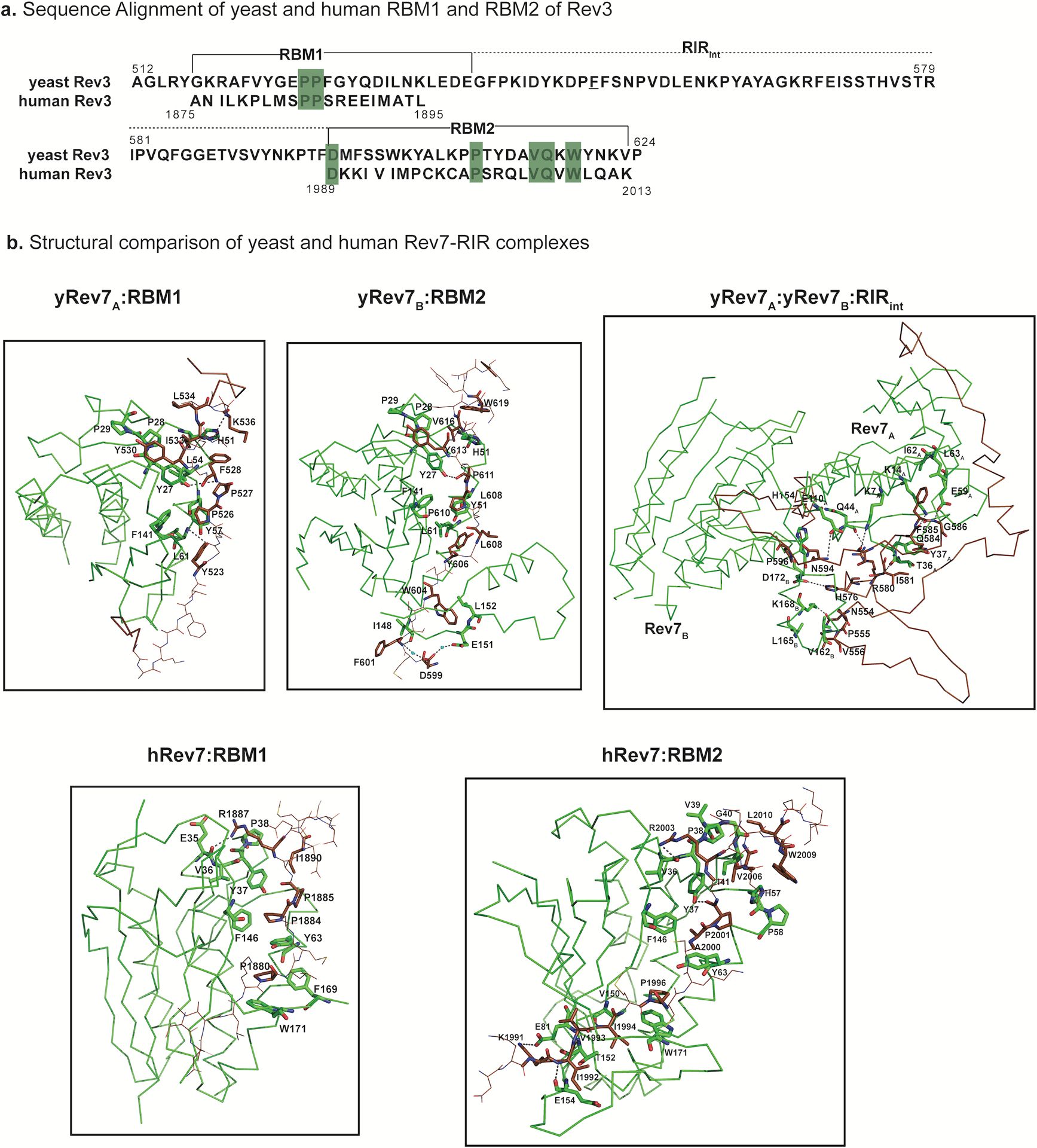
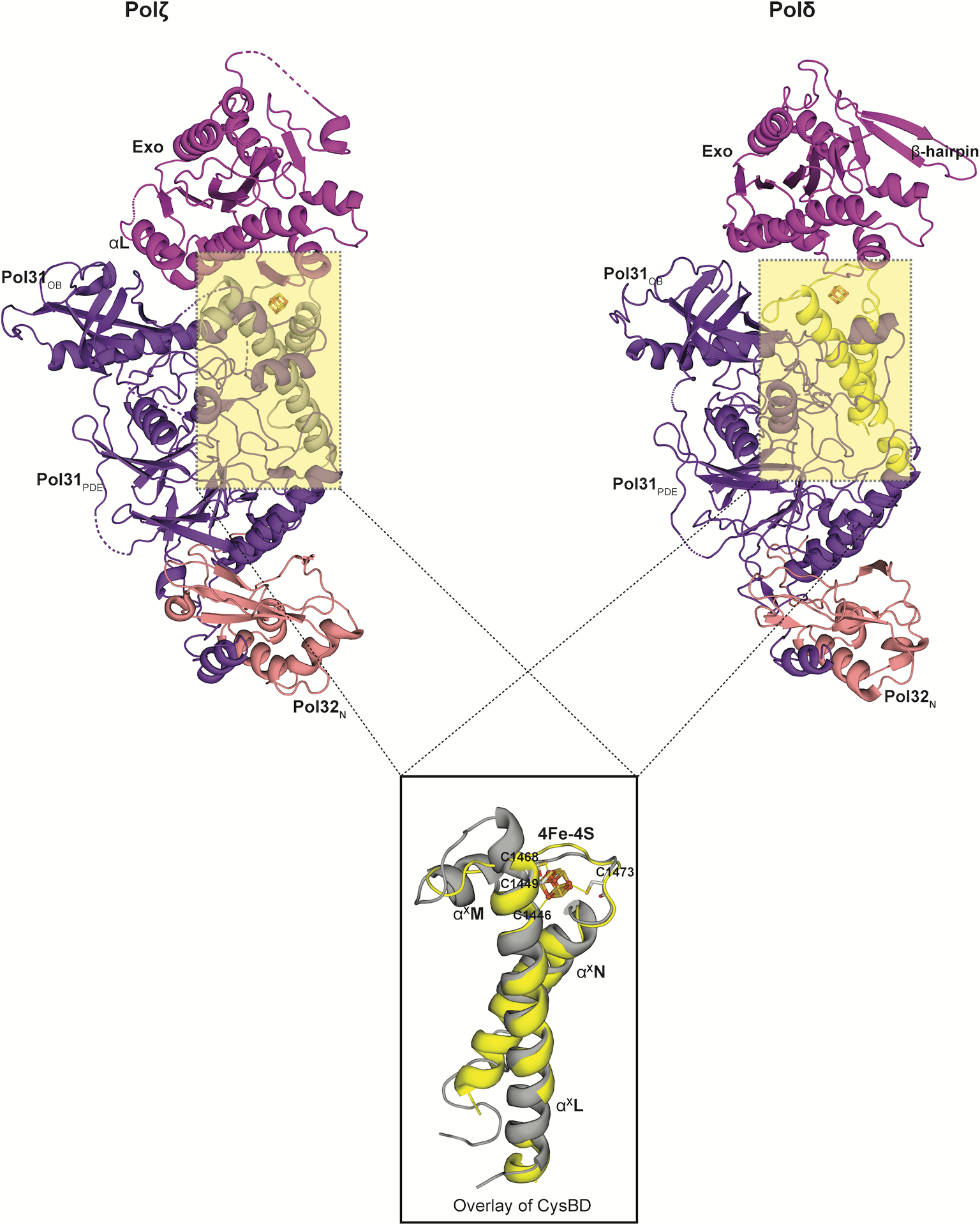
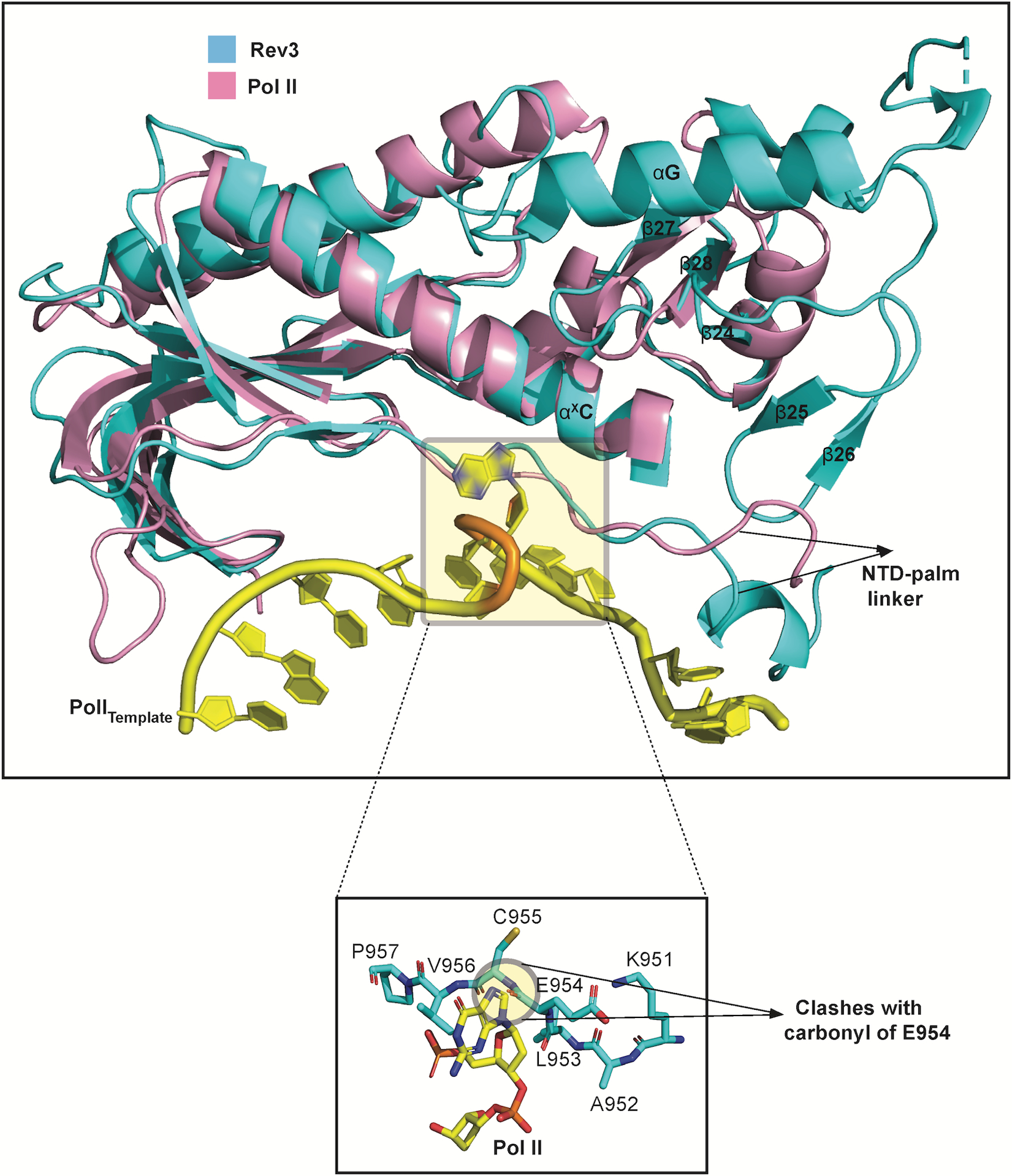
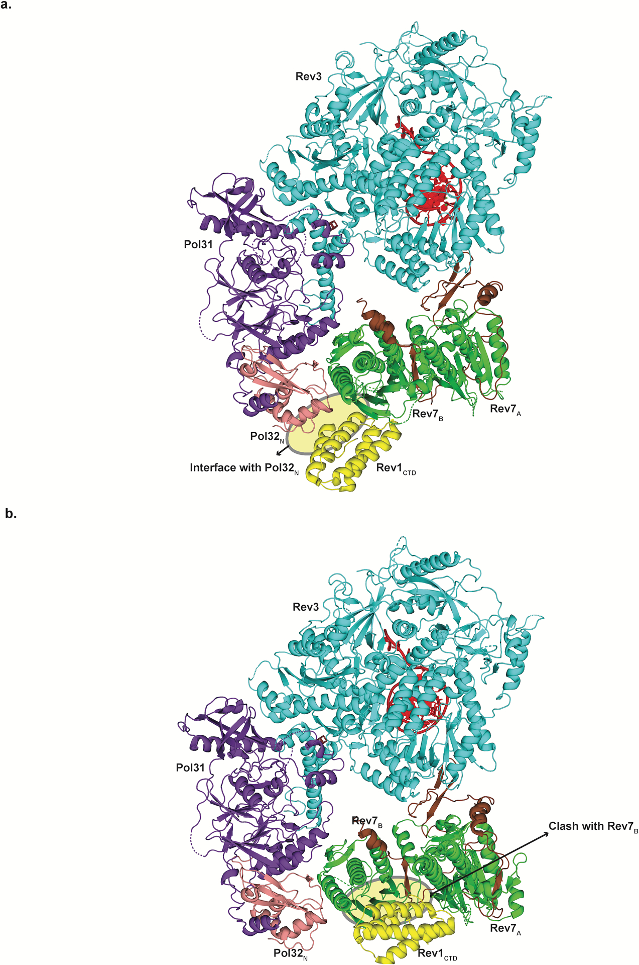
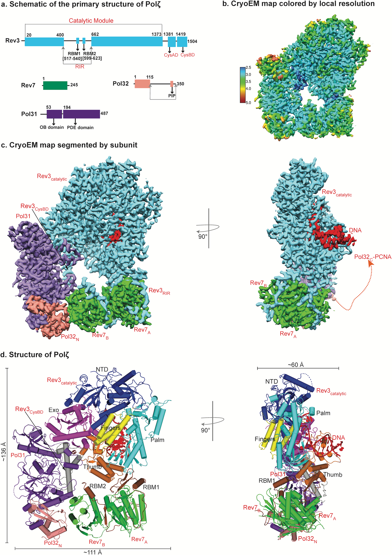
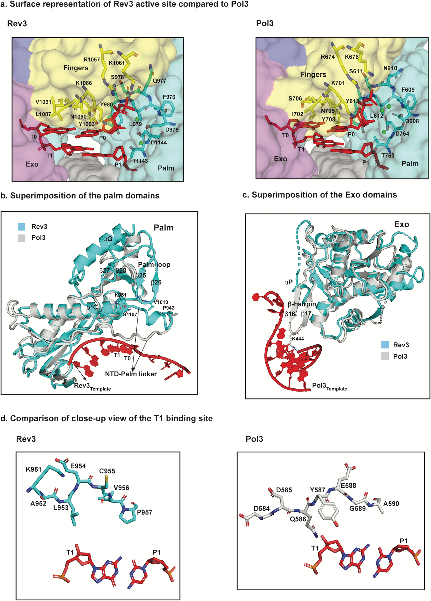
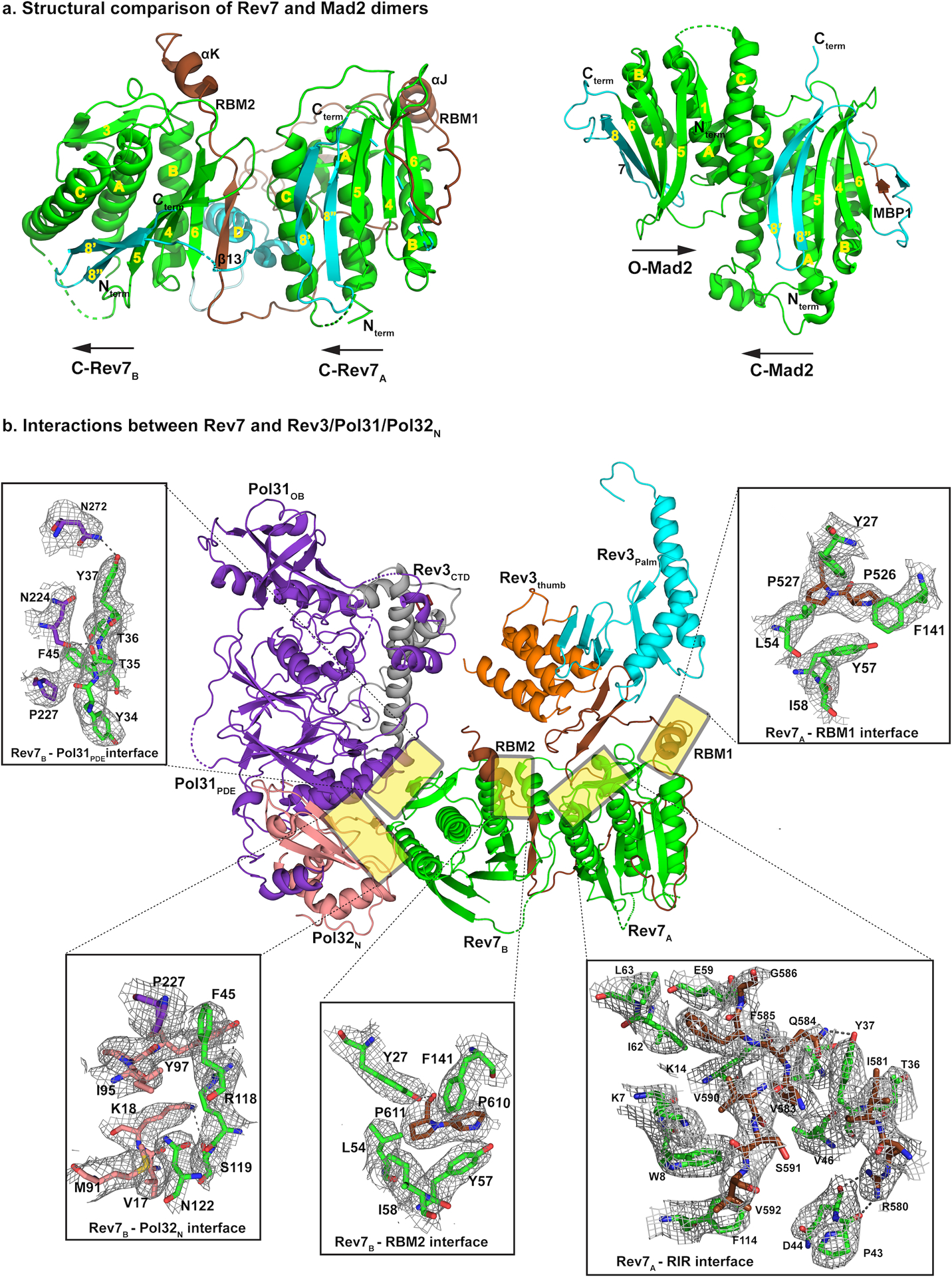
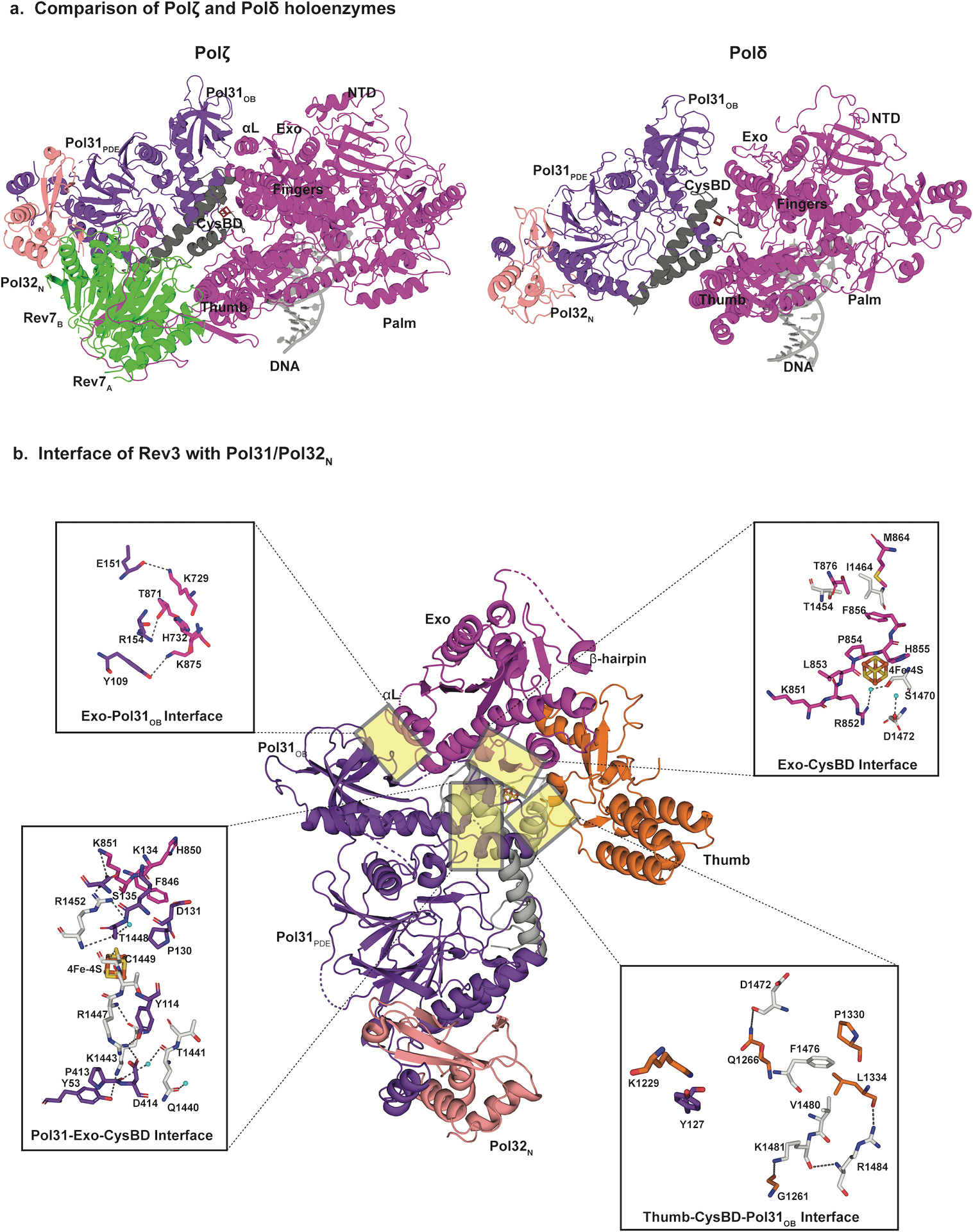
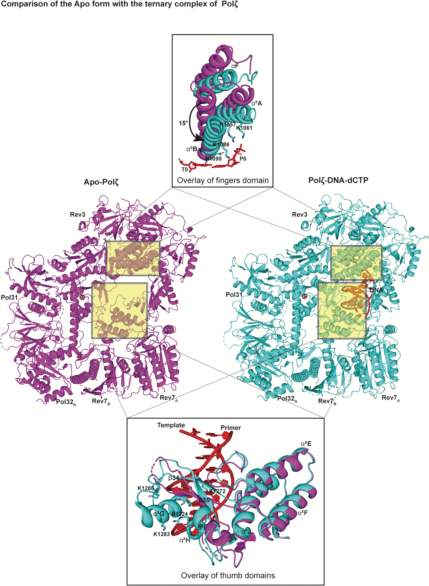
DNA polymerase ζ (Polζ) belongs to the same B-family as high-fidelity replicative polymerases, yet is specialized for the extension reaction in translesion DNA synthesis (TLS). Despite its importance in TLS, the structure of Polζ is unknown. We present cryo-EM structures of the Saccharomyces cerevisiae Polζ holoenzyme in the act of DNA synthesis (3.1 Å) and without DNA (4.1 Å). Polζ displays a pentameric ring-like architecture, with catalytic Rev3, accessory Pol31' Pol32 and two Rev7 subunits forming an uninterrupted daisy chain of protein-protein interactions. We also uncover the features that impose high fidelity during the nucleotide-incorporation step and those that accommodate mismatches and lesions during the extension reaction. Collectively, we decrypt the molecular underpinnings of Polζ's role in TLS and provide a framework for new cancer therapeutics.


Conflict of interest statement

Competing interests

The authors declare no competing interests.

Figures

Extended Data Fig. 1
Extended Data Fig. 1. Preferred specimen orientation
a, Data collected at 0° stage angle resulted in disproportionally low number of classes for side-views of the ternary complex of Polζ depicted in the 2D class averages. This resulted in a ‘smeared 3D model’ as shown by the anisotropic 3DFSC plot. Scale bar = 123 Å. b, Data collected at a stage angle of 0° for the apo-state of Polζ also had preferred set of views as shown in the 2D class averages. The final construction was anisotropic as depicted by the directional FSC plot. Scale bar = 123 Å..
Extended Data Fig. 2
Extended Data Fig. 2. Cryo-EM data collection and processing of Polζ-DNA-dCTP complex
a, Data were collected on Chameleon grids and particles from one session were picked with template based picker (FindEM) and processed in cryoSPARC to give a consensus map with a FSC0.143 of 3.57 Å. Major stages of processing are shown schematically and particles involved at each stage are highlighted in green. Scale bar = 137 Å. b, Final particles from two sessions were merged and used to train Topaz. Data processing from Topaz picked particles in cryoSPARC2 improved the sphericity. A schematic representation of the improved consensus map displaying a FSC0.143 of 3.2 Å is shown. Scale bar = 137 Å. c, Focused refinement of the final volume was done in cryoSPARC2. Masks were created (along the blue dashed line) for 3D refinement of Rev3 and accessory subunits separately to give consensus maps at 3.02 Å and 3.08 Å, respectively..
Extended Data Fig. 3
Extended Data Fig. 3. Cryo-EM data collection and processing for Polζ apo state
a, Data were collected at a 40° tilt angle and processed in cryoSPARC to give a good distribution of particles (green) with different views depicted in the 2D class averages. The final 3D reconstruction displaying a FSC0.143 of 4.1 Å showed an isotropic map amenable for model building. Scale bar = 123 Å. b, Per- particle CTF refinement of the map improved the sphericity further as shown by the 3DFSC plot..
Extended Data Fig. 4
Extended Data Fig. 4. Comparison of the NTD of Rev3 and Pol3
The NTD in Rev3 and Pol3, is composed of three motifs (I, II, III) but is much more elaborate and extended in Rev3. Loop 1 and Loop 2 contact all three motifs and connect the NTD to the fingers and palm domains, respectively..
Extended Data Fig. 5
Extended Data Fig. 5. Surface representation of the T1 binding site
Residues around the T1 site are shown (sticks) for Rev3 (left) and Pol3 (right). Surface for the palm domains and DNA are shown in cyan and grey, respectively. The T1 base (red) and the key residues are highlighted in dots..
Extended Data Fig. 6
Extended Data Fig. 6. Comparison of yeast and human Rev7-RIR complexes
a, Sequence alignment of yeast and human RBM1 and RBM2 regions of Rev3. Conserved prolines within RBM1 and RBM2 are highlighted in green. Also, highlighted are the conserved residues among the yeast and human homologs within the RIR region. b, Structural comparison of the yeast and human RBM1 and RBM2. Individual structures of human Rev7 with RBM1 peptide (hRev7:RBM1; PDB ID: 3ABD) and RBM2 peptide (hRev7:RBM2; PDB ID: 6BC8) are compared to the corresponding sub-regions (yRev7A:RBM1; yRev7B:RBM2) in the yeast Polζ holoenzyme. The protein residues involved in the interactions are highlighted in green and the RIR is shown in brown. The interactions of Rev7A and Rev7B with the RIR segment connecting RBM1 and RBM2 (yRev7A:yRev7B:RIRint) is also depicted..
Extended Data Fig. 7
Extended Data Fig. 7. Comparison between the CysBD of Polζ and Polδ
A superimposition of the CysBD of the Polζ (left; grey in color) and Polδ (right; yellow in color; PDB ID: 6P1H) shows conservation in its overall topology. Notably, helix αXM in Polζ CysBD has been substituted by a loop in Polδ (PDB ID: 6P1H). All the four cysteines interacting with the 4Fe-4S cluster in Rev3 are also highlighted..
Extended Data Fig. 8
Extended Data Fig. 8. Comparison of Rev3 and Pol II
Overlay of the palm domains of Rev3 and Pol II show a similar trajectory for the NTD-palm linker. In Rev3, this trajectory is coupled to interactions with the Palm-loop. The Pol II template DNA strand is shown in yellow (PDB ID: 3K5M). A close-up view of the looped-out abasic lesion and the adjoining 5’ guanine nucleotide. Notably, the guanine base clashes with the backbone carbonyl of E954 in Rev3..
Extended Data Fig. 9
Extended Data Fig. 9. Docking of Rev1 CTD on the Polζ holoenzyme
a, Superimposition of human Rev7-RBM1-Rev1CTD (PDB ID: 4EXT) on Rev7B shows close proximity to Pol32N (shown in yellow), highlighting the importance of Pol32N in stabilizing this interaction. b, Superimposition of the human Rev7-RBM1-Rev1CTD on Rev7A shows clashes of Rev1CTD with various secondary structure elements of Rev7B (shown in yellow)..
Fig. 1.
Fig. 1.
The architecture of DNA bound Polζ holoenzyme. a, Schematic of the primary structure of S. Cerevisiae Polζ subunits. Different colors denote each subunit. b, Near atomic resolution cryo-EM density map of DNA bound Polζ holoenzyme colored by local resolution. c, The three-dimensional reconstruction of Polζ holoenzyme viewed (left) perpendicular and parallel (right) to the DNA axis. A red dashed connector represents disordered Pol32c and the arrowhead marks the putative interaction location with PCNA. d, Cryo-EM structure of DNA bound Polζ colored by domain, and viewed from the same orientations as in (c).
Fig. 2.
Fig. 2.
Structure and cryo-EM density details of Rev3. a, Close-up view of the active site of Rev3 depicting key residues forming the T0-P0 binding site, including ligands and metal ions. Highlighted on the right is the well-resolved density for the T0 and T1 positions (red) and the sequence of the palindromic DNA employed to form the ternary complex. The region of the template-primer duplex enclosed in the box was built into the final model. b, Structure of Rev3 colored by domain. Dark blue, brown, magenta, cyan, yellow, orange and grey denote, respectively, the N-terminal, RIR, exonuclease, palm, fingers, thumb, and C-terminal domains of Rev3. Cryo-EM density for selected regions of Rev3 that highlight the differences in sequence with Pol3 (PDB ID: 3IAY), including residues in the inactive exonuclease domain, residues in close proximity to the CysBD, and the near absence of the β-hairpin region in Rev3. Also shown are close-up views of the coordination around the 4Fe-4S cluster, and the interfaces between the RIR and the palm and thumb domains.
Fig. 3.
Fig. 3.
Structural basis for fidelity and mismatch extension. a, Surface representation of a close-up view of the active sites of Rev3 and Pol3 depicting conserved residues interacting with the incoming nucleotide (dCTP) as well as the templating base (G). b, Superimposition of the palm domain of Rev3 and Pol3. The Rev3 template DNA strand is shown in red and the T1 base is highlighted. In comparison to Pol3, the palm-loop (β25 and β26) is a unique Rev3 structural element that interacts with the NTD-palm linker. Key residues in the NTD-palm linker are shown as sticks interacting with the palm-loop residues as well as the αXC palm helix. Another unique structural element, αG, which interacts with β27 and β28 is also highlighted. c, Superimposition of the exonuclease domains of Rev3 and Pol3. Overlay of the exonuclease domains of Rev3 and Pol3 shows a much shorter and disordered helical loop in Rev3 in comparison to a well-defined β-hairpin in Pol3. The Pol3 DNA is highlighted in red. d, Comparison of the T1 binding site in Rev3 and Pol3, showing details of the interaction between the NTD-palm linker region and the T1 base. The residue E954 in Rev3 points away from the T1 base, whereas residue Y587 stacks against the sugar in Pol3. L953 in Rev3 is the only residue close to the T1 base, resulting in a less constrained pocket in comparison to Pol3.
Fig. 4:
Fig. 4:
Rev7 dimer is the organizing center of Polζ holoenzyme. a, Rev7 dimer presents a novel head-to-tail arrangement (left) in comparison to other HORMA family members (right, PDB ID: 2V64). The C-terminal seatbelt region is highlighted in cyan. Both Rev7 subunits are in the closed state with Rev7A binding RBM1 and Rev7B binding RBM2. b, Both Rev7 subunits are required for assembly of the Polζ holoenzyme. Structure and cryo-EM density details of the Rev7 dimer interface with Rev3, Pol31 and Pol32N. RBM1 and RBM2 share a consecutive proline-proline motif that interacts with similar set of residues (Y27, F141, L54, Y57, for example) from both monomers. Interactions at the Rev7A and RIR interface that further stabilize the Rev7A:Rev7B dimer are shown. In addition, interactions of Rev7B with Pol31PDE–Pol32N depicted here aid in restricting the movement of Pol31–Pol32N subcomplex relative to Rev3.
Fig. 5:
Fig. 5:
Rigidity of Polζ holoenzyme. a, Comparison of the cryo-EM structures of DN bound Polζ (left) and Polδ (right, PDB ID: 6P1H) holoenzymes viewed parallel to the DNA axis. Compared to their positioning relative to the catalytic subunit in Polδ, the Pol31–Pol32N subcomplex pivots away from Rev3 through a combination of rotation and translation. This movement helps accommodate the Rev7 dimer. In addition, in Polζ the interface between the exonuclease domain and Pol31OB includes helix αL, and is more compact than in Polδ. b, Detailed views of selected interaction sites between Rev3 and Pol31 and Pol32N interface. Mobility of the Pol31-Pol32N subcomplex is further restricted due to extensive contacts at the interface between exonuclease, CysBD and Pol31. The 4Fe-4S cluster is shown as a box with orange and yellow sticks.
Fig. 6:
Fig. 6:
Conformational changes upon DNA binding. Structures of the ‘open’ apo state of Polζ (left, colored in magenta) in comparison to its ‘closed’ ternary complex (right, colored in cyan) viewed perpendicular to the DNA axis. An overlay of the fingers and the thumb domain of both states highlight significant conformational changes. In addition to the inward rotation of the fingers domain, various structural elements and loops (including αxG highlighted here) in the thumb domain become ordered upon DNA binding.

Similar articles

Cited by

References

    1. Prakash S, Johnson RE & Prakash L Eukaryotic translesion synthesis DNA polymerases: specificity of structure and function. Annu Rev Biochem 74, 317–353 (2005). - PubMed
    1. Jain R, Aggarwal AK & Rechkoblit O Eukaryotic DNA polymerases. Curr Opin Struct Biol 53, 77–87 (2018). - PubMed
    1. Makarova AV & Burgers PM Eukaryotic DNA polymerase zeta. DNA Repair (Amst) 29, 47–55 (2015). - PMC - PubMed
    1. Martin SK & Wood RD DNA polymerase zeta in DNA replication and repair. Nucleic Acids Res 47, 8348–8361 (2019). - PMC - PubMed
    1. Johnson RE, Washington MT, Haracska L, Prakash S & Prakash L Eukaryotic polymerases iota and zeta act sequentially to bypass DNA lesions. Nature 406, 1015–1019 (2000). - PubMed

Publication types

MeSH terms