Review
. 2020 Oct 16;15(20):3180-3208.
doi: 10.1002/asia.202000847. Epub 2020 Sep 21.

Metal Nanoparticles-Enhanced Biosensors: Synthesis, Design and Applications in Fluorescence Enhancement and Surface-enhanced Raman Scattering

Affiliations
Review

Metal Nanoparticles-Enhanced Biosensors: Synthesis, Design and Applications in Fluorescence Enhancement and Surface-enhanced Raman Scattering

Mohammad Tavakkoli Yaraki et al. Chem Asian J. .

Abstract

Metal nanoparticles (NP) that exhibit localized surface plasmon resonance play an important role in metal-enhanced fluorescence (MEF) and surface-enhanced Raman scattering (SERS). Among the optical biosensors, MEF and SERS stand out to be the most sensitive techniques to detect a wide range of analytes from ions, biomolecules to macromolecules and microorganisms. Particularly, anisotropic metal NPs with strongly enhanced electric field at their sharp corners/edges under a wide range of excitation wavelengths are highly suitable for developing the ultrasensitive plasmon-enhanced biosensors. In this review, we first highlight the reliable methods for the synthesis of anisotropic gold NPs and silver NPs in high yield, as well as their alloys and composites with good control of size and shape. It is followed by the discussion of different sensing mechanisms and recent advances in the MEF and SERS biosensor designs. This includes the review of surface functionalization, bioconjugation and (directed/self) assembly methods as well as the selection/screening of specific biorecognition elements such as aptamers or antibodies for the highly selective bio-detection. The right combinations of metal nanoparticles, biorecognition element and assay design will lead to the successful development of MEF and SERS biosensors targeting different analytes both in-vitro and in-vivo. Finally, the prospects and challenges of metal-enhanced biosensors for future nanomedicine in achieving ultrasensitive and fast medical diagnostics, high-throughput drug discovery as well as effective and reliable theranostic treatment are discussed.

Keywords: Assay design; Fluorescence detection; Metal nanoparticles; Plasmonic enhancement; SERS Biosensors.


Conflict of interest statement

The authors declare no conflict of interest.

Figures

Figure 1
Figure 1
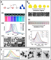
a) Comparison of the extinction spectra of different polyhedral Au nanostructures with average diameter of 90 nm,23 b) The role of the amount of Br in the formation of Au nanocube (top). TEM images of Au nanostructures synthesized at different concentration of Br ions (middle). The extinction spectra of Au nanocubes with different edge to curvature radius ratio (bottom). R refers to round edges/corners and S refers to sharp edges/corners,24 c) Schematic illustration of the purification process of Au bipyramids (AuBPs) by growing silver shell (top left), TEM images of purified AuBPs with different sizes (bottom left) and their corresponding extinction spectra (right).29
Figure 2
Figure 2
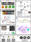
a) Triangular Au nanoplates synthesized by oxidative etching method: (i) Schematic diagram showing the formation mechanism of triangular Au nanoplates, (ii) solution color, (iii) LSPR spectrum and (iv) TEM images of Au nanoplates of different edge lengths.32 b) Synthesis of Au nanodiscs using HCl and H2O2 as etching agents: (i) TEM images and solution color of Au nanoplates at different stages of oxidation process. (ii) The extinction spectra of final product (i. e., spherical nanodiscs) as a function of added H2O2 volume in mL [37]. c) Synthesis of Au nanostars with multiple sharp tips using different HEPES/Au3+ ratio: (left) LSPR spectrum and (right) TEM images of Au nanostars.48a
Figure 3
Figure 3
a) Shape evolution of Ag polyhedral nanostructures as function of Cl ions concentration,56c b) Solution color (top) of Ag nanocubes with different edge length synthesized by polyol approach and their corresponding absorbance spectra (bottom). The number on each spectrum refers to the amount of added HCl solution into the reaction mixture .58 c) Synthesis (top) and formation mechanism (bottom) of Ag nanocubes with sharp edges/corners using CF3COOAg as precursor.59a d) Formation of right silver bipyramids through seed‐mediated growth coupled with in‐situ oxidation approach.5c e) (i) Synthesis of Ag nanoplates in ethanol,67 (ii) Photo‐mediated synthesis of triangular Ag nanoplates using H2O2.69 f) (i) Synthesis of AuAg nanoboxes using AgCl as the sacrificial template and etching agent. (ii) TEM images show the formation of thinner/porous AuAg nanoboxes by increasing the galvanic replacement time [74].
Figure 4
Figure 4
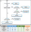
a) Decision map to select the right biorecognition element for biosensor designs [91], b) A comparison of different recognition elements including antibodies, aptamers, small molecules and polymers.93
Figure 5
Figure 5
a) Hybridization of two complementary single‐stranded DNAs, b) Bridge formation between the two DNA‐grafted particles with sticky ends or complementary sequences,96 c) dsDNA with short complementary stick ends to form the segmented dsDNA‐modified AuNPs for colorimetric biosensor development,95a d) Deep‐coating approach for the layer‐by‐layer (LBL) assembly of differently charged polyelectrolytes on the planar substrate,102 e) Steps in the LBL assembly of the oppositely charged polyelectrolytes in forming the multi‐layered particle.103
Figure 6
Figure 6
Jablonski diagram illustrating the principles of metal‐enhanced fluorescence for a classical fluorophore.
Figure 7
Figure 7
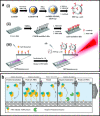
a) Schematic illustration of (i) surface‐enhanced fluorescence (SEF) tag ‐pAb preparation using shell‐isolated gold nanoparticle with a layer of Nile blue and another layer of silica followed by conjugation with antibody, (ii) Surface functionalization of glass slide by (3‐triethoxysilyl) propylsuccinic anhydride (TEPSA) to obtain carboxylic group on the surface, followed by antibody conjugation, (iii) Sandwich‐like structure for detection of Immunoglobulin‐M in the milk through metal‐enhanced fluorescence,125 b) Schematic illustration of the experimental procedure and assay construction for ultrasensitive trypsin detection using silvered 96 well plates.123
Figure 8
Figure 8
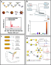
a) DNA‐functionalized gold nanobipyramids (AuNBPs) for the detection of ATP. Interaction between the DNA and ATP results in adjustment of distance between the AuBP and Cy‐7 for fluorescence enhancement,116 b) Ag@Silica nanoparticles (Ag@SiO2) for metal‐enhanced fluorescence detection of 2‐aminoanthracene,108 c) dsDNA‐functionalized Ag@SiO2 probes for MEF detection of ATP through a competition assay design. As the ATP molecule interacts with the aptamer leaving the original dsDNA structure, the PicoGreen (PG) dye cannot bind to the cDNA−Ag@SiO2, leading to weaker fluorescence,117 d) Schematic illustration of (A) the synthesis of Au nanocube@Silica@dye MEF biosensor. (B) Detection of pyrophosphate (PPi). (C) Detection of point mutation based on change in the fluorescence of MEF sensor in the presence of PPi or Cu2+,107 e) Schematic illustration of the “turn‐on” MEF probe for detecting γ‐glutamyl transpeptidase,120 f) Antibody‐functionalized AuNPs as MEF probe for fluorescence imaging. The confocal fluorescence images shows the detection of megalin in the tubules was coloured by the Alexa Fluor 488 (green) while the podocin in the glomeruli was coloured by the Alexa Fluor 647 (red),127 g) Schematic illustration of ultrasensitive ELISA based MEF biosensor for simultaneous multicolour detection of pathogens.121
Figure 9
Figure 9
a) (i) Schematic representation of aggregation‐based MEF biosensors using Au@Ag nanoparticles and RITC dye, ii) Fluorescence spectra and enhancement factor (inset) for cysteine detection and iii) Selectivity of cysteine in comparison with other amino acids using the Au@Ag enhanced biosensors.131 b) Schematic illustration of the sensing principle based on aggregation‐induced hot‐spots where target DNA can bridge two ssDNA‐functionalized metal nanoparticles and enhance the fluorescence of Cy5 dye molecules in the gap between plasmonic NPs.130
Figure 10
Figure 10
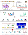
a) Different approaches for the development of selective SERS probes F,93 b) Developing planar SERS substrate using antibody‐functionalized Au nanocages (in the solution) and Au nanostars (self‐assembled on the surface of ITO glass). The sandwich structure enhances the Raman signal from Raman reporter molecules on the surface of Au nanocages and Au nanostars,171 c) Fabrication of planar SERS substrate by the deposition of Au bipyramid (AuBPs) inside the nanoholes of anodic aluminium oxide for detection of detect the aflatoxin B1.153
Figure 11
Figure 11
a) Schematic illustration of colloidal biosensor using DNA‐functionalized Au@AgAg nanorod as SERS probe (top) for the detection of gene HPV‐16 using magnetic bead (bottom),158 b) Synthesis of anisotropic Au nanorod/Polyaniline nanoparticles by a microfluidics approach (top) and change in the Raman intensity in the presence of different ions (bottom),163 c) development of polymer‐coated Au nanorod for simultaneous detection of different Raman active analytes (MO, FITC and R6G) via electrostatic interaction or hydrogen binding,182 d) Simultaneous detection of ochratoxin A and aflatoxin B1 by self‐assembly of Au nanoparticles around magnetic nanoparticle via the hybridization of designed aptamers and their complementary DNAs.192
Figure 12
Figure 12
a) Schematic illustration of the preparation folic acid‐functionalized gold bipyramid nanoparticles (AuNBPs) loaded by Raman reporter and its application in SERS detection of MCF‐7 breast cancer cells,187 b) Antibody‐functionalized Ag@Fe3O4 nanoparticles for multiplex detection of different strains of bacteria,185 c) Au nanobone functionalized with aptamer and Raman reporter for the detection of E.coli pathogen. The top right images shows the enhanced electric field around the Au nanobone in single and dimer configurations.178

References

    1. None
    1. Yu H., Peng Y., Yang Y., Li Z.-Y., npj Computat. Mater. 2019, 5, 45;
    1. Wang X., Huang S.-C., Hu S., Yan S., Ren B., Nat. Rev. Phys. 2020, 2, 253–271.
    1. None
    1. Tayebi M., Tavakkoli Yaraki M., Ahmadieh M., Tahriri M., Vashaee D., Tayebi L., Colloid Polym. Sci. 2016, 294, 1453–1462;

LinkOut - more resources