

Functional principles of baobab fruit pedicels - anatomy and biomechanics
- PMID: 32808645
- PMCID: PMC7684697
- DOI: 10.1093/aob/mcaa149
Functional principles of baobab fruit pedicels - anatomy and biomechanics
Abstract
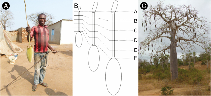
Background and aims: Fruit pedicels have to deal with increasing loads after pollination due to continuous growth of the fruits. Thus, they represent interesting tissues from a mechanical as well as a developmental point of view. However, only a few studies exist on fruit pedicels. In this study, we unravel the anatomy and structural-mechanical relationships of the pedicel of Adansonia digitata, reaching up to 90 cm in length.
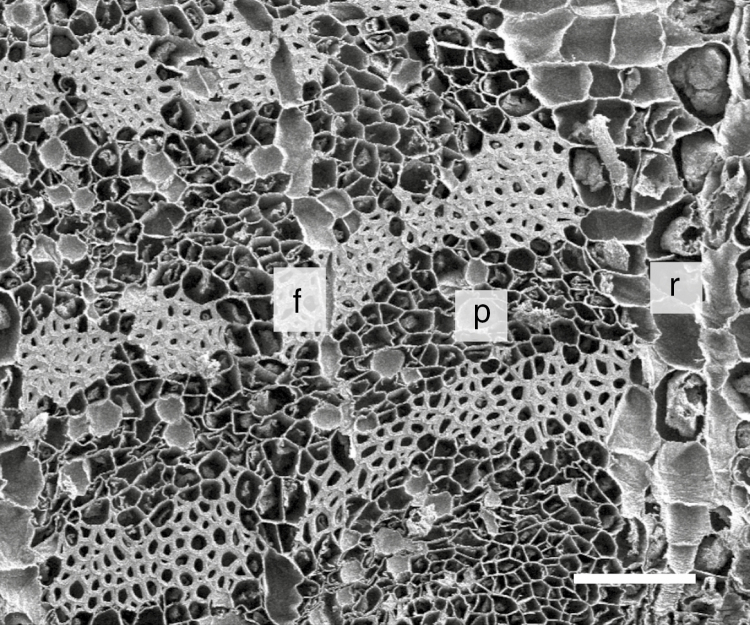
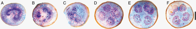
Methods: Morphological and anatomical analyses included examination of stained cross-sections from various positions along the stalk as well as X-ray microtomography and scanning electron microscopy. For mechanical testing, fibre bundles derived from the mature pedicels were examined via tension tests. For establishing the structural-mechanical relationships, the density of the fibre bundles as well as their cellulose microfibril distribution and chemical composition were analysed.
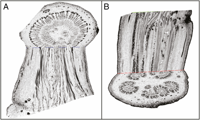
Key results: While in the peduncle the vascular tissue and the fibres are arranged in a concentric ring-like way, this organization shifts to the polystelic structure of separate fibre bundles in the pedicel. The polystelic pedicel possesses five vascular strands that consist of strong bast fibre bundles. The fibre bundles have a Young's modulus of up to 5 GPa, a tensile strength of up to 400 MPa, a high density (>1 g cm-3) and a high microfibril angle of around 20°.
Conclusions: The structural arrangement as well as the combination of high density and high microfibril angle of the bast fibre bundles are probably optimized for bearing considerable strain in torsion and bending while at the same time allowing for carrying high-tension loads.
Keywords: Adansonia digitata L; Angola; biomechanics; composite material; density; fibre characteristics; mechanical stresses; pedicel; specific Young’s modulus; strengthening tissue.
© The Author(s) 2020. Published by Oxford University Press on behalf of the Annals of Botany Company. All rights reserved. For permissions, please e-mail: journals.permissions@oup.com.
Figures

Similar articles
-
A materials perspective of Martyniaceae fruits: Exploring structural and micromechanical properties.Acta Biomater. 2015 Dec;28:13-22. doi: 10.1016/j.actbio.2015.10.002. Epub 2015 Oct 9. Acta Biomater. 2015. PMID: 26441125
-
Plant material features responsible for bamboo's excellent mechanical performance: a comparison of tensile properties of bamboo and spruce at the tissue, fibre and cell wall levels.Ann Bot. 2014 Dec;114(8):1627-35. doi: 10.1093/aob/mcu180. Epub 2014 Sep 1. Ann Bot. 2014. PMID: 25180290 Free PMC article.
-
Ontogenetic tissue modification in Malus fruit peduncles: the role of sclereids.Ann Bot. 2014 Jan;113(1):105-18. doi: 10.1093/aob/mct262. Epub 2013 Nov 27. Ann Bot. 2014. PMID: 24287811 Free PMC article.
-
Phytochemical Profile, Antioxidant and Antidiabetic Activities of Adansonia digitata L. (Baobab) from Mali, as a Source of Health-Promoting Compounds.Molecules. 2018 Nov 27;23(12):3104. doi: 10.3390/molecules23123104. Molecules. 2018. PMID: 30486448 Free PMC article.
-
Adansonia digitata L. (Baobab) Bioactive Compounds, Biological Activities, and the Potential Effect on Glycemia: A Narrative Review.Nutrients. 2023 May 1;15(9):2170. doi: 10.3390/nu15092170. Nutrients. 2023. PMID: 37432337 Free PMC article. Review.
References
-
- Baum DA. 1995. Systematic revision of Adansonia (Bombacaceae). Annals of the Missouri Botanical Garden 82: 440–471.
-
- Biagiotti J, Puglia D, Kenny JM. 2004. A review on natural fibre-based composites-Part I. Journal of Natural Fibers 1: 37–68.
-
- Brink M, Achigan-Dako EG. 2012. Fibres. Wageningen: PROTA Foundation.
-
- Bustan A, Erner V, Goldschmidt EE. 1995. Interactions between developing citrus fruits and their supportive vascular system. Annals of Botany 76: 657–666.
-
- Cave ID. 1968. The anisotropic elasticity of the plant cell wall. Wood Science and Technology 2: 268–278.
Publication types
MeSH terms
LinkOut - more resources
Full Text Sources
