Review
. 2020 Dec;25(6):522-544.
doi: 10.1177/2472630320950248. Epub 2020 Aug 18.

The Virological, Immunological, and Imaging Approaches for COVID-19 Diagnosis and Research

Affiliations
Review

The Virological, Immunological, and Imaging Approaches for COVID-19 Diagnosis and Research

An Sen Tan et al. SLAS Technol. 2020 Dec.

Abstract

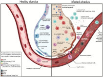
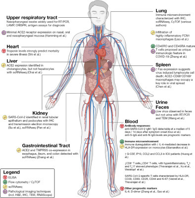
In 2019, a novel coronavirus (SARS-CoV-2) was found to cause a highly contagious disease characterized by pneumonia. The disease (COVID-19) quickly spread around the globe, escalating to a global pandemic. In this review, we discuss the virological, immunological, and imaging approaches harnessed for COVID-19 diagnosis and research. COVID-19 shares many clinical characteristics with other respiratory illnesses.Accurate and early detection of the infection is pivotal to controlling the outbreak, as this enables case identification, isolation, and contact tracing. We summarize the available literature on current laboratory and point-of-care diagnostics, highlight their strengths and limitations, and describe the emerging diagnostic approaches on the horizon.We also discuss the various research techniques that are being used to evaluate host immunity in laboratory-confirmed patients. Additionally, pathological imaging of tissue samples from affected patients has a critical role in guiding investigations on this disease. Conventional techniques, such as immunohistochemistry and immunofluorescence, have been frequently used to characterize the immune microenvironment in COVID-19. We also outline the emerging imaging techniques, such as the RNAscope, which might also aid in our understanding of the significance of COVID-19-specific biomarkers, such as the angiotensin-converting enzyme 2 (ACE2) cellular receptor.Overall, great progress has been made in COVID-19 research in a short period. Extensive, global collation of our current knowledge of SARS-CoV-2 will provide insights into novel treatment modalities, such as monoclonal antibodies, and support the development of a SARS-CoV-2 vaccine.

Keywords: COVID-19; diagnostics; immunology; pathology; specific T cells.


Conflict of interest statement

Declaration of Conflicting Interests

The authors declared no potential conflicts of interest with respect to the research, authorship, and/or publication of this article.

Figures

Figure 1.
Figure 1.
Loop-mediated isothermal amplification (LAMP). (A) LAMP begins when the forward inner primer (FIP) binds to the A2(C) region while the forward primer (A1) binds to A1(C), which displaces the FIP complementary strand. (B) The backward inner primer (BIP) binds B2(C) while the backward primer (B3) binds B3(C) and displaces the BIP complementary strand. (C) A complementary sequence that initiates loop formation is produced. (D) Loop structures are formed that allow for LAMP with the use of loop primers.
Figure 2.
Figure 2.
CRISPR technique. Viral RNA is converted to dsDNA using RT-RPA (recombinase polymerase amplification). (A) The CAS12a nuclease enzyme is activated upon complex binding to the target sequence, resulting in cleavage of the target sequence and the fluorescent RNA reporter. (B) T7 transcription converts DNA to complementary RNA. Cas13 nuclease enzyme activity is activated upon complex binding to the target sequence, resulting in a similar cleavage of the target sequence and the fluorescent RNA reporter.
Figure 3.
Figure 3.
Lateral flow immunoassay (LFIA). (A) Serum sample deposited on the sample pad. (B) Anti-SARS-CoV-2 antibodies in the sample will bind to the target antigen with a labeled tag. (C) Immobilized anti-human IgM antibodies will capture the SARS-CoV-2 antibody–antigen complex. (D) Control antibodies are captured by immobilized antibodies in the control line.
Figure 4.
Figure 4.
Chemiluminescence enzyme immunoassay (CLIA). SARS-CoV-2 antigens will capture IgM and IgG antibodies from the sample serum. Secondary antibodies that are conjugated with horseradish peroxidase (HRP) bind to the captured primary IgM and IgG antibodies and react with a chemiluminescent substrate to generate a strong chemiluminescent signal that is measured in terms of relative light units (RLU).
Figure 5.
Figure 5.
The alveolar immune microenvironment of patients with severe COVID-19 infection—comparison between healthy alveolus (left) and infected alveolus (right). As part of the SARS-CoV-2 antiviral response, pulmonary recruitment involves immune cells such as, but not limited to, (i) activated T cells, identifiable based on the expression of HLA-DR, CD38, CD69, CD44, and CD25; (ii) CD16+CD107a+ NK cells; (iii) CD11b+CD16+ neutrophils; (iv) FCN1+ macrophages; and (v) CD14+CD276+ dendritic cells. Recruitment of these pro-inflammatory immune cells results in a cytokine storm within the lung, as reported by elevated levels of IL-1β, IL-6, IL-8, and GM-CSF. This overall hyperinflammatory environment, when fueled by dysregulation of macrophage and lymphocyte populations in the lung, is a contributing factor to the observed lung function failure. In the blood immune microenvironment, despite consistent reports of lymphopenia, higher populations of CD14+CD16+ monocytes were observed. This is accompanied by a cytokine storm involving IL-1β, IL-2, IL-6, IL-8, TNFα, IFNγ, GM-CSF, and granzyme B, as well as an increase in functionally exhausted PD-1+Tim3+ T cells.
Figure 6.
Figure 6.
Selected findings by organ system. In the liver, scRNAseq has identified ACE2 expression predominantly on cholangiocytes, and cholangiocyte dysfunction has been speculated to explain liver injury. In the kidney, evidence of SARS-CoV-2 within renal tubular epithelium and podocytes suggests acute kidney injury as a primary element of severe COVID-19 infection. Within the gastrointestinal tract, ACE2 expression as well as detectable live virus in fecal samples indicates that fecal–oral transmission should be considered a possible route of transmission. In the lung, studies have characterized the immune microenvironment using pathological imaging approaches as well as scRNAseq and CyTOF. CD45RO and CD45RA mature T cells have been proposed as a unique immunologic feature in COVID-19. In the spleen and lymph nodes, ACE2CD68+CD169+ macrophages are postulated to mediate SARS-CoV-2 translocation. CyTOF and scRNAseq approaches have also elucidated the extensive immune dysregulation at the heart of COVID-19. Immunological techniques like ELISA have also identified prognostic markers in serum such as troponin, IL-6, and D-dimer.

Similar articles

Cited by

References

    1. Wang C., Horby P.W., Hayden F.G., et al. A Novel Coronavirus Outbreak of Global Health Concern. Lancet. 2020;395:470–473. - PMC - PubMed
    1. Zhu N., Zhang D., Wang W., et al. A Novel Coronavirus from Patients with Pneumonia in China, 2019. N. Engl. J. Med. 2020;382:727–733. - PMC - PubMed
    1. World Health Organization. Coronavirus Disease 2019 (COVID-19) Situation Report—162. https://www.who.int/docs/default-source/coronaviruse/20200630-covid-19-s... (accessed Aug 8, 2020).
    1. Zhou F., Yu T., Du R., et al. Clinical Course and Risk Factors for Mortality of Adult Inpatients with COVID-19 in Wuhan, China: A Retrospective Cohort Study. Lancet. 2020;395:1054–1062. - PMC - PubMed
    1. Remuzzi A., Remuzzi G. COVID-19 and Italy: What Next? Lancet. 2020;395:1225–1228. - PMC - PubMed