

Review
doi: 10.1007/s40123-020-00291-5.
Epub 2020 Aug 18.
Retinal Pigment Epithelial Detachment in Age-Related Macular Degeneration
Affiliations
- PMID: 32809132
- PMCID: PMC7708599
- DOI: 10.1007/s40123-020-00291-5
Item in Clipboard
Review
Retinal Pigment Epithelial Detachment in Age-Related Macular Degeneration
Ophthalmol Ther.
2020 Dec.
Abstract
Retinal pigment epithelial detachment is defined as a separation of the retinal pigment epithelium from the inner collagenous layer of Bruch's membrane. It is a common manifestation in both dry and wet types of age-related macular degeneration. This review aims to provide a comprehensive guide to the pathophysiology, clinical and imaging characteristics, natural course and treatment of the various types of pigment epithelial detachments in order to assist in diagnosis and management of this important feature of age-related macular degeneration.
Keywords: Age-related macular degeneration; Drusenoid; Fibrovascular; Haemorrhagic; Pigment epithelial detachment; Serous.
Figures
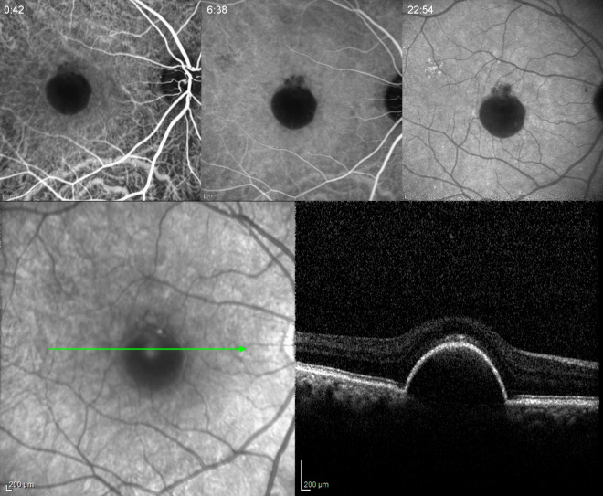
Serous PED in a 58-year-old woman. Early, mid- and late phase of ICGA showing hypofluorescence in all phases. Note the absence of a hot spot. Spectral-domain OCT (SD-OCT) did not demonstrate any IRF or SRF
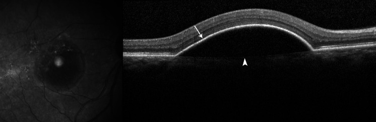
SD-OCT of a serous PED. The PED appears as a dome-shaped elevation of the RPE (arrow) over a uniform hypo-reflective space. Bruch’s membrane is seen at the base of the PED (arrowhead)
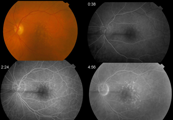
Drusenoid PED in a 76-year-old woman. Color fundus photograph with multiple soft drusen, RPE changes and a central drusenoid PED. Early, mid- and late phase of FA showing early hyperfluorescence corresponding to the area of drusen and PED as well as late staining with no leakage
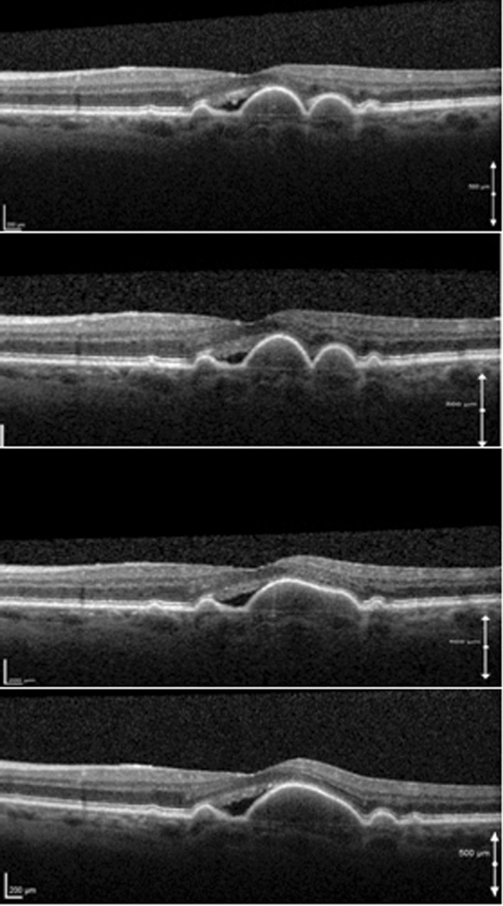
Serial SD-OCT scans of a 68-year-old woman with soft drusen and an adjacent hypo-reflective space who was followed up for 3 years. OCT scans were taken on yearly intervals. Note the gradual coalescence of soft drusen and the development of a drusenoid PED. The PED has a homogenous, mildly hyper-reflective interior, and Bruch’s membrane is seen at its base. During the observation period the hypo-reflective space and visual acuity remained stable
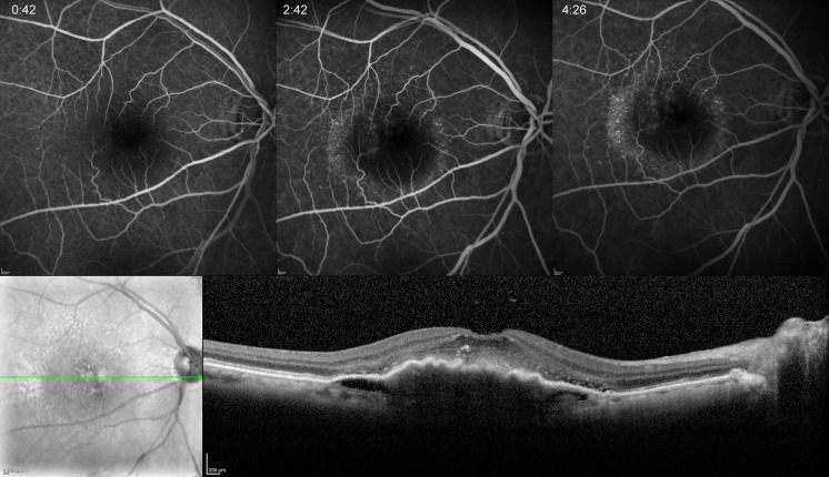
Occult CNV in a 76-year-old woman. Early, mid- and late frames of FA demonstrating stippled hyperfluorescence in mid- and late phases. SD-OCT shows an irregular elevation of the RPE with a non-uniform hyper-reflective interior. There is also mild SRF and hyper-reflective foci in the neuroretina
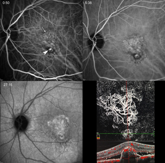
IPCV in a 70-year-old man. Early, mid- and late phase of ICGA showing the polyp (arrowhead) as well as associated branching vascular network (arrow) that is more clearly delineated on en-phase OCTA (lower right panel). Cross-sectional OCTA demonstrates a steep, convex PED with non-uniform hyper-reflective interior, vascular flow at its apex and surrounding SRF
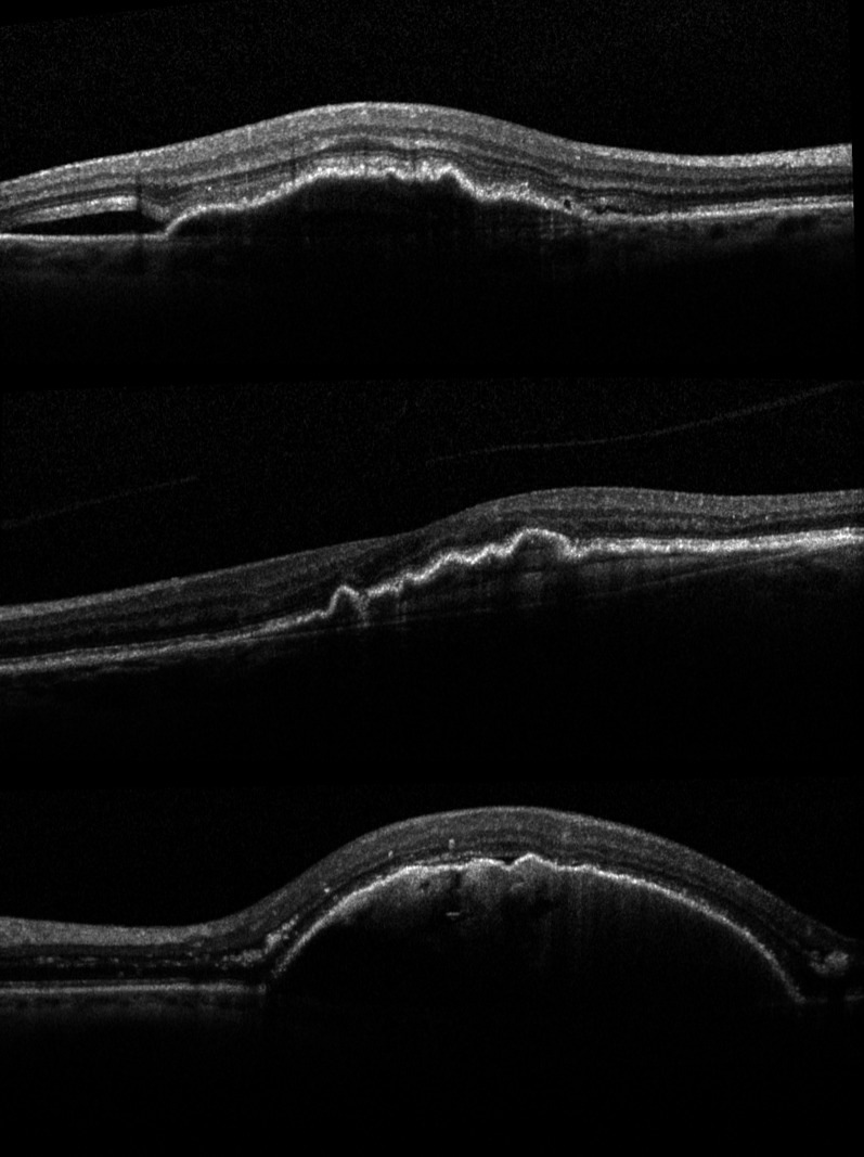
SD-OCT scans of different cases of fibrovascular PEDs. Note the irregular elevation of the RPE, the inhomogeneous hyper-reflective interior, and the presence of SRF (top, bottom) as well as hyper-reflective neuroretinal foci (top, bottom)
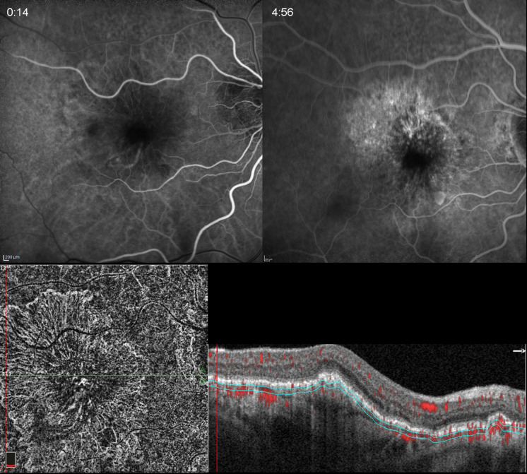
Fibrovascular PED in an 89-year-old woman who had anti-VEGF treatment despite the absence of SRF or IRF in cross-sectional OCTA. Early and late phases of FA showing mild late staining. En-phase OCTA shows the CNV, which in this case demonstrates signs of maturity (prominent trunk vessels, pruning). This case was considered a “silent CNV” due to the absence of exudative signs throughout follow-up
Similar articles
-
[Avascular retinal pigment epithelium detachments in age-related macular degeneration].Vestn Oftalmol. 2020;136(4. Vyp. 2):284-288. doi: 10.17116/oftalma2020136042284. Vestn Oftalmol. 2020. PMID: 32880152 Russian.
-
Clinicopathological correlation of retinal pigment epithelial tears in exudative age related macular degeneration: pretear, tear, and scarred tear.Br J Ophthalmol. 2001 Apr;85(4):454-60. doi: 10.1136/bjo.85.4.454. Br J Ophthalmol. 2001. PMID: 11264137 Free PMC article.
-
A hyporeflective space between hyperreflective materials in pigment epithelial detachment and Bruch's membrane in neovascular age-related macular degeneration.BMC Ophthalmol. 2014 Dec 16;14:159. doi: 10.1186/1471-2415-14-159. BMC Ophthalmol. 2014. PMID: 25515712 Free PMC article.
-
[Drusen in Bruch's membrane. Their significance for the pathogenesis and therapy of age-associated macular degeneration].Ophthalmologe. 1992 Oct;89(5):363-86. Ophthalmologe. 1992. PMID: 1304217 Review. German.
-
Drusen in age-related macular degeneration: pathogenesis, natural course, and laser photocoagulation-induced regression.Surv Ophthalmol. 1999 Jul-Aug;44(1):1-29. doi: 10.1016/s0039-6257(99)00072-7. Surv Ophthalmol. 1999. PMID: 10466585 Review.
Cited by
-
Treat & extend in neovascular age-related macular degeneration: how we got here and where do we go next?Eye (Lond). 2023 Mar;37(4):581-583. doi: 10.1038/s41433-022-02221-0. Epub 2022 Sep 5. Eye (Lond). 2023. PMID: 36064769 Free PMC article. No abstract available.
-
A New Generation of Gene Therapies as the Future of Wet AMD Treatment.Int J Mol Sci. 2024 Feb 17;25(4):2386. doi: 10.3390/ijms25042386. Int J Mol Sci. 2024. PMID: 38397064 Free PMC article. Review.
-
The Role of Medical Image Modalities and AI in the Early Detection, Diagnosis and Grading of Retinal Diseases: A Survey.Bioengineering (Basel). 2022 Aug 4;9(8):366. doi: 10.3390/bioengineering9080366. Bioengineering (Basel). 2022. PMID: 36004891 Free PMC article. Review.
-
High-accuracy 3D segmentation of wet age-related macular degeneration via multi-scale and cross-channel feature extraction and channel attention.Biomed Opt Express. 2024 Jan 26;15(2):1115-1131. doi: 10.1364/BOE.513619. eCollection 2024 Feb 1. Biomed Opt Express. 2024. PMID: 38404340 Free PMC article.
-
Comparative efficacy of aflibercept and ranibizumab in the treatment of age-related macular degeneration with retinal pigment epithelial detachment: a systematic review and network meta-analysis.BMC Ophthalmol. 2023 Nov 21;23(1):473. doi: 10.1186/s12886-023-03214-7. BMC Ophthalmol. 2023. PMID: 37990182 Free PMC article.
References
-
- Gass JD, Norton EW, Justice J., Jr Serous detachment of the retinal pigment epithelium. Trans Am Acad Ophthalmol Otolaryngol. 1966;70:990–1015. - PubMed
-
- Bird AC. Doyne Lecture: Pathogenesis of retinal pigment epithelial detachment in the elderly: the relevance of Bruch’s membrane change. Eye. 1991;5:1–12. - PubMed
-
- Bird AC, Marshall J. Retinal pigment epithelial detachments in the elderly. Trans Ophthalmol Soc U K. 1986;105(Pt 6):674–682. - PubMed
Publication types
LinkOut - more resources
Full Text Sources
Other Literature Sources
