. 2020 Nov;36(11):1344-1354.
doi: 10.1007/s12264-020-00557-9. Epub 2020 Aug 18.

Spinal CCL2 Promotes Pain Sensitization by Rapid Enhancement of NMDA-Induced Currents Through the ERK-GluN2B Pathway in Mouse Lamina II Neurons

Affiliations

Spinal CCL2 Promotes Pain Sensitization by Rapid Enhancement of NMDA-Induced Currents Through the ERK-GluN2B Pathway in Mouse Lamina II Neurons

Hui Zhang et al. Neurosci Bull. 2020 Nov.

Abstract

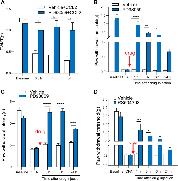
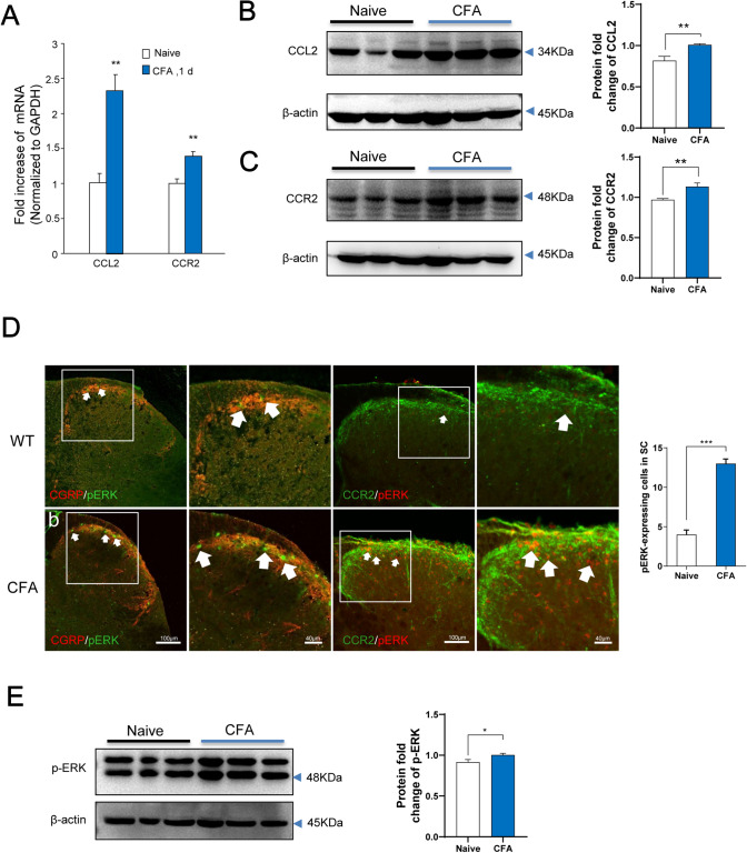
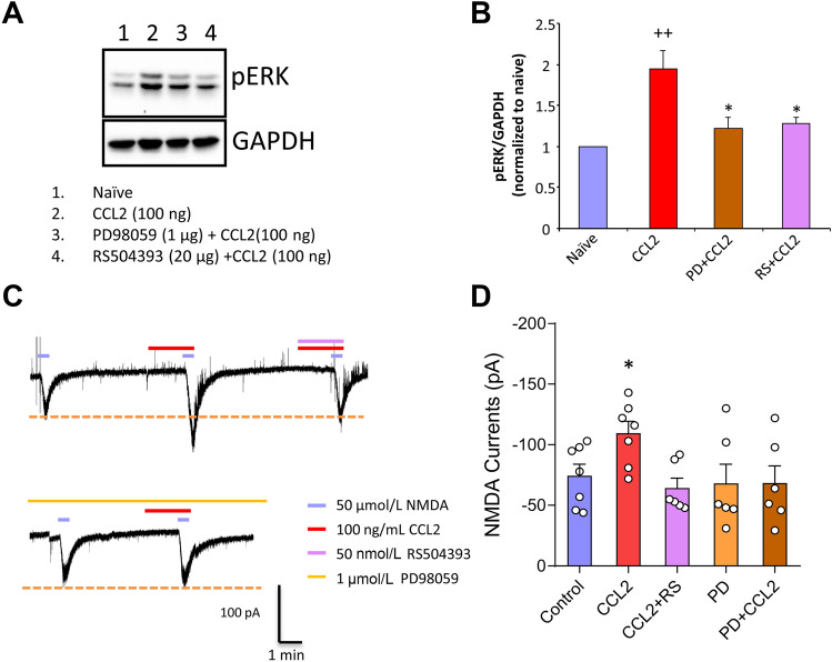
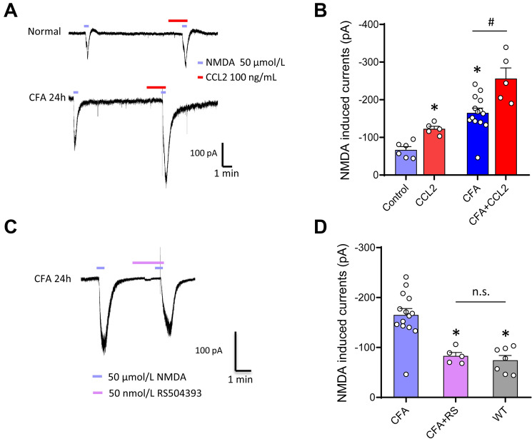
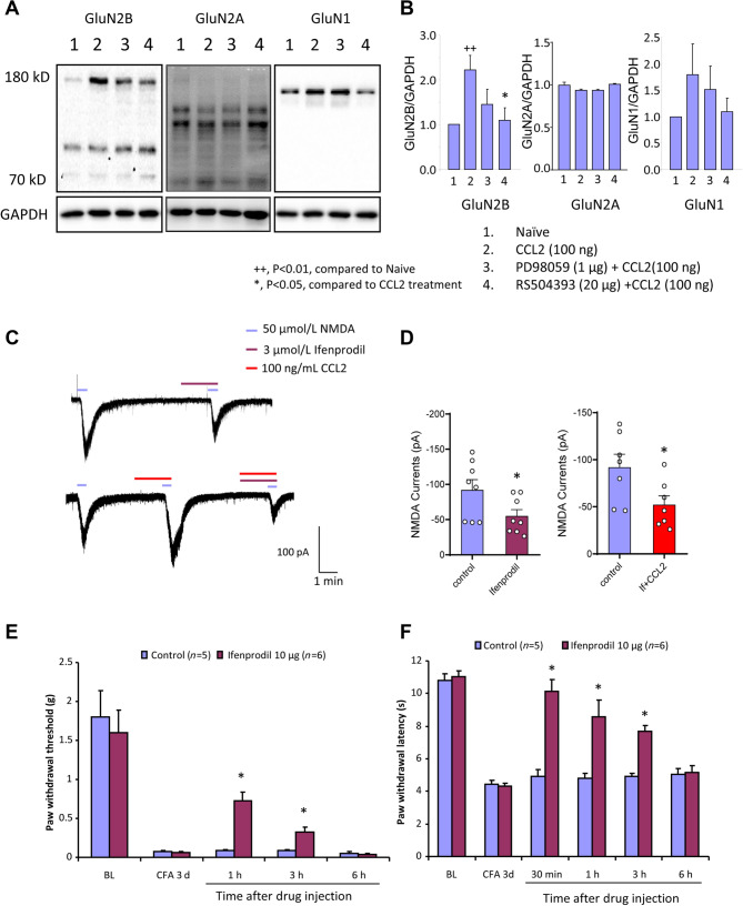
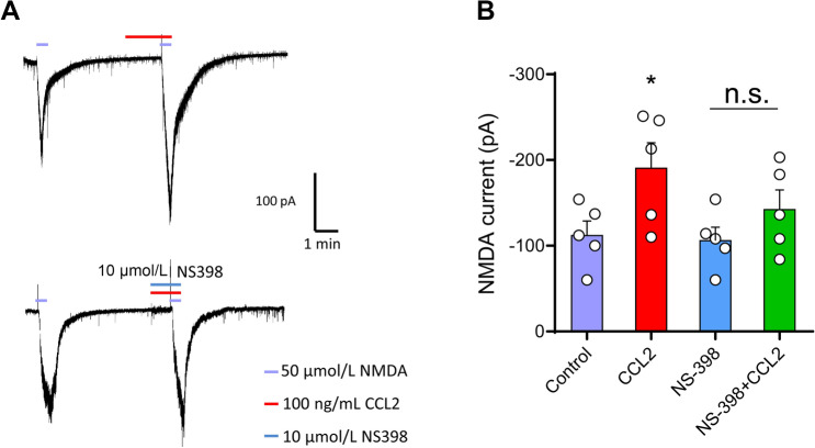
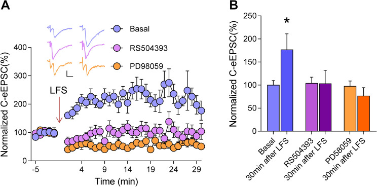
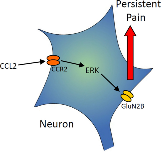
Previous studies have shown that CCL2 (C-C motif chemokine ligand 2) induces chronic pain, but the exact mechanisms are still unknown. Here, we established models to explore the potential mechanisms. Behavioral experiments revealed that an antagonist of extracellular signal-regulated kinase (ERK) inhibited not only CCL2-induced inflammatory pain, but also pain responses induced by complete Freund's adjuvant. We posed the question of the intracellular signaling cascade involved. Subsequent experiments showed that CCL2 up-regulated the expression of phosphorylated ERK (pERK) and N-methyl D-aspartate receptor [NMDAR] subtype 2B (GluN2B); meanwhile, antagonists of CCR2 and ERK effectively reversed these phenomena. Whole-cell patch-clamp recordings revealed that CCL2 enhanced the NMDAR-induced currents via activating the pERK pathway, which was blocked by antagonists of GluN2B and ERK. In summary, we demonstrate that CCL2 directly interacts with CCR2 to enhance NMDAR-induced currents, eventually leading to inflammatory pain mainly through the CCL2-CCR2-pERK-GluN2B pathway.

Keywords: C-C motif chemokine ligand 2; Extracellular signal-regulated kinase; Monocyte chemoattractant protein 1; Neuron-glial interaction.


Conflict of interest statement

The authors claim that there are no conflicts of interest.

Figures

Fig. 1
Fig. 1
Inhibition of CCL2- and CFA-induced mechanical hyperalgesia by intrathecal injection of RS504393 or PD98059. A Prevention of CCL2 (100 ng, i.t.)-induced mechanical hyperalgesia by PD98059 (1 g, i.t.). B Reversal of CFA-induced mechanical hyperalgesia by PD98059 (1 μg, i.t.) given 1 day after CFA injection. C Reversal of CFA-induced thermal hyperalgesia by PD98059 (1 μg, i.t.) given 1 day after CFA injection. D Reversal of CFA-induced mechanical hyperalgesia by RS504393 (20 μg, i.t.) given 1 day after CFA injection. *P < 0.05, **P < 0.01, ***P < 0.001, ****P < 0.0001, n = 5–6.
Fig. 2
Fig. 2
CFA induces upregulation of CCL2, CCR2, and ERK activation in the spinal dorsal horn. A Real-time PCR reveals distinct regulation of CCL2 and CCR2 mRNA expression in the spinal cord following inflammation. Both CCL2 and CCR2 are upregulated after inflammatory pain. CFA induces a transient increase in CCL2 and CCR2 mRNA levels on day 1 (**P < 0.01, n = 6–7). B, C Western blots showing that CFA leads to a significant increase in CCL2 and CCR2 expression. D Immunofluorescence staining showing an increase in pERK-immunoreactive (pERK-ir) cells in CCR2-expressing neurons in the ipsilateral (injured) dorsal horn at 24 h after CFA (right panels, enlargement of the white frames; arrows, pERK-ir cells in the superficial dorsal horn). E Western blot showing that CFA results in a significant increase in pERK expression.
Fig. 3
Fig. 3
CCL2 enhances NMDA currents via ERK activation. A, B Western blots showing CCL2 directly increases neuronal pERK levels, and this effect is blocked by intracellular PD98059 (pERK blocker) and extracellular RS504393 (CCR2 inhibitor) (++P < 0.01 vs Naïve, *P < 0.05 vs CCL2, n = 6–7). C, D Superfusion of NMDA (50 μmol) at a holding potential of –40 mV, induces a robust inward current in WT mice. Superfusion of CCL2 at 100 ng/mL increases NMDA-induced currents (*P < 0.05, n = 6–7), and this is abolished by the antagonist RS504393. The ERK blocker PD98059 in the pipette solution abolishes the enhancement effect of CCL2 on NMDA-induced currents on lamina IIo neurons.
Fig. 4
Fig. 4
Role of endogenous CCR2 and effects of CCR2 antagonist on NMDA currents after inflammation. A, B NMDA-induced currents are enhanced in spinal lamina IIo neurons in both naïve (A, upper panels) and inflammatory (A, lower panels) states. C, D Superfusion of the CCR2 antagonist RS504393 eliminates the facilitation of NMDA-induced currents by CFA inflammation in spinal lamina IIo neurons. *P < 0.05, n = 5–14.
Fig. 5
Fig. 5
CCL2 enhances NMDA currents via GluN2B activation. A, B Western blots showing that CCL2 induces a significant increase in GluN2B expression. C, D Ifenprodil, a GluN2B blocker, partially inhibits NMDAR-induced currents in lamina IIo neurons and abolishes the CCL2-induced enhancement of these currents. E, F Behavioral tests showing that CFA-induced mechanical allodynia and thermal hyperalgesia are reversed by i.p. Ifenprodil. *P < 0.05, ++P < 0.01, n = 6–8.
Fig. 6
Fig. 6
Enhancement of NMDA currents by CCL2/CCR2 via the Cox-2 pathway. A, B Perfusion of NMDA (50 μmol) at a holding potential of –40 mV induces a robust inward current which is increased by perfusion with CCL2. The enhancement of NMDA currents by CCL2 is partly blocked by NS398 (10 μm), a Cox-2 inhibitor. *P < 0.05, n = 5.
Fig. 7
Fig. 7
Elimination of LTP of C-fiber eEPSCs in dorsal horn neurons by RS504393 and PD98059. A Spinal LTP is induced by low-frequency stimulation (240 pulses at 2 Hz,) of C-fiber intensity in C57/Bl6 mice. Addition of RS504393 or PD58059 to the pipette solution largely inhibits the induction of LTP. Scale bars, 50 pA and 10 ms. B Quantitative summary of experiments as in A. *P < 0.05, one-way ANOVA, n = 5–6 neurons/group.
Fig. 8
Fig. 8
Diagram of the model. Following peripheral inflammation, directly released CCL2 acts on CCR2 on superficial dorsal horn neurons; this activates the ERK signaling cascade, leading to up-regulation of GluN2B on the cell membrane, and inducing chronic pain.

Similar articles

Cited by

References

    1. Basbaum AI, Bautista DM, Scherrer G, Julius D. Cellular and molecular mechanisms of pain. Cell. 2009;139:267–284. - PMC - PubMed
    1. Ji RR, Xu ZZ, Gao YJ. Emerging targets in neuroinflammation-driven chronic pain. Nat Rev Drug Discov. 2014;13:533–548. - PMC - PubMed
    1. Gold MS, Gebhart GF. Nociceptor sensitization in pain pathogenesis. Nat Med. 2010;16:1248–1257. - PMC - PubMed
    1. Kiguchi N, Kobayashi Y, Kishioka S. Chemokines and cytokines in neuroinflammation leading to neuropathic pain. Curr Opin Pharmacol. 2012;12:55–61. - PubMed
    1. White FA, Jung H, Miller RJ. Chemokines and the pathophysiology of neuropathic pain. Proc Natl Acad Sci U S A. 2007;104:20151–20158. - PMC - PubMed

MeSH terms