Review
. 2020 Sep;9(17):e016760.
doi: 10.1161/JAHA.120.016760. Epub 2020 Aug 15.

Impact of Interatrial Shunts on Invasive Hemodynamics and Exercise Tolerance in Patients With Heart Failure

Affiliations
Review

Impact of Interatrial Shunts on Invasive Hemodynamics and Exercise Tolerance in Patients With Heart Failure

Jan M Griffin et al. J Am Heart Assoc. 2020 Sep.

Abstract

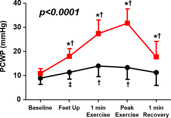
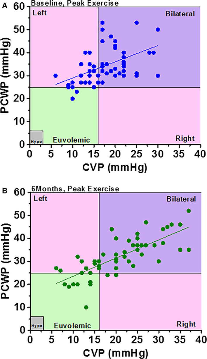
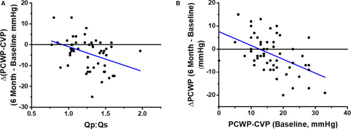
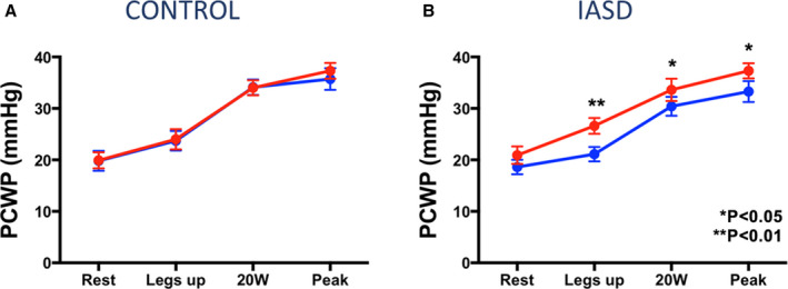
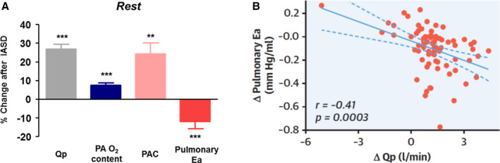
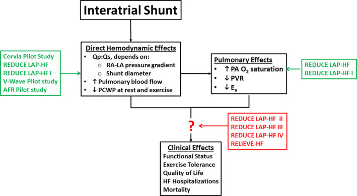
Approximately 50% of patients with heart failure have preserved ejection fraction. Although a wide variety of conditions cause or contribute to heart failure with preserved ejection fraction, elevated left ventricular filling pressures, particularly during exercise, are common to all causes. Acute elevation in left-sided filling pressures promotes lung congestion and symptoms of dyspnea, while chronic elevations often lead to pulmonary vascular remodeling, right heart failure, and increased risk of mortality. Pharmacologic therapies, including neurohormonal modulation and drugs that modify the nitric oxide/cyclic GMP-protein kinase G pathway have thus far been limited in reducing symptoms or improving outcomes in patients with heart failure with preserved ejection fraction. Hence, alternative means of reducing the detrimental rise in left-sided heart pressures are being explored. One proposed method of achieving this is to create an interatrial shunt, thus unloading the left heart at rest and during exercise. Currently available studies have shown 3- to 5-mm Hg decreases of pulmonary capillary wedge pressure during exercise despite increased workload. The mechanisms underlying the hemodynamic changes are just starting to be understood. In this review we summarize results of recent studies aimed at elucidating the potential mechanisms of improved hemodynamics during exercise tolerance following interatrial shunt implantation and the current interatrial shunt devices under investigation.

Keywords: exercise; exercise capacity; interatrial; shunt.


Conflict of interest statement

D.B. reports hemodynamic core laboratory fees from Corvia Medical, Inc. J.K. is an employee of Corvia Medical. Inc. F.G. reports consulting fees from Carmat, Abbott, Pfizer, and Boehringer‐Ingelheim, and speakers’ fees from Astra‐Zeneca, Orion Pharma. and Novartis. S.J.S. reports receiving grants from the National Institutes of Health (R01 HL140731, R01 HL120728, R01 HL107577, and R01 HL149423), the American Heart Association (#16SFRN28780016 and #15CVGPSD27260148), Actelion, AstraZeneca, Corvia, and Novartis; and has received consulting fees from Actelion, Amgen, AstraZeneca, Bayer, Boehringer‐Ingelheim, Cardiora, Eisai, Ionis, Ironwood, Merck, Novartis, Pfizer, Sanofi, and United Therapeutics. The remaining authors have no disclosures to report.

Figures

Figure 1
Figure 1. Changes in pulmonary capillary wedge pressure (PCWP) during exercise in patients with heart failure with preserved ejection fraction (red squares) vs controls (black circles).
*P<0.001 for change in PCWP (vs control); †P<0.001 vs baseline (within group); ‡P<0.01 vs baseline (within group). Reproduced in part from Borlaug et al 11 with permission. Copyright ©2010, Wolters Kluwer Health, Inc.
Figure 2
Figure 2. Relationship between central venous pressure (CVP) and pulmonary capillary wedge pressure (PCWP).
Patients with heart failure with preserved ejection fraction (HFpEF; blue dots), controls (open red circles), and age‐matched controls (red circles with black dots). A, Resting data. B, Data at peak exercise for patients with HFpEF and at submaximal exercise for controls. C, Data at peak exercise for both patients with HFpEF and controls. Bilateral refers to left‐ and right‐sided congestion, Left refers to left‐sided congestion only, and Right refers to right‐sided congestion only. Hypo indicates hypovolemic. Adapted from Wessler et al 31 .
Figure 3
Figure 3. Hemodynamic effects of interatrial shunt device (IASD) based on cardiovascular simulation models.
A, Mean right (blue) and left (red) atrial pressures as a function of shunt diameter based on hemodynamics of an average patient with heart failure with preserved ejection fraction (HFpEF). B, Simulations of right and left atrial pressure waves at rest (left) and during exercise (right) from the average patient with HFpEF before simulated insertion of an IASD. C, Right and left atrial pressure waves following simulated insertion of an IASD. D, Blood flow waveform across the IASD at rest and during exercise showing continuous left‐to‐right flow. LA indicates left atrium; and RA, right atrium. Reproduced in part from Kaye et al 28 with permission. Copyright ©2014, Elsevier.
Figure 4
Figure 4. Interatrial shunt devices (IASDs).
A, IASD II system (Corvia Medical, Inc.); B, V‐Wave device (V‐Wave Ltd.); C, Atrial flow regulator (AFR) Occlutech; D, Edwards Lifesciences Corporation transcatheter atrial shunt system; and E, NoYA adjustable interatrial shunt system (NoYA Global). LA indicates left atrial; and RA, right atrial.
Figure 5
Figure 5. Relationship between central venous pressure (CVP) and pulmonary capillary wedge pressure (PCWP) in patients with heart failure with preserved ejection fraction at peak exercise (A) at baseline and (B) at 6 months following interatrial shunt device implantation.
Bilateral refers to left‐ and right‐sided congestion, Left refers to left‐sided congestion only, and Right refers to right‐sided congestion only. Hypo indicates hypovolemic. Reproduced in part from Wessler et al 13 with permission. Copyright ©2018, Wolters Kluwer Health, Inc.
Figure 6
Figure 6. Correlates of interatrial shunt efficacy.
A, Dependence of reduction in pressure gradient on the magnitude of shunt flow ratio of pulmonary to systemic blood flow (Qp:Qs). B, Dependence of reduction in pulmonary capillary wedge pressure (PCWP) on baseline PCWP‐central venous pressure (CVP), which is the initial driving pressure for shunt flow. Reproduced in part from Wessler et al 13 with permission. Copyright ©2018, Wolters Kluwer Health, Inc.
Figure 7
Figure 7. Pulmonary capillary wedge pressure (PCWP) at different stages of exercise in (A) patients who underwent sham procedure (control group) and (B) patients who received an interatrial shunt device (IASD).
Comparison time points: baseline (red) and 1‐month postprocedure (blue). Reproduced in part from Feldman et al 37 with permission. Copyright ©2017, Wolters Kluwer Health, Inc.
Figure 8
Figure 8. Impact of interatrial shunt on pulmonary vascular properties.
(A) Changes in pulmonary hemodynamics and mechanics 6 months following interatrial shunt device (IASD) implantation. (B) Relationship between changes in pulmonary effective arterial elastance (Ea) to the change of pulmonary flow (ΔQp) in response to the IASD. **P<0.01 and ***P<0.001 baseline vs follow‐up visit. PAO2 indicates pulmonary arterial oxygen concentration; PAC, pulmonary arterial compliance; and Qp, pulmonary blood flow. Reproduced in part from Obokata et al 43 with permission. Copyright ©2019, Elsevier.
Figure 9
Figure 9
Summary of the current understanding of the mechanisms of interatrial shunts and the studies that generated data to support these findings.AFR indicates atrial flow regulator; HF, heart failure; LA, left atrial; O2, oxygen; PA, pulmonary artery; PCWP, pulmonary capillary wedge pressure; PVR, pulmonary vascular resistance; REDUCE LAP‐HF, Reduce Elevated Left Atrial Pressure in Patients With Heart Failure; Qp:Qs, ratio of pulmonary to systemic blood flow; RAP, right atrial; and RELIEVE‐HF, Reducing Lung Congestion Symptoms in Advanced Heart Failure. Refer to Table 1 for study details and references.

Similar articles

Cited by

References

    1. Obokata M, Olson TP, Reddy YNV, Melenovsky V, Kane GC, Borlaug BA. Haemodynamics, dyspnoea, and pulmonary reserve in heart failure with preserved ejection fraction. Eur Heart J. 2018;39:2810–2821. - PMC - PubMed
    1. Reddy YNV, Obokata M, Wiley B, Koepp KE, Jorgenson CC, Egbe A, Melenovsky V, Carter RE, Borlaug BA. The haemodynamic basis of lung congestion during exercise in heart failure with preserved ejection fraction. Eur Heart J. 2019;40:3721–3730. - PMC - PubMed
    1. Pfeffer MA, Shah AM, Borlaug BA. Heart failure with preserved ejection fraction in perspective. Circ Res. 2019;124:1598–1617. - PMC - PubMed
    1. Triposkiadis F, Butler J, Abboud FM, Armstrong PW, Adamopoulos S, Atherton JJ, Backs J, Bauersachs J, Burkhoff D, Bonow RO, et al. The continuous heart failure spectrum: moving beyond an ejection fraction classification. Eur Heart J. 2019;40:2155–2163. - PMC - PubMed
    1. Shibata S, Hastings JL, Prasad A, Fu Q, Bhella PS, Pacini E, Krainski F, Palmer MD, Zhang R, Levine BD. Congestive heart failure with preserved ejection fraction is associated with severely impaired dynamic Starling mechanism. J Appl Physiol. 1985;2011:964–971. - PMC - PubMed

Publication types

MeSH terms

Substances