. 2020 Aug 18;16(8):e1008990.
doi: 10.1371/journal.pgen.1008990. eCollection 2020 Aug.

Polo kinase recruitment via the constitutive centromere-associated network at the kinetochore elevates centromeric RNA

Affiliations

Polo kinase recruitment via the constitutive centromere-associated network at the kinetochore elevates centromeric RNA

Guðjón Ólafsson et al. PLoS Genet. .

Abstract

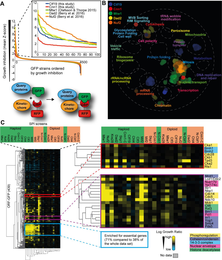
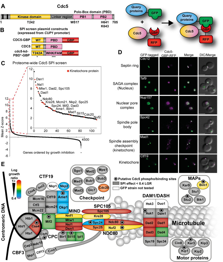
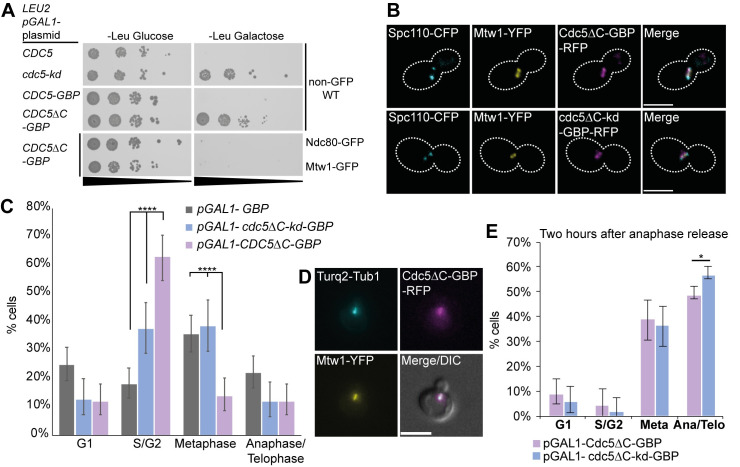
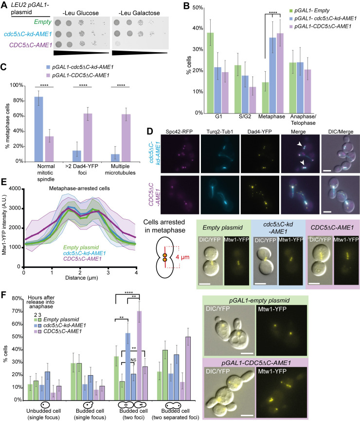
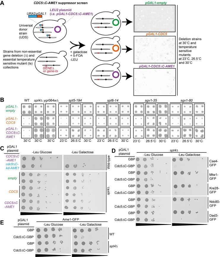
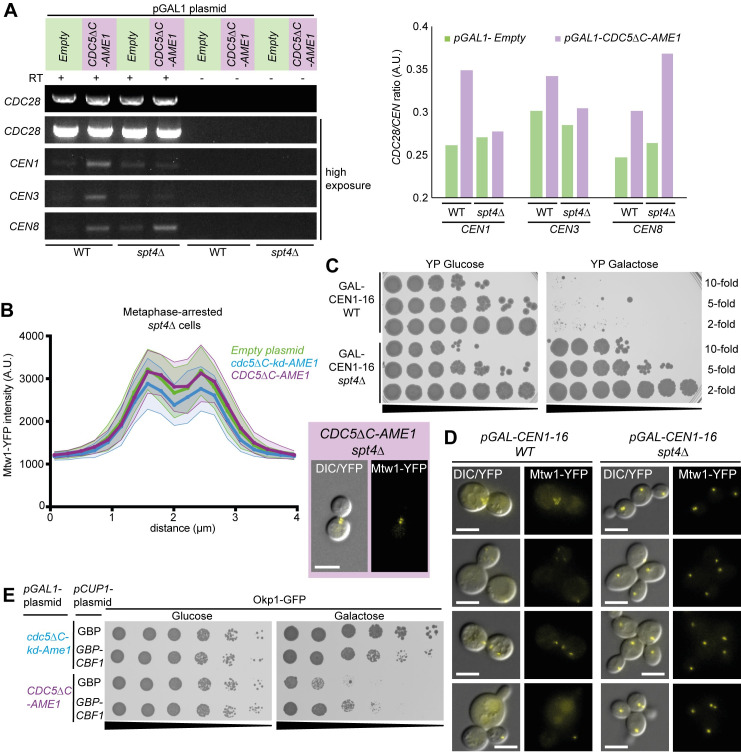
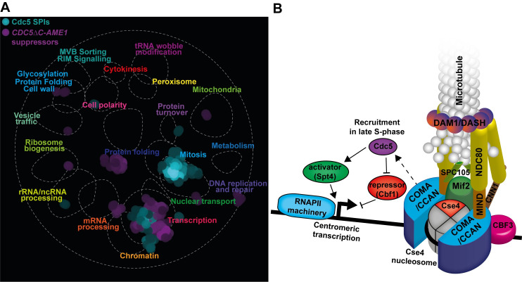
The kinetochore, a multi-protein complex assembled on centromeres, is essential to segregate chromosomes during cell division. Deficiencies in kinetochore function can lead to chromosomal instability and aneuploidy-a hallmark of cancer cells. Kinetochore function is controlled by recruitment of regulatory proteins, many of which have been documented, however their function often remains uncharacterized and many are yet to be identified. To identify candidates of kinetochore regulation we used a proteome-wide protein association strategy in budding yeast and detected many proteins that are involved in post-translational modifications such as kinases, phosphatases and histone modifiers. We focused on the Polo-like kinase, Cdc5, and interrogated which cellular components were sensitive to constitutive Cdc5 localization. The kinetochore is particularly sensitive to constitutive Cdc5 kinase activity. Targeting Cdc5 to different kinetochore subcomplexes produced diverse phenotypes, consistent with multiple distinct functions at the kinetochore. We show that targeting Cdc5 to the inner kinetochore, the constitutive centromere-associated network (CCAN), increases the levels of centromeric RNA via an SPT4 dependent mechanism.


Conflict of interest statement

The authors have declared that no competing interests exist.

Figures

Fig 1
Fig 1. Identification of kinetochore regulators using Synthetic Physical Interactions.
(A) Data from five proteome-wide kinetochore SPI screens are plotted in order of growth inhibition. Three SPI screens were performed previously (Mtw1, Nuf2 and Dad2) and two were performed in this study (Ctf19 and Cnn1). Growth inhibition caused by forced binary protein interactions are indicated on the y-axis is a mean Z-score (see Materials and Methods for more details). We took a Z-score of ≥ 2 as a cutoff for a significant growth defect. The Z-score ≥ 2 corresponds to a two-fold or greater difference in colony sizes. Most forced interactions do not cause a growth defect (Z-score ≈ 0) whereas SPI screens such as Nuf2 and Dad2 have many SPIs (>100 strains with Z-score ≥ 2). The Ctf19 and Cnn1 SPI data are listed in S1 Data. (B) The data from the five proteome-wide kinetochore SPI screens were mapped onto the global similarity network using spatial analysis of functional enrichment (SAFE) on thecellmap.org, which shows that the kinetochore SPI data is enriched for specific biological processes or cellular compartments; mainly mitosis, nuclear transport, mRNA processing and chromatin. (C) SPI data from 12 screens (in both haploid and heterozygous diploid strains) for 439 GFP-tagged strains were analyzed using Cluster software and visualized using Java TreeView. The strength of the growth inhibition (log growth ratio) is shown using a yellow-blue color scale where yellow is a strong growth defect compared to controls, black indicates no effect, and blue indicates growth enhancement. The clustering analysis distinctly clusters together the haploid SPI screens to the left (indicated in green) and diploid screens to the right (indicated in orange), with the exception of GBP-Cnn1 and GBP-Cbf1 screens. Furthermore, the cluster analysis clusters together GFP strains that are similarly affected by the GBP-tagged kinetochore proteins. Three distinct clusters are highlighted in the inset (see Materials and Methods and text for details).
Fig 2
Fig 2. Proteome-wide Cdc5 SPI screen is enriched for kinetochore proteins.
(A) The key domains of Polo kinase Cdc5 are shown. The N-terminal kinase domain contains a threonine 242 residue when substituted to alanine creates a catalytic-inactive mutant. The C-terminal non-catalytic domain contains two polo-boxes (PB1 and PB2), together called the polo-box domain (PBD) which binds to previously phosphorylated sites which targets the kinase domain of Cdc5 to its substrates and facilitates the phosphorylation at another site on the same substrate or surrounding substrates. A flexible linker region joins the kinase domain and PBD. (B) Schematic of the Cdc5-GBP constructs and controls used in the Cdc5 SPI assays (left). Expression of all constructs are controlled by CUP1 promoter. The cdc5-kd-PBD*-GBP control contains a mutated form of PBD (W517F, H641A, K643M). The GBP includes an RFP tag. The inset on the right shows a cartoon displaying the Cdc5-GBP interaction with a query GFP-tagged protein. (C) Data from the proteome-wide Cdc5 SPI screen are plotted as in Fig 1A. The inset shows that many kinetochore proteins (red dots) are among the top 100 Cdc5 SPIs. (D) Live imaging of cells containing different GFP-tagged proteins and expressing Cdc5-GBP shows that Cdc5-GBP can be constitutively recruited to many different subcellular locations as judged by colocalization. Scale bars are 5μm. (E) An illustration of the Cdc5 kinetochore SPIs, mapped onto a cartoon representation of the kinetochore. The color-coded map is based on log growth ratios of Cdc5-GBP compared with the average of both kinase-dead controls. The strength of the growth inhibition caused by Cdc5 association is indicated by a color-coded scale with high log growth ratios (LGR > 1) shown in red. Forced interactions that do not produce a growth phenotype are shown in grey (LGR < 0.4). Proteins that contain phosphosites that fit Cdc5 consensus for either phosphorylation or binding (E/D/Q)-X-(pS/T)-X and S-pS/T-P, respectively) are indicated with black dots.
Fig 3
Fig 3. Forcing Cdc5 association with Mtw1 disrupts cell-cycle progression prior to mitosis.
(A) 10-fold serial-dilution spot assay shows that pGAL1-driven expression of CDC5 in wild-type (WT) cells is lethal on galactose containing media whereas CDC5ΔC-GBP is not. However, expressing CDC5ΔC-GBP in GFP-tagged kinetochore strains (Mtw1 and Ndc80) is lethal. (B) Live imaging of cells containing Mtw1-YFP and Spc110-CFP (PT257) and expressing Cdc5ΔC-GBP-RFP (or cdc5ΔC-kd-GBP-RFP) shows that Cdc5-GBP sufficiently colocalizes with YFP foci and also between two SPB foci confirming kinetochore localization. Note that GBP does not have an affinity for CFP. Scale bar represents 5μm. (C) Cell-cycle analysis of Mtw1-YFP cells (T607) containing mTurquoise2-tagged tubulin (Turq2-Tub1) was performed using fluorescence microscopy. Cells from asynchronous cultures containing pGAL1-CDC5ΔC-GBP (n = 153), pGAL1-cdc5ΔC-kd-GBP (n = 120) or pGAL1-GBP (n = 219) were imaged after 4 hours of growth in 2% galactose media to induce expression. Cells that did not show RFP/YFP colocalization were excluded from this analysis. Cells expressing Cdc5ΔC-GBP-RFP were significantly increased in S/G2 phase compared to controls. Fishers exact statistical test; p-values **** = p < 10−4. Error bars indicate 95% binomial confidence intervals (C.I.). (D) Representative cropped image from the analysis in (C) showing a CDC5ΔC-GBP expressing cell in S/G2-phase. Scale bar represents 5μm. (E) Mtw1-YFP cells expressing CDC5ΔC-GBP (n = 319) during mitosis are able to progress into anaphase/telophase although at a slightly lower frequency than cells expressing cdc5ΔC-kd-GBP (n = 211). The metaphase-arrested cells in S5B Fig were released into anaphase by resuspending the cell cultures in galactose media lacking methionine. Two hours after release the cell-cycle stage of these cells was analyzed and revealed that about half of the cells had progressed into anaphase/telophase. Statistical analysis was done using Fishers exact test; p-values * = p < 0.05. Error bars indicate 95% binomial C.I.
Fig 4
Fig 4. Association of Cdc5 with Ame1 disrupts Mtw1 foci and mitotic progression.
(A) 10-fold serial-dilution spot assay showing that pGAL1-driven expression of CDC5ΔC-AME1 prevents growth whereas cdc5ΔC-kd-AME1 does not. (B) Cell-cycle analysis as in Fig 3C but with Dad4-YFP Turq2-Tub1 cells (T692) also containing RFP-tagged SPB (Spc42-RFP) and expressing pGAL1-CDC5ΔC-AME1 (n = 223), pGAL1-cdc5ΔC-kd-AME1 (n = 173) or empty plasmid (n = 249). Cell expressing either CDC5ΔC-AME1 or cdc5ΔC-kd-AME1 were significantly increased in metaphase compared to empty plasmid control. Fishers exact statistical test; p-values **** = p < 10−6. Error bars indicate 95% binomial confidence intervals. (C) The metaphase cells in (B) were categorized based upon the cell and spindle morphology observed using fluorescence imaging for both pGAL1-CDC5ΔC-AME1 (n = 93), pGAL1-cdc5ΔC-kd-AME1 (n = 62). Normal spindle was categorized as two separated Spc42-RFP and Dad4-YFP foci with microtubule signal in between. Cells expressing CDC5ΔC-AME1 had significantly fewer normal spindles and increased Dad4-YFP foci and microtubules. Fishers exact statistical test; p-values **** = p < 10−4. Error bars indicate 95% binomial confidence intervals. (D) Representative cropped images from the analysis in (C) showing cdc5ΔC-kd-AME1 expressing cells (top) and CDC5ΔC-AME1 expressing cells (bottom). Normal mitotic spindles are indicated with white arrows. Scale bar indicates 5μm. (E) Mtw1-YFP cells (PT63-12B) were arrested in metaphase using Cdc20 depletion and the Mtw1-YFP fluorescence intensity was quantified along the axis of the mitotic spindle after inducing CDC5ΔC-AME1 or controls for two hours (see S5A Fig for further description of experimental setup). A 4 μm line with 19 points (illustrated in the inset on the right) was used to include background signal and to cover the spread YFP signal phenotype in cells expressing CDC5ΔC-AME1 (40 randomly selected mitotic spindles were measured for each condition). The shadowed area indicates standard deviation. Cells expressing CDC5ΔC-AME1 have a more dispersed arrangement of Mtw1-YFP, as indicated by the flatter profile. Representative cropped images are shown on the right. (F) The metaphase-arrested cells in (E) were released into anaphase and imaged two and three hours later. Two hours after anaphase release (non-outlined bars) cells expressing CDC5ΔC-AME1 (n = 129) were still arrested in metaphase compared to cdc5ΔC-kd-AME1 (n = 154) and empty plasmid (n = 149) controls and displayed abnormal Mtw1-YFP foci shown on the right. Three hours after anaphase release (black-outlined bars) there was no statistical difference between cells expressing CDC5ΔC-AME1 (n = 220) and cdc5ΔC-kd-AME1 (n = 190) but cells containing empty plasmid control (n = 198) showed significantly reduced number of budded cells with two kinetochore foci compared to CDC5ΔC-AME1. Fishers exact statistical test; p-values ** = p < 0.005, **** = p < 10−5. Error bars indicate 95% binomial C.I. All scale bars indicate 5μm.
Fig 5
Fig 5. Suppressors of the forced Cdc5-Ame1 interaction phenotype.
(A) Diagram showing the experimental layout of the suppressor screen. Selective ploidy ablation (SPA) was used to introduce pGAL1-CDC5ΔC-AME1, pGAL1-CDC5 and pGAL1-empty plasmids into the deletion (Δ) and temperature-sensitive mutant (ts) collections. The Δ screen was performed at 30°C and the ts screen at 23°C, 26.5°C and 30°C. The agar plates from the final selection step were scanned after three days and representative examples are shown (see methods for further details). (B) A selection of cropped images of colonies from the suppressor screens showing that growth defects caused by pGAL1-CDC5ΔC-AME1 or pGAL1-CDC5 are suppressed by genetic deletions or mutations. (C) 10-fold serial dilutions spot assay with wild-type and spt4Δ strains containing pGAL1-CDC5ΔC-AME1 and control plasmids shows that spt4Δ suppresses growth defect caused by CDC5ΔC-AME1 expression. (D) 10-fold serial dilutions spot assay with GFP-tagged kinetochore strains (Cse4-GFP (internally tagged), Mtw1-GFP, Kre28-GFP, Ndc80-GFP and Dad3-GFP) containing pGAL1-CDC5ΔC-GBP and pGAL1-GBP plasmids shows that spt4Δ is not sufficient to suppress the growth defect caused by constitutive localization of Cdc5 to these proteins. (E) 10-fold serial dilutions spot assay with wild-type and spt4Δ strains encoding Ame1-GFP and pGAL1-CDC5ΔC-GBP and pGAL1-GBP plasmids showing that spt4Δ suppressed growth defect caused by constitutive Cdc5 association with Ame1.
Fig 6
Fig 6. Association of Cdc5 with Ame1 increases centromeric RNA levels which largely depends on Spt4.
(A) Reverse transcription PCR (RT-PCR) was performed after five hours of induction of CDC5ΔC-AME1 in wild-type and spt4Δ cells. Reactions without reverse transcription were used to control for potential contaminating DNA. Due to very faint CEN signal the exposure was increased to visualize the CEN RNA bands (see Materials and methods for further details). Quantification of the relative CEN RNA levels is shown in the graph on the right. Ratio of the CDC28 RNA control to the CEN RNAs was calculated. (B) Live imaging of metaphase-arrested Mtw1-YFP cells expressing CDC5ΔC-AME1 or controls and the kinetochore foci analysis was performed as in Fig 4 but in a spt4Δ strain (T739). The scattered Mtw1-YFP foci phenotype seen in Fig 4A was absent in spt4Δ cells (represented in the inset on the right). (C) A serial dilutions spot assay (10-, 5- and 2-fold) was performed with a GALCEN1-16 strain in both wild-type (W8164-2B) and spt4Δ background (T682). GALCEN1-16 spt4Δ strain grew equally well on glucose and galactose after two days at 30°C, in contrast to GALCEN1-16 WT which did not grow on galactose. (D) GALCEN1-16 Mtw1-YFP strains in either a WT (T360) or spt4Δ (T738) background were grown in galactose media for 16 hours and then imaged with fluorescence microscopy to investigate the kinetochore phenotype. In WT cells we found a range of phenotypes with cells showing scattered/fractured kinetochores or some with a diffused Mtw1-YFP signal whereas in spt4Δ cells this was not the case. We note that after 4–6 hours in galactose the cells did not show a clear phenotype. All scale bars are 5μm. (E) 5-fold serial dilutions spot assay of Okp1-GFP cells coexpressing CDC5ΔC-AME1 and GBP-CBF1 or GBP shows that additional Cbf1 recruitment partially suppresses the growth defect caused by CDC5ΔC-AME1 expression.
Fig 7
Fig 7. Cdc5 recruitment to the CCAN influences centromeric transcription.
(A) SAFE analysis as in Fig 1B of the Cdc5 SPIs and the CDC5ΔC-AME1 suppressor data identified highly dense regions that correspond to shared functions (mitosis and transcription for Cdc5 SPIs and transcription and mRNA processing for suppressors of CDC5ΔC-AME1). (B) A cartoon depicting Cdc5 recruitment to the kinetochore COMA complex (CCAN) resulting in increased production of CEN RNAs. We hypothesize that Cdc5 recruitment in late S-phase may influence CEN transcription by inhibiting a repressor such as Cbf1 and/or promote an activator such as Spt4 (see text for further details).

References

    1. Biggins S. The composition, functions, and regulation of the budding yeast kinetochore. Genetics. 2013;194: 817–846. 10.1534/genetics.112.145276 - DOI - PMC - PubMed
    1. Yan K, Yang J, Zhang Z, McLaughlin SH, Chang L, Fasci D, et al. Structure of the inner kinetochore CCAN complex assembled onto a centromeric nucleosome. Nature. 2019;574: 278–282. 10.1038/s41586-019-1609-1 - DOI - PMC - PubMed
    1. Hinshaw SM, Harrison SC. The structure of the ctf19c/ccan from budding yeast. Elife. 2019;8: 1–21. 10.7554/eLife.44239 - DOI - PMC - PubMed
    1. Hamilton G, Dimitrova Y, Davis TN. Seeing is believing: our evolving view of kinetochore structure, composition, and assembly. Curr Opin Cell Biol. 2019;60: 44–52. 10.1016/j.ceb.2019.03.016 - DOI - PMC - PubMed
    1. Kitamura E, Tanaka K, Kitamura Y, Tanaka TU. Kinetochore-microtubule interaction during S phase in Saccharomyces cerevisiae. Genes Dev. 2007;21: 3319–3330. 10.1101/gad.449407 - DOI - PMC - PubMed

Publication types

MeSH terms

LinkOut - more resources