

Development and evaluation of loop-mediated isothermal amplification for detection of Yersinia pestis in plague biological samples
- PMID: 32810167
- PMCID: PMC7437451
- DOI: 10.1371/journal.pone.0237655
Development and evaluation of loop-mediated isothermal amplification for detection of Yersinia pestis in plague biological samples
Abstract
Background: Several tests are available for plague confirmation but bacteriological culture with Yersinia pestis strain isolation remains the gold standard according to the World Health Organization. However, this is a time consuming procedure; requiring specific devices and well-qualified staff. In addition, strain isolation is challenging if antibiotics have been administered prior to sampling. Here, we developed a loop-mediated isothermal amplification (LAMP) technique, a rapid, simple, sensitive and specific technique that would be able to detect Y. pestis in human biological samples.
Methods: LAMP primers were designed to target the caf1 gene which is specific to Y. pestis. The detection limit was determined by testing 10-fold serial dilution of Y. pestis DNA. Cross-reactivity was tested using DNA extracts from 14 pathogens and 47 residual samples from patients suffering from non-plague diseases. Specificity and sensitivity of the LAMP caf1 were assessed on DNA extracts of 160 human biological samples. Then, the performance of the LAMP caf1 assay was compared to conventional PCR and bacteriological culture.
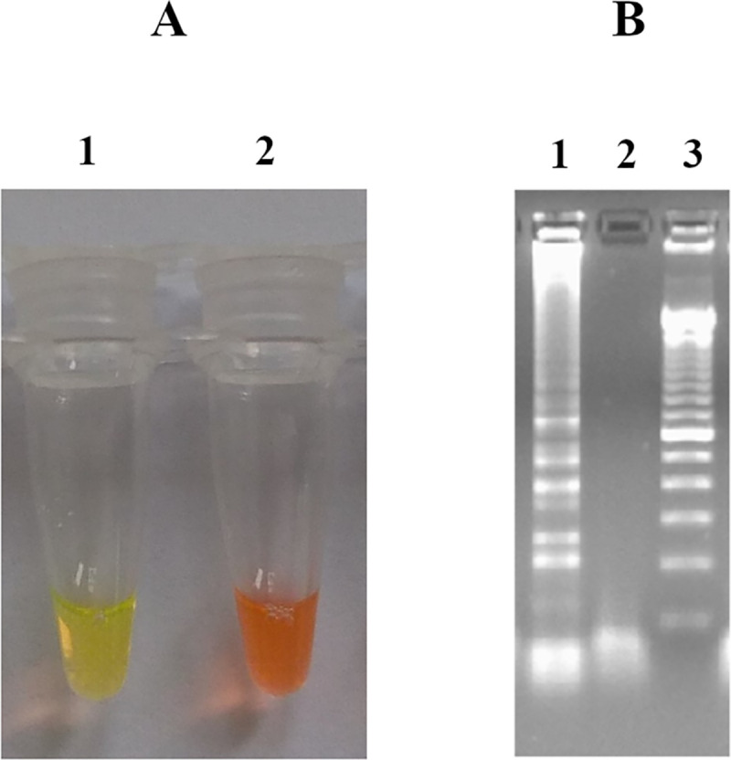
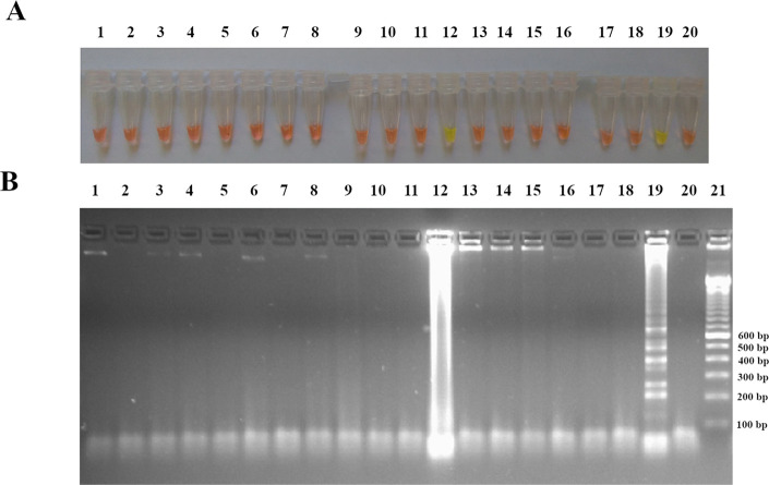
Results: The detection limit of the developed Y. pestis LAMP assay was 3.79 pg/μl, similar to conventional PCR. The result could be read out within 45 min and as early as 35 minutes in presence of loop primer, using a simple water bath at 63°C. This is superior to culture with respect to time (requires up to 10 days) and simplicity of equipment compared to PCR. Furthermore, no cross-reactivity was found when tested on DNA extracts from other pathogens and human biological samples from patients with non-plague diseases. Compared to the gold standard, LAMP sensitivity and specificity were 97.9% (95% CI: 89.1%-99.9%) and 94.6% (95% CI: 88.6%-97.9%), respectively.
Conclusion: LAMP detected Y. pestis effectively with high sensitivity and specificity in human plague biological samples. It can potentially be used in the field during outbreaks in resource limited countries such as Madagascar.
Conflict of interest statement
The authors have declared that no competing interests exist.
Figures
Similar articles
-
Development of a pair of real-time loop mediated isothermal amplification assays for detection of Yersinia pestis, the causative agent of plague.Mol Cell Probes. 2020 Dec;54:101670. doi: 10.1016/j.mcp.2020.101670. Epub 2020 Oct 22. Mol Cell Probes. 2020. PMID: 33132200
-
Yersinia pestis detection by loop-mediated isothermal amplification combined with magnetic bead capture of DNA.Braz J Microbiol. 2018 Jan-Mar;49(1):128-137. doi: 10.1016/j.bjm.2017.03.014. Epub 2017 Aug 26. Braz J Microbiol. 2018. PMID: 28887007 Free PMC article.
-
Development and evaluation of a multi-target droplet digital PCR assay for highly sensitive and specific detection of Yersinia pestis.PLoS Negl Trop Dis. 2024 May 3;18(5):e0012167. doi: 10.1371/journal.pntd.0012167. eCollection 2024 May. PLoS Negl Trop Dis. 2024. PMID: 38701065 Free PMC article.
-
Review of genotyping methods for Yersinia pestis in Madagascar.PLoS Negl Trop Dis. 2024 Jun 27;18(6):e0012252. doi: 10.1371/journal.pntd.0012252. eCollection 2024 Jun. PLoS Negl Trop Dis. 2024. PMID: 38935608 Free PMC article. Review.
-
[Methods of diagnosis and differentiation of plague pathogen: approaches to detection of atypical strains of Yersinia pestis by molecular biology. Part I].Mol Gen Mikrobiol Virusol. 2006;(1):3-6. Mol Gen Mikrobiol Virusol. 2006. PMID: 16512602 Review. Russian.
Cited by
-
A Novel Loop-Mediated Isothermal Amplification Assay for Rapid Detection of Yersinia pestis.Front Microbiol. 2022 Apr 7;13:863142. doi: 10.3389/fmicb.2022.863142. eCollection 2022. Front Microbiol. 2022. PMID: 35464914 Free PMC article.
-
The surveillance of plague among rodents and dogs in Western Iran.PLoS Negl Trop Dis. 2023 Nov 10;17(11):e0011722. doi: 10.1371/journal.pntd.0011722. eCollection 2023 Nov. PLoS Negl Trop Dis. 2023. PMID: 37948337 Free PMC article.
-
Screening and identification of DNA nucleic acid aptamers against F1 protein of Yersinia pestis using SELEX method.Mol Biol Rep. 2024 Jun 3;51(1):722. doi: 10.1007/s11033-024-09561-y. Mol Biol Rep. 2024. PMID: 38829419
-
Yersinia pestis antibiotic resistance: a systematic review.Osong Public Health Res Perspect. 2022 Feb;13(1):24-36. doi: 10.24171/j.phrp.2021.0288. Epub 2022 Feb 18. Osong Public Health Res Perspect. 2022. PMID: 35255676 Free PMC article.
References
-
- WHO. Plague around the world, 2010–2015. Wkly Epidemiol Rec. 2016;91(8):89–93. - PubMed
Publication types
MeSH terms
Substances
LinkOut - more resources
Full Text Sources
Medical
