. 2020;42(1):72-82.
doi: 10.1159/000509365. Epub 2020 Aug 18.

Caffeine Restores Background EEG Activity Independent of Infarct Reduction after Neonatal Hypoxic Ischemic Brain Injury

Affiliations

Caffeine Restores Background EEG Activity Independent of Infarct Reduction after Neonatal Hypoxic Ischemic Brain Injury

Haiyan Sun et al. Dev Neurosci. 2020.

Abstract

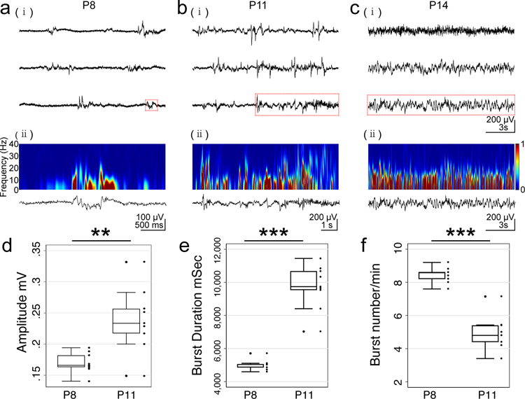
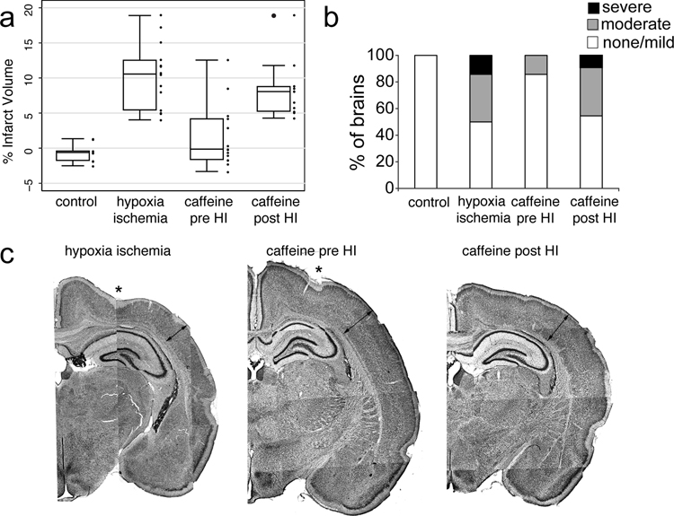
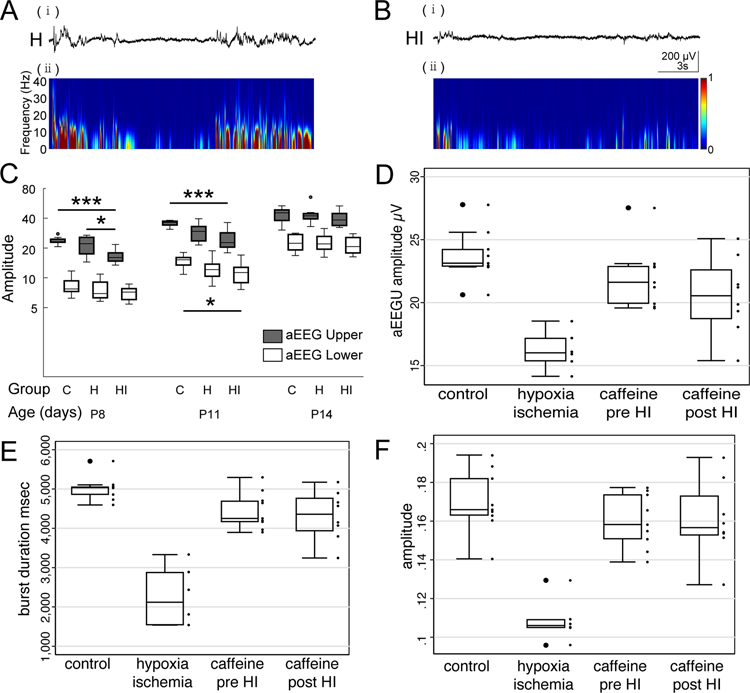
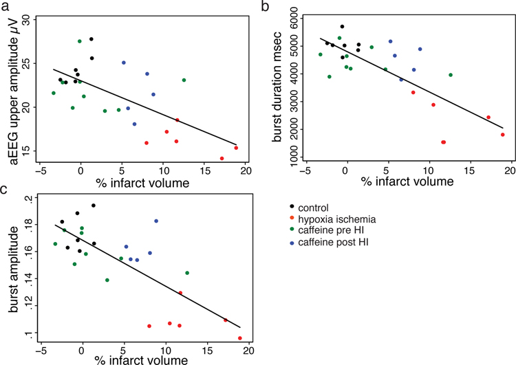
In human preterm newborns, caffeine increases brain activity and improves neurodevelopmental outcomes. In animal models of hypoxic ischemic brain injury, caffeine pretreatment reduces infarct volume. We studied the relationship between tissue neuroprotection and brain activity after injury to further understand caffeine neuroprotection. Rat dams received caffeine prior to birth or on postnatal day 3 (P3) through P16. Caffeine-treated and -untreated pups underwent the Vannucci procedure (unilateral carotid ligation, global hypoxia) on P2. A subset had EEG recordings. Brain hemispheric infarct volume was measured on P16. P2 hypoxic ischemia (HI) results in histologic brain injury (mean ± standard deviation infarct volume 10.3 ± 4.6%) and transient suppression of EEG activity. Caffeine pretreatment reduces brain injury (mean ± standard deviation infarct volume 1.6 ± 4.5%, p < 0.001) and improves amplitude-integrated EEG (aEEG) and EEG burst duration and amplitude. Caffeine treatment after HI does not reduce infarct volume (mean ± standard deviation 8.3 ± 4.1%, p = 1.0). However, caffeine posttreatment was equally effective at restoring aEEG amplitude and EEG burst duration and amplitude. Thus, caffeine supports brain background electrical activity independent of tissue neuroprotection.

Keywords: Activity-dependent brain development; Injury; Neuroprotection.


Conflict of interest statement

Disclosure Statement: The authors declare no conflict of interest, nor financial support beyond grant funding listed below.

Figures

Figure 1.
Figure 1.
Developmental changes in EEG traces are shown for P8 (a), P11 (b) and P14 (c). Raw EEG (i) shows typical discontinuous activity with bursts followed by periods of no activity. Activity increases in both amplitude and burst frequency, becoming continuous around P14. Frequency spectrogram (ii) for a single burst of activity (highlighted in red in raw traces) is shown for each age. The activity includes both low frequency (0.5 – 2 Hz) activity and nested higher frequencies (8 – 20 Hz) characteristic of a spindle burst. Power is normalized from 0 to 1 (color scale). Quantification of developmental changes in EEG activity with increase in burst amplitude (d) and burst duration (e) with corresponding decrease in burst frequency (f) from P8 to P11. Bursts cannot be quantified by P14 as activity has become continuous.
Figure 2.
Figure 2.
Quantification of infarct volume after hypoxia ischemia at P2 (P2 HI) with and without caffeine treatment given before (pre HI) and after hypoxia ischemia (post HI) (a). Summary of qualitative brain injury grade after P2 HI with and without caffeine treatment (b). Representative low magnification montage images of injury pattern and extent are shown (c) for HI, caffeine pre HI and caffeine post HI treatments. Thinning of neocortex (lines with arrows) and disruption of layer V1b/subplate can be appreciated in the HI image but not caffeine pre HI image. Intermediate effects are seen in caffeine post HI image.
Figure 3.
Figure 3.
Early hypoxia ischemia results in decreased EEG bursts with reduced power across all frequency bands (a,b). Quantification of amplitude integrated EEG (aEEG) (c), in control, hypoxia and hypoxia ischemia hemispheres. HI results in a transient decrease in upper aEEG margin at P8 and P11 and a decrease in lower aEEG margin at P11. Activity returns to normal by P14. Caffeine pretreatment restores aEEG upper margins at P8 (d). Quantification of burst duration (e) and amplitude (f) in control animals compared with hypoxia ischemia and caffeine treated hypoxia ischemia hemispheres at P8. Hypoxia ischemia reduces burst duration and amplitude. Caffeine pre- and post-treatment completely restores burst duration and amplitude.
Figure 4.
Figure 4.
Correlation between infarct volume (x-axis) and aEEG upper margin (a), burst duration (b) and burst amplitude (c). All groups are plotted by color coded symbol – control (black), hypoxia ischemia (red), caffeine before HI (green) and caffeine after HI (blue). A strong negative relationship exists between infarct volume and all measures of brain activity.

References

    1. Stoll BJ, Hansen NI, Bell EF, Walsh MC, Carlo WA, Shankaran S, et al. Trends in Care Practices, Morbidity, and Mortality of Extremely Preterm Neonates, 1993–2012. JAMA. 2015. September;314(10):1039–51. - PMC - PubMed
    1. Rogers EE, Hintz SR. Early neurodevelopmental outcomes of extremely preterm infants. Semin Perinatol. 2016;40(8):497–509. - PubMed
    1. Fleiss B, Gressens P. Tertiary mechanisms of brain damage: a new hope for treatment of cerebral palsy? Lancet Neurol. 2012. June;11(6):556–66. - PubMed
    1. Parikh P, Juul SE. Neuroprotective Strategies in Neonatal Brain Injury. J Pediatr. 2018;192:22–32. - PubMed
    1. Davis AS, Berger VK, Chock VY. Perinatal Neuroprotection for Extremely Preterm Infants. Am J Perinatol. 2016. February;33(3):290–6. - PubMed

Publication types