

Cervicofacial and mediastinal emphysema following minor dental procedure: a case report and review of the literature
- PMID: 32811562
- PMCID: PMC7433085
- DOI: 10.1186/s40463-020-00455-0
Cervicofacial and mediastinal emphysema following minor dental procedure: a case report and review of the literature
Abstract
Background: Subcutaneous cervical emphysema is a clinical sign associated with many conditions, including laryngotracheal trauma, pneumothorax and necrotizing deep tissue infections.
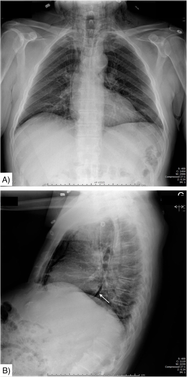
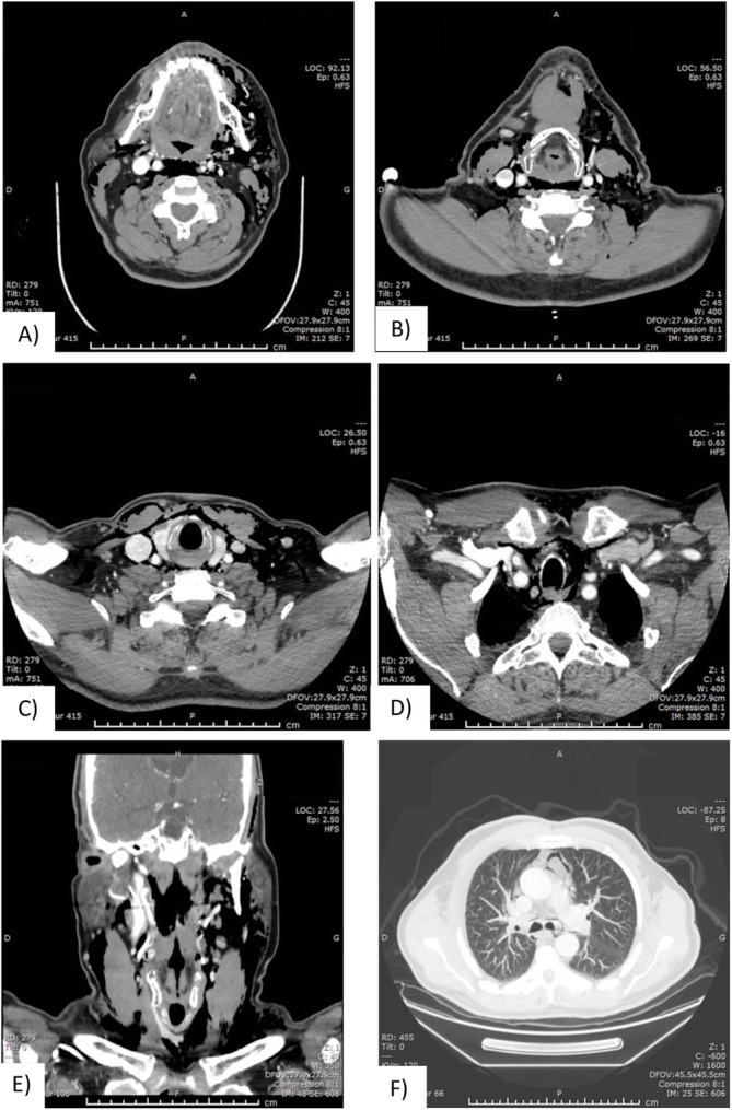
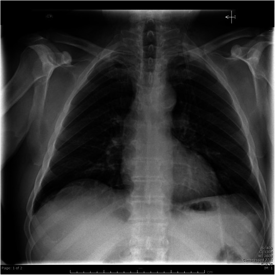
Case presentation: We discuss a case of a 76-year-old man presenting with extensive cervical emphysema a few hours after a minor dental filling procedure. The CT-scan revealed a significant amount of air within the cervical and mediastinal spaces, reaching lobar bronchi. Vitals were within normal values Bloodwork demonstrated an elevation of creatinine kinase (3718; normal < 150) and mild leukocytosis (WBC = 11.6). We decided to proceed to an urgent cervical exploration to exclude necrotizing fasciitis. This revealed air but no tissue necrosis nor abnormal fluid. The patient improved clinically and was discharged two days later with oral antibiotics. Although cervicofacial subcutaneous emphysema following dental procedures has been reported, it is usually less extensive and involving more invasive procedures using air-driven handpieces.
Conclusion: As an otolaryngologist confronted with extensive subcutaneous emphysema following a potential entry route for an aggressive infection, given the seriousness of this diagnosis, the decision of whether or not to perform a diagnostic surgical exploration should remain.
Keywords: Dental restoration; Necrotizing fasciitis; Pneumomediastinum; Subcutaneous emphysema.
Conflict of interest statement
The authors declare no competing interests.
Figures
References
-
- Mather AJ. Cervicofacial and Mediastinal Emphysema Complicating a Dental Procedure. J Can Dent Assoc. 2006;72(6)565–8. - PubMed
-
- Heyman SN, Babayofk I. Emphysematous complications in dentistry, 1960–1993: An illustrative case and review of the literature. Quintessence Int. 1995;26:535–43. - PubMed
-
- McKenzie WS, Rosenberg M. Iatrogenic subcutaneous emphysema of dental and surgical origin: a literature review. J Oral Maxillofac Surg. 2009;67(6):1265–1268. - PubMed
-
- Arai I, Aoki T, Yamazaki H, Ota Y, Kaneko A. Pneumomediastinum and subcutaneous emphysema after dental extraction detected incidentally by regular medical checkup: a case report. Oral Surg Oral Med Oral Pathol Oral Radiol Endodontology. 2009;107(4):e33–e38. - PubMed
Publication types
MeSH terms
LinkOut - more resources
Full Text Sources
Medical
Research Materials
