. 2020 Aug 18;10(1):13951.
doi: 10.1038/s41598-020-70912-3.

Tissue-targeted R-spondin mimetics for liver regeneration

Affiliations

Tissue-targeted R-spondin mimetics for liver regeneration

Zhengjian Zhang et al. Sci Rep. .

Abstract

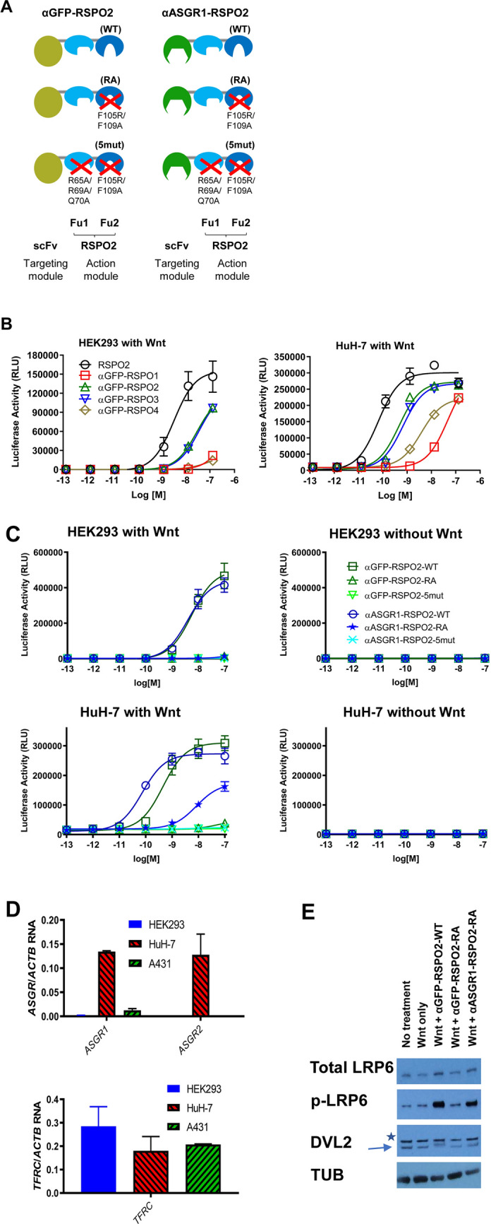
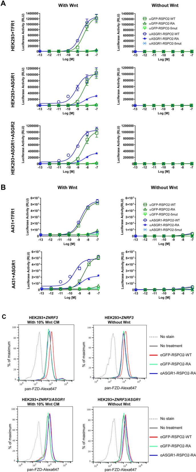
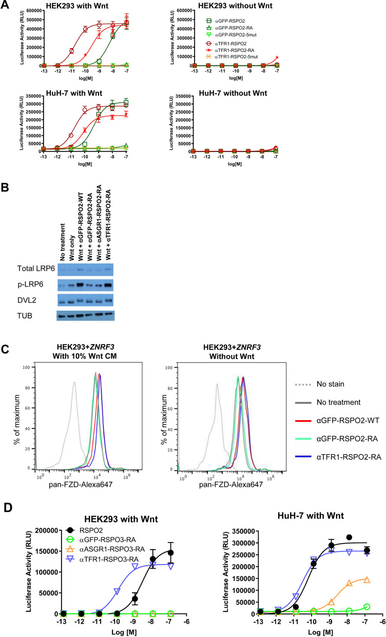
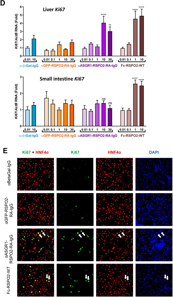
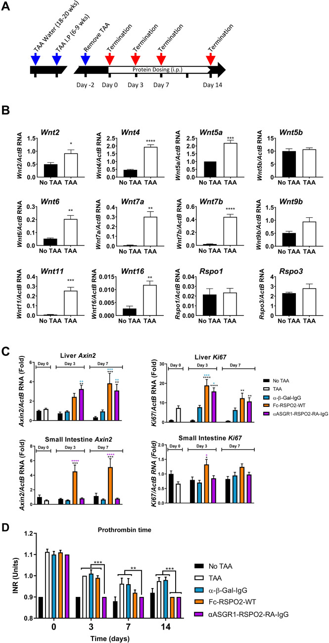
R-spondin (RSPO) proteins amplify Wnt signaling and stimulate regeneration in a variety of tissues. To repair tissue in a tissue-specific manner, tissue-targeted RSPO mimetic molecules are desired. Here, we mutated RSPO (RSPO2 F105R/F109A) to eliminate LGR binding while preserving ZNRF3/RNF43 binding and targeted the mutated RSPO to a liver specific receptor, ASGR1. The resulting bi-specific molecule (αASGR1-RSPO2-RA) enhanced Wnt signaling effectively in vitro, and its activity was limited to ASGR1 expressing cells. Systemic administration of αASGR1-RSPO2-RA in mice specifically upregulated Wnt target genes and stimulated cell proliferation in liver but not intestine (which is more responsive to non-targeted RSPO2) in healthy mice, and improved liver function in diseased mice. These results not only suggest that a tissue-specific RSPO mimetic protein can stimulate regeneration in a cell-specific manner, but also provide a blueprint of how a tissue-specific molecule might be constructed for applications in a broader context.


Conflict of interest statement

All authors are current or former full-time employees and shareholders of Surrozen, Inc.

Figures

Figure 1
Figure 1
Design and characterization of cell type specific Wnt signaling enhancing mimetic molecules. (A) Scheme of designed molecules. Fragments of RSPO2 spanning the Fu1 and Fu2 domains (action module) were fused the C-terminus of cell type specific or control scFv (targeting module). Specific mutations used are indicated. (B) In vitro STF activity of four RSPO proteins. Furin domains of human RSPO1-4 were tested as fusions to an anti-GFP scFv. RSPO2 furin domain alone (N37-E143) was used as a control. Luciferase activity unit is arbitrary (RLU). (C) STF activity of RSPO mimetics on HEK293 (top) or HuH-7 (bottom) in the presence (left) or absence (right) of an exogenous Wnt source (30% Wnt3a conditioned media). (D) Quantitative PCR analysis of ASGR1, ASGR2 and TFRC gene expression in HEK293, HuH-7 and A431 cells. The signals were relative to ACTB. (E) Western Blot analysis on LRP6 receptor and DVL2 phosphorylation in HuH-7 cells. Asterisk indicates a non-specific band that is detected in HuH-7 by the antibody used. The lower bands indicated by the arrow correspond to DVL2 with and without phosphorylation. Tubulin (TUB) is used as the loading control. 10% Wnt3a conditioned media was used as the exogenous Wnt source. Images were cropped for clarity. The uncropped images are presented in Fig. S2.
Figure 2
Figure 2
Confirmation of receptor-specific activation of Wnt signaling. (A) STF activity in HEK293 cells transiently transfected with TFR1 (control, top), ASGR1 (middle), and ASGR1 together with ASGR2 cDNAs (bottom), either in the presence (left) or absence (right) of exogenous Wnt source (30% Wnt3a conditioned media). (B) STF activity in A431 cells with TFR1 or ASGR1 overexpression as specified. (C) Flow cytometry analysis of cell surface level of FZD proteins. HEK293 cells were transiently transfected with either ZNRF3 alone (top) or ASGR1 and ZNRF3 cDNAs (bottom), treated with RSPO mimetics as specified at 10 nM, then stained by the pan-FZD antibody 18R5.
Figure 3
Figure 3
Application of the targeted RSPO mimetic design to a transferrin receptor and RSPO3. (A) STF activity of specified proteins on HEK293 (top) or HuH-7 (bottom) in the presence (left) or absence (right) of exogenous Wnt source (30% Wnt3a conditioned media). (B) Western Blot analysis for LRP6 receptor expression and DVL2 phosphorylation in HEK293 cells. Tubulin (TUB) is used as the loading control. Images were cropped for clarity. The uncropped images are presented in Fig. S2. (C) Flow cytometry analysis of cell surface level of FZD proteins. (D) STF activity of RSPO3-based mimetic molecules in HEK293 (left) and HuH-7 (right) cells, in the presence of 30% Wnt3a conditioned media.
Figure 4
Figure 4
In vivo tissue-specific enhancement of Wnt signaling. (A) STF activity of the RSPO mimetic molecules in the appended-IgG format. (B) BLI analysis of the ASGR1 antibody (expressed as a Fab) on human and mouse ASGR1. The affinity (Kd) determined by steady-state fitting is indicated. (C) Axin2 and (D) Ki67 expression in liver (upper panel) and small intestine (lower panel) were analyzed by quantitative PCR, 48 h after i.p. injection of proteins as specified (n = 8 mice per group). Statistical analysis was performed using 1-way ANOVA: (ns) not significant, **p < 0.01, ****p < 0.0001. (E) Immunofluorescence staining of liver samples for Ki67 and HNF4α from 10 mg/kg treatment groups. White arrows denote some double-positive cells as examples. DAPI was used to stain nuclei.
Figure 4
Figure 4
In vivo tissue-specific enhancement of Wnt signaling. (A) STF activity of the RSPO mimetic molecules in the appended-IgG format. (B) BLI analysis of the ASGR1 antibody (expressed as a Fab) on human and mouse ASGR1. The affinity (Kd) determined by steady-state fitting is indicated. (C) Axin2 and (D) Ki67 expression in liver (upper panel) and small intestine (lower panel) were analyzed by quantitative PCR, 48 h after i.p. injection of proteins as specified (n = 8 mice per group). Statistical analysis was performed using 1-way ANOVA: (ns) not significant, **p < 0.01, ****p < 0.0001. (E) Immunofluorescence staining of liver samples for Ki67 and HNF4α from 10 mg/kg treatment groups. White arrows denote some double-positive cells as examples. DAPI was used to stain nuclei.
Figure 5
Figure 5
Effects of liver targeted RSPO mimetic in a TAA mouse model of chronic liver disease. (A) Study design. (B) Quantitative PCR analysis of Wnt and Rspo RNA in the livers of normal and TAA treated mice. Genes below detection limit were not shown. (C) Quantitative PCR analysis of Axin2 (left) and Ki67 (right) RNA in liver (top) and small intestine (bottom; n = 10 mice per group). (D) INR of prothrombin time during treatment. Statistical analysis was performed using 1-way ANOVA: *p < 0.05, **p < 0.01, ***p < 0.001, ****p < 0.0001 (n = 10 mice per group).

References

    1. Clevers H, Loh KM, Nusse R. Stem cell signalling. An integral program for tissue renewal and regeneration: Wnt signaling and stem cell control. Science. 2014;346:1248012. doi: 10.1126/science.1248012. - DOI - PubMed
    1. Knight MN, Hankenson KD. R-spondins: novel matricellular regulators of the skeleton. Matrix Biol. 2014;37:157–161. doi: 10.1016/j.matbio.2014.06.003. - DOI - PubMed
    1. Hao HX, Jiang X, Cong F. Control of Wnt receptor turnover by R-spondin-ZNRF3/RNF43 signaling module and its dysregulation in cancer. Cancers (Basel) 2016 doi: 10.3390/cancers8060054. - DOI - PMC - PubMed
    1. De Lau W, Peng WC, Gros P, Clevers H. The R-spondin module: regulator of Wnt signal strength. Genes Dev. 2014;28:305–316. doi: 10.1101/gad.235473.113. - DOI - PMC - PubMed
    1. Zebisch M, et al. Structural and molecular basis of ZNRF3/RNF43 transmembrane ubiquitin ligase inhibition by the Wnt agonist R-spondin. Nat. Commun. 2013;4:2787. doi: 10.1038/ncomms3787. - DOI - PMC - PubMed

MeSH terms