

Automatic identification of atypical clinical fMRI results
- PMID: 32812070
- PMCID: PMC7666675
- DOI: 10.1007/s00234-020-02510-z
Automatic identification of atypical clinical fMRI results
Erratum in
-
Correction to: Automatic identification of atypical clinical fMRI results.Neuroradiology. 2020 Dec;62(12):1723. doi: 10.1007/s00234-020-02565-y. Neuroradiology. 2020. PMID: 32986199 Free PMC article.
Abstract
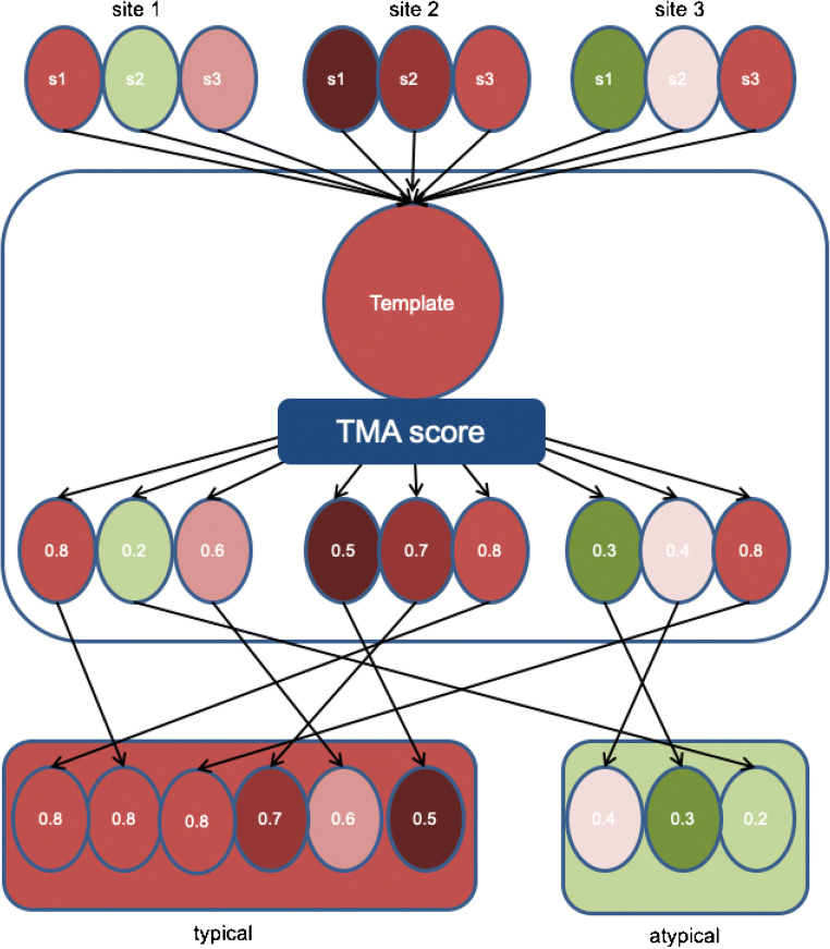
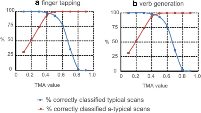
Purpose: Functional MRI is not routinely used for neurosurgical planning despite potential important advantages, due to difficulty of determining quality. We introduce a novel method for objective evaluation of fMRI scan quality, based on activation maps. A template matching analysis (TMA) is presented and tested on data from two clinical fMRI protocols, performed by healthy controls in seven clinical centers. Preliminary clinical utility is tested with data from low-grade glioma patients.
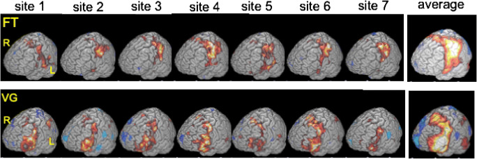
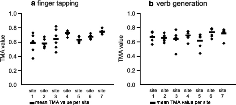
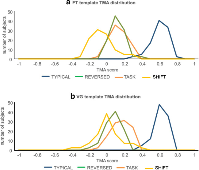
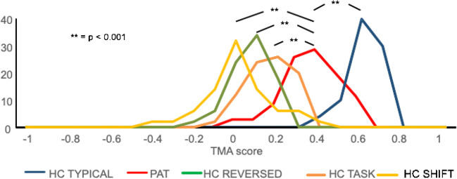
Methods: Data were collected from 42 healthy subjects from seven centers, with standardized finger tapping (FT) and verb generation (VG) tasks. Copies of these "typical" data were deliberately analyzed incorrectly to assess feasibility of identifying them as "atypical." Analyses of the VG task administered to 32 tumor patients assessed sensitivity of the TMA method to anatomical abnormalities.
Results: TMA identified all atypical activity maps for both tasks, at the cost of incorrectly classifying 3.6 (VG)-6.5% (FT) of typical maps as atypical. For patients, the average TMA was significantly higher than atypical healthy scans, despite localized anatomical abnormalities caused by a tumor.
Conclusion: This study supports feasibility of TMA for objective identification of atypical activation patterns for motor and verb generation fMRI protocols. TMA can facilitate the use and evaluation of clinical fMRI in hospital settings that have limited access to fMRI experts. In a clinical setting, this method could be applied to automatically flag fMRI scans showing atypical activation patterns for further investigation to determine whether atypicality is caused by poor scan data quality or abnormal functional topography.
Keywords: Brain function; Clinical fMRI; Functional MRI; Language; Motor cortex.
Conflict of interest statement
Author NR declares a conflict of interest. He is the director, and owns stock, of startup company Braincarta BV. All other authors declare that they have no conflict of interest.
Figures
Similar articles
-
Tumor Tissue Detection using Blood-Oxygen-Level-Dependent Functional MRI based on Independent Component Analysis.Sci Rep. 2018 Jan 19;8(1):1223. doi: 10.1038/s41598-017-18453-0. Sci Rep. 2018. PMID: 29352123 Free PMC article.
-
Real-time functional magnetic resonance imaging (rt-fMRI) in patients with brain tumours: preliminary findings using motor and language paradigms.Br J Neurosurg. 2005 Feb;19(1):25-32. doi: 10.1080/02688690500089621. Br J Neurosurg. 2005. PMID: 16147579
-
Functional Magnetic Resonance Imaging Activation Optimization in the Setting of Brain Tumor-Induced Neurovascular Uncoupling Using Resting-State Blood Oxygen Level-Dependent Amplitude of Low Frequency Fluctuations.Brain Connect. 2019 Apr;9(3):241-250. doi: 10.1089/brain.2017.0562. Epub 2019 Feb 28. Brain Connect. 2019. PMID: 30547681 Free PMC article.
-
Is preoperative functional magnetic resonance imaging reliable for language areas mapping in brain tumor surgery? Review of language functional magnetic resonance imaging and direct cortical stimulation correlation studies.Neurosurgery. 2010 Jan;66(1):113-20. doi: 10.1227/01.NEU.0000360392.15450.C9. Neurosurgery. 2010. PMID: 19935438 Review.
-
Magnetic resonance spectroscopy imaging (MRSI) and brain functional magnetic resonance imaging (fMRI) for radiotherapy treatment planning of glioma.Technol Cancer Res Treat. 2008 Oct;7(5):349-62. Technol Cancer Res Treat. 2008. PMID: 18783284 Review.
Cited by
-
7 T and beyond: toward a synergy between fMRI-based presurgical mapping at ultrahigh magnetic fields, AI, and robotic neurosurgery.Eur Radiol Exp. 2024 Jul 1;8(1):73. doi: 10.1186/s41747-024-00472-y. Eur Radiol Exp. 2024. PMID: 38945979 Free PMC article. Review.
-
Large-scale fMRI dataset for the design of motor-based Brain-Computer Interfaces.Sci Data. 2025 May 16;12(1):804. doi: 10.1038/s41597-025-05134-1. Sci Data. 2025. PMID: 40379686 Free PMC article.
-
Methodological Recommendations for Studies on the Daily Life Implementation of Implantable Communication-Brain-Computer Interfaces for Individuals With Locked-in Syndrome.Neurorehabil Neural Repair. 2022 Nov;36(10-11):666-677. doi: 10.1177/15459683221125788. Epub 2022 Sep 20. Neurorehabil Neural Repair. 2022. PMID: 36124975 Free PMC article.
References
-
- Castellano A, Cirillo S, Bello L, Riva M, Falini A. Functional MRI for surgery of gliomas. Curr Treat Options Neurol. 2017;19:34. - PubMed
-
- Dimou S, Battisti RA, Hermens DF, Lagopoulos J. A systematic review of functional magnetic resonance imaging and diffusion tensor imaging modalities used in presurgical planning of brain tumour resection. Neurosurg Rev. 2013;36:205–214. - PubMed
-
- Rutten GJ, Ramsey NF. The role of functional magnetic resonance imaging in brain surgery. Neurosurg Focus. 2010;28:E4. - PubMed
-
- Wang L, Chen D, Olson J, Ali S, Fan T, Mao H. Re-examine tumor-induced alterations in hemodynamic responses of BOLD fMRI: implications in presurgical brain mapping. Acta Radiol. 2012;53:802–811. - PubMed
-
- Bick AS, Mayer A, Levin N. From research to clinical practice: implementation of functional magnetic imaging and white matter tractography in the clinical environment. J Neurol Sci. 2012;312:158–165. - PubMed
Publication types
MeSH terms
LinkOut - more resources
Full Text Sources
Medical
