

Calcified brain metastases may be more frequent than normally considered
- PMID: 32812176
- PMCID: PMC7813689
- DOI: 10.1007/s00330-020-07164-2
Calcified brain metastases may be more frequent than normally considered
Abstract
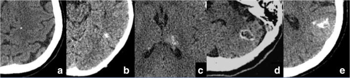
Objectives: To verify the incidence of calcified brain metastases (CBM), illustrating the different presentation patterns and histology of primary tumor.
Methods: A series of 1002 consecutive brain computed tomography (CT) scans of patients with known primary tumors was retrospectively assessed. CBM were defined by the presence of calcification within intra-axial-enhancing lesions; identification of CBM was based on visual examination and ROI analysis (> 85 Hounsfield units). Also, calcifications in the primary tumor of all patients with brain metastases were evaluated. In CBM patients, we investigated the type of calcifications (punctate, nodular, cluster, ring, coarse), the histology of primary tumor, and if a previous RT was performed.
Results: Among 190 (18.9%) patients with brain metastatic disease, 34 presented with CBM (17.9%). Sixteen patients were previously treated with RT, while 18 presented calcifications ab initio (9.5% of all brain metastases). The majority of patients with CBM had a primitive lung adenocarcinoma (56%), followed by breast ductal invasive carcinoma (20%) and small cell lung carcinoma (11.8%). CBM were single in 44.1% of patients and multiple in 55.9%. With regard to the type of calcifications, the majority of CBM were punctate, without specific correlations between calcification type and histology of primary tumor. No patients with ab initio CBM had calcifications in primary tumor.
Conclusion: In conclusion, our data show that CBM are more common than usually thought, showing an incidence of 9.5% ab initio in patients with brain metastases. This study underlines that neuroradiologists should not overlook intraparenchymal brain calcifications, especially in oncologic patients.
Key points: • Among the differential diagnosis of brain intraparenchymal calcifications, metastases are considered uncommon and found predominantly in patients treated with radiotherapy (RT). • Our data show that CBM are more common than usually thought, showing an incidence of 9.5% ab initio in patients with brain metastases. • A proportion of intraparenchymal brain calcifications, especially in oncologic patients, might represent evolving lesions and neuroradiologists should not overlook them to avoid a delay in diagnosis and treatment.
Keywords: Brain neoplasms; Calcification; Incidence; Metastases; Tomography X-ray computed.
Conflict of interest statement
The authors of this manuscript declare no relationships with any companies whose products or services may be related to the subject matter of the article.
Figures


References
-
- Lee KF, Suh JH (1977) CT evidence of grey matter calcification secondary to radiation therapy. Comput Tomogr 1(1):103–110 - PubMed
MeSH terms
LinkOut - more resources
Full Text Sources
Medical
