Review
. 2020 Dec;21(6):833-854.
doi: 10.1007/s40257-020-00553-9.

Immune-Mediated Dermatoses in Patients with Haematological Malignancies: A Comprehensive Review

Affiliations
Review

Immune-Mediated Dermatoses in Patients with Haematological Malignancies: A Comprehensive Review

Roberto Maglie et al. Am J Clin Dermatol. 2020 Dec.

Abstract

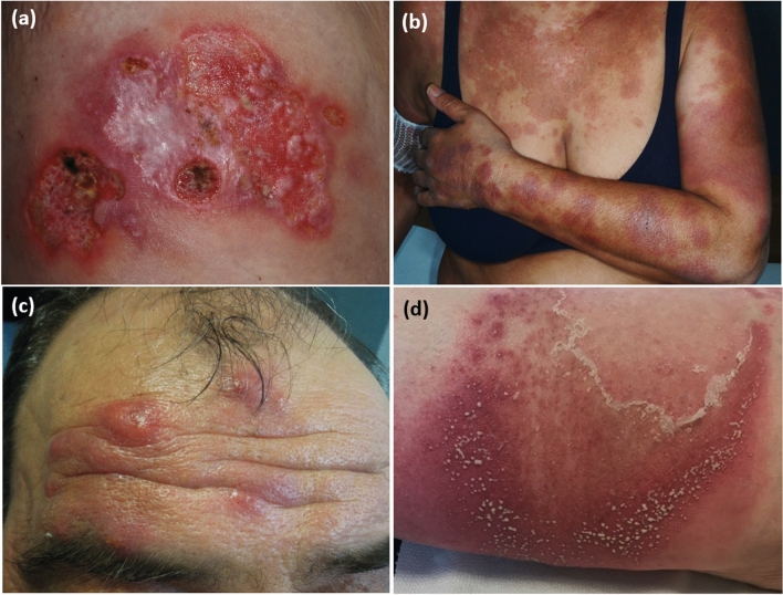
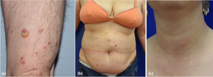
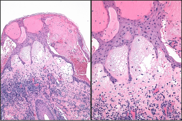
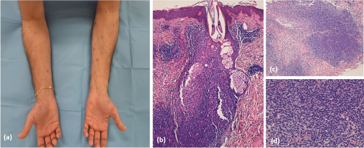
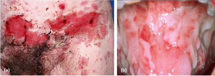
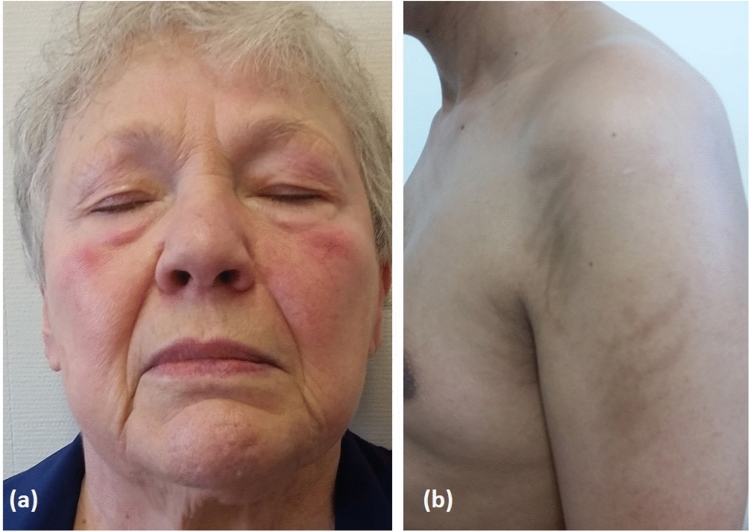
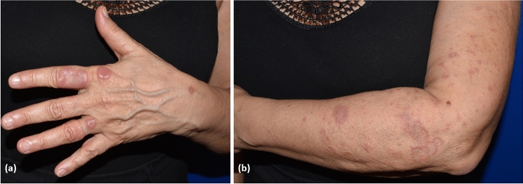
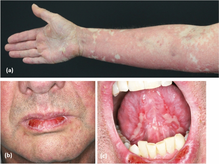
Haematological malignancies induce important alterations of the immune system, which account for the high frequency of autoimmune complications observed in patients. Cutaneous immune-mediated diseases associated with haematological malignancies encompass a heterogeneous group of dermatoses, including, among others, neutrophilic and eosinophilic dermatoses, autoantibody-mediated skin diseases, vasculitis and granulomatous dermatoses. Some of these diseases, such as paraneoplastic pemphigus, are associated with an increased risk of death; others, such as eosinophilic dermatoses of haematological malignancies, run a benign clinical course but portend a significant negative impairment on a patient's quality of life. In rare cases, the skin eruption reflects immunological alterations associated with an unfavourable prognosis of the associated haematological disorder. Therapeutic management of immune-mediated skin diseases in patients with haematological malignancies is often challenging. Systemic corticosteroids and immunosuppressive drugs are considered frontline therapies but may considerably augment the risk of serious infections. Indeed, developing a specific targeted therapeutic approach is of crucial importance for this particularly fragile patient population. This review provides an up-to-date overview on the immune-mediated skin diseases most frequently encountered by patients with onco-haematological disorders, discussing new pathogenic advances and therapeutic options on the horizon.


Conflict of interest statement

Roberto Maglie, Giovanni Genovese, Farzan Solimani, Alba Guglielmo, Alessandro Pileri, Francesca Portelli, Michael Hertl, Angelo Valerio Marzano and Emiliano Antiga have no conflicts of interest that are directly relevant to the content of this article.

Figures

Fig. 1
Fig. 1
Spectrum of neutrophilic dermatoses: a pyoderma gangrenosum; b Sweet syndrome; c erythema elevatum diutinum; and d subcorneal pustular dermatosis
Fig. 2
Fig. 2
Main clinical pattern of eosinophilic dermatosis of haematological malignancies: a bullous pemphigoid-like pattern; b insect bite-like pattern; and c eosinophilic cellulitis-like pattern
Fig. 3
Fig. 3
a Histopathological presentation of eosinophilic dermatoses of haematological malignancies, showing intense dermal oedema and a mixed inflammatory infiltrate in the dermis with numerous eosinophils and flame figures; intra-epidermal blistering is also visible (haematoxylin and eosin staining ×20) and b detail of the flame figures (haematoxylin and eosin staining ×40)
Fig. 4
Fig. 4
a Clinical presentation of T-cell papulosis associated with B-cell malignancies in a patient with B-cell chronic lymphocytic leukaemia and b–d microscopic examination showed a prominent lymphocytic inflammatory infiltrate with perifollicular distribution resembling folliculotropic mycosis fungoides (haematoxylin and eosin staining ×20)
Fig. 5
Fig. 5
Paraneoplastic pemphigus a with extensive haemorrhagic multiform blisters and erosions of the trunk and b extensive fibrinous erosions of the tongue
Fig. 6
Fig. 6
Palpable purpura consistent with cutaneous leukocytoclastic vasculitis in a patient with follicular non-Hodgkin’s lymphoma
Fig. 7
Fig. 7
a Heliotrope rash in a patient with dermatomyositis who developed non-Hodgkin’s lymphoma 2 years after the diagnosis and b flagellate erythema involving the upper arms in a young boy with Hodgkin’s lymphoma receiving a bleomycin-containing regimen
Fig. 8
Fig. 8
a, b Widespread annular granuloma in a female patient with chronic myelomonocytic leukaemia
Fig. 9
Fig. 9
Cutaneous graft-vs-host disease a with extensive multiform erythematous plaques and palmar hyperkeratosis, b lichenoid plaques on the lower lip, and c erosions and small bullae on the base of the tongue

Similar articles

Cited by

References

    1. Swerdlow SH, Campo E, Pileri SA, Harris NL, Stein H, Siebert R, et al. The 2016 revision of the World Health Organization classification of lymphoid neoplasms. Blood. 2016;127:2375–2390. doi: 10.1182/blood-2016-01-643569. - DOI - PMC - PubMed
    1. Arber DA, Orazi A, Hasserjian R, Thiele J, Borowitz MJ, Le Beau MM, et al. The 2016 revision to the World Health Organization classification of myeloid neoplasms and acute leukemia. Blood. 2016;127:2391–2405. doi: 10.1182/blood-2016-03-643544. - DOI - PubMed
    1. Moia R, Patriarca A, Schipani M, Ferri V, Favini C, Sagiraju S, et al. Precision medicine management of chronic lymphocytic leukemia. Cancers (Basel) 2020;12(3):642. doi: 10.3390/cancers12030642. - DOI - PMC - PubMed
    1. Zappasodi P, Forno C, Corso A, Lazzarino M. Mucocutaneous paraneoplastic syndromes in hematologic malignancies. Int J Dermatol. 2006;45:14–22. doi: 10.1111/j.1365-4632.2005.02668.x. - DOI - PubMed
    1. Li AW, Yin ES, Stahl M, Kim TK, Panse G, Zeidan AM, et al. The skin as a window to the blood: cutaneous manifestations of myeloid malignancies. Blood Rev. 2017;31:370–388. doi: 10.1016/j.blre.2017.07.003. - DOI - PubMed

MeSH terms

Substances