. 2021:2174:143-170.
doi: 10.1007/978-1-0716-0759-6_10.

Extracellular Vesicles and Their Roles in Cancer Progression

Affiliations

Extracellular Vesicles and Their Roles in Cancer Progression

Wen-Hsuan Chang et al. Methods Mol Biol. 2021.

Abstract

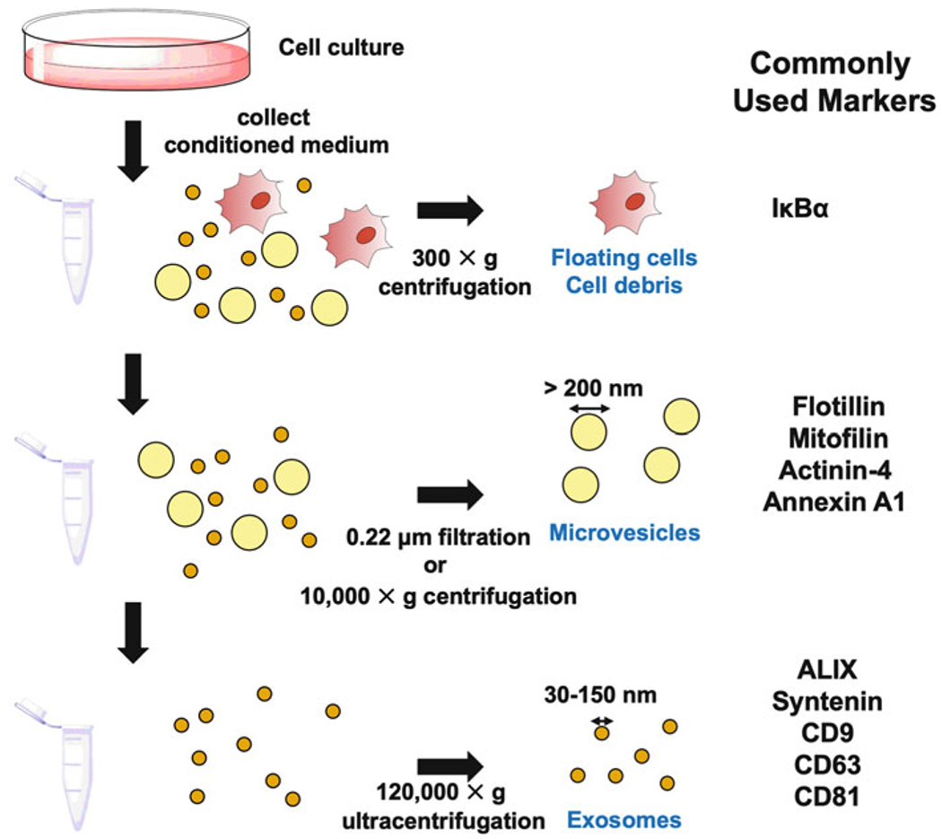
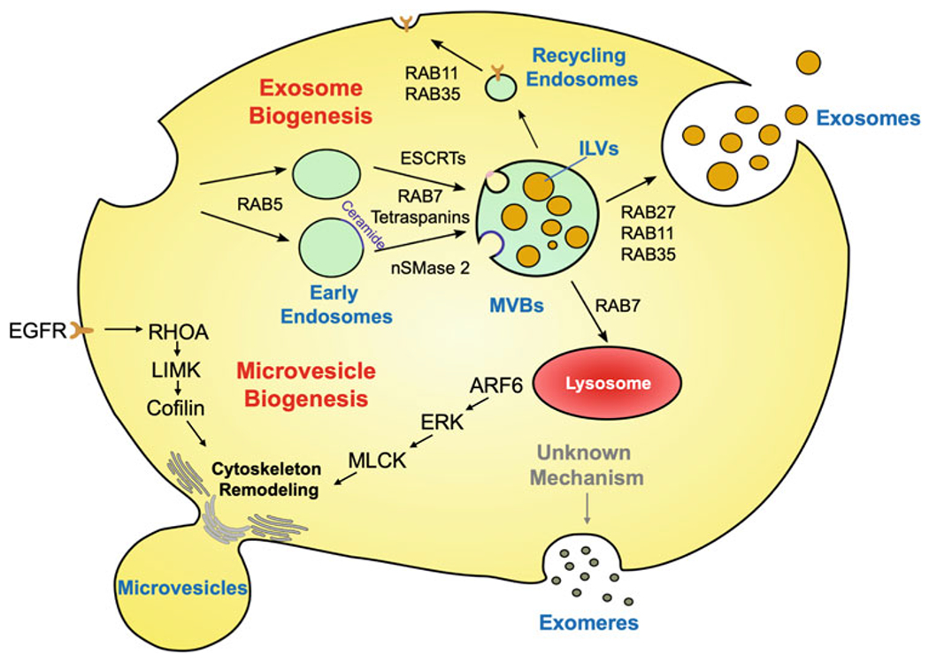
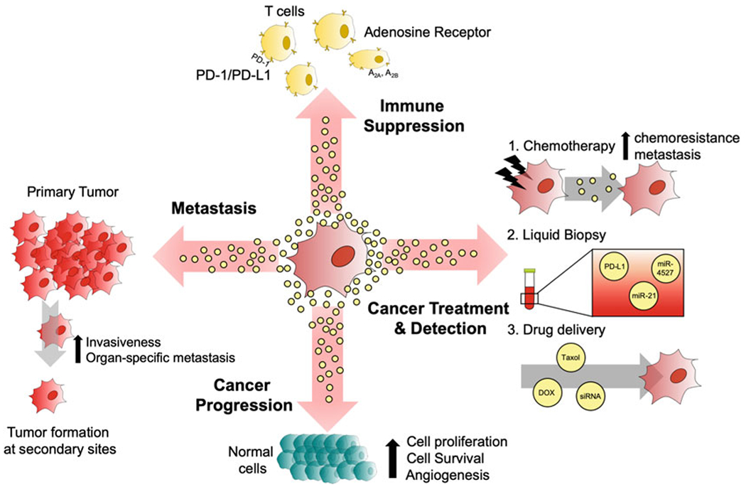
Extracellular vesicles (EVs) produced by cancer cells function as a unique form of intercellular communication that can promote cell growth and survival, help shape the tumor microenvironment, and increase invasive and metastatic activity. There are two major classes of EVs, microvesicles (MVs) and exosomes, and they differ in how they are formed. MVs are generated by the outward budding and fission of the plasma membrane. On the other hand, exosomes are derived as multivesicular bodies (MVBs) fuse with the plasma membrane and release their contents. What makes EVs especially interesting is how they mediate their effects. Both MVs and exosomes have been shown to contain a wide-variety of bioactive cargo, including cell surface, cytosolic, and nuclear proteins, as well as RNA transcripts, micro-RNAs (miRNAs), and even fragments of DNA. EVs, and their associated cargo, can be transferred to other cancer cells, as well as to normal cell types, causing the recipient cells to undergo phenotypic changes that promote different aspects of cancer progression. These findings, combined with those demonstrating that the amounts and contents of EVs produced by cancer cells can vary depending on their cell of origin, stage of development, or response to therapies, have raised the exciting possibility that EVs can be used for diagnostic purposes. Moreover, the pharmaceutical community is aggressively pursuing the use of EVs as a potential drug delivery platform. Here, in this chapter, we will highlight what is currently known about how EVs are generated, how they impact cancer progression, and the different ways they are being exploited for clinical applications.

Keywords: Exosomes; Extracellular vesicles; Intercellular communication; Microvesicles; Multivesicular bodies; Therapy deliver system; Tumor microenvironment.


Figures

Fig 1.
Fig 1.
Common EV isolation approaches and markers. Biological liquids, that is, the conditioned medium collected from cells in culture are subjected to a low-speed centrifugation (300 × g) to remove any intact floating cells and cell debris (top panel). Microvesicles can be purified by either filtering the conditioned medium through a 0.22 μm pore size filter, or by centrifugation at 10,000 × g (middle panel). The flow through or supernatant is then subjected to ultracentrifugation (120,000 × g) to pellet exosomes (bottom panel). The cells, microvesicles, and exosomes can be probed for the proteins listed under Commonly Used Markers
Fig 2.
Fig 2.
Scheme depicting the biogenesis of different classes of EVs. Exosomes originate from the endolysosomal pathway. The plasma membrane invaginates to form early endosomes in a RAB5-dependent manner. The endosomes are further trafficked and processed by ESCRTs, RAB7, tetraspanins, and nSMase 2 to generate multivesicular bodies (MVBs) containing intraluminal vesicles (ILVs). The MVBs are either trafficked by RAB7 to lysosomes and degraded, or directed to the plasma membrane by RAB27, RAB11, or RAB35. The MVBs then fuse with the plasma membrane and their contents are released. Microvesicles are formed as an outcome of an outward budding and fission from the plasma membrane. This process is mediated by RHOA or ARF6 signaling events that regulate cytoskeletal remodeling. Exomeres and nonvesicle fractions are considered as unique forms of non-membrane enclosed structures that are copurified with exosomes. The mechanisms underlying their formation are still unknown
Fig 3.
Fig 3.
Illustration showing some of the major roles and potential clinical applications for cancer cell-derived EVs. The EVs generated by cancer cells have the ability to suppress the immune system of the host by activating PD-1/PD-L1 or adenosine receptor signaling pathways. EVs have also been shown to promote several other aspects of cancer progression, including cell growth, survival, and angiogenesis through a variety of mechanisms. EVs also play an important role in metastasis, as they have been shown to increase the rates of organ-specific metastatic spread and the overall invasiveness of tumors. Clinically, it has been shown that cancer cells treated with chemotherapy generate EVs that can promote chemoresistance. The contents of EVs can be used for diagnostic purposes, and EVs can serve as therapeutic vehicles to deliver drugs or siRNA into cancer cells

Similar articles

Cited by

References

    1. Wilson KJ, Mill C, Lambert S et al. (2012) EGFR ligands exhibit functional differences in models of paracrine and autocrine signaling. Growth Factors 30:107–116. 10.3109/08977194.2011.649918 - DOI - PMC - PubMed
    1. Latifkar A, Hur YH, Sanchez JC et al. (2019) New insights into extracellular vesicle biogenesis and function. J Cell Sci 132:jcs222406. 10.1242/jcs.222406 - DOI - PMC - PubMed
    1. Maas SLN, Breakefield XO, Weaver AM (2017) Extracellular vesicles: unique intercellular delivery vehicles. Trends Cell Biol 27:172–188. 10.1016/j.tcb.2016.11.003 - DOI - PMC - PubMed
    1. Hessvik NP, Llorente A (2018) Current knowledge on exosome biogenesis and release. Cell Mol Life Sci 75:193–208. 10.1007/s00018-017-2595-9 - DOI - PMC - PubMed
    1. Van Niel G, D’Angelo G, Raposo G (2018) Shedding light on the cell biology of extracellular vesicles. Nat Rev Mol Cell Biol 19:213–228. 10.1038/nrm.2017.125 - DOI - PubMed

MeSH terms