

Differentiating novel coronavirus pneumonia from general pneumonia based on machine learning
- PMID: 32814568
- PMCID: PMC7436068
- DOI: 10.1186/s12938-020-00809-9
Differentiating novel coronavirus pneumonia from general pneumonia based on machine learning
Abstract
Background: Chest CT screening as supplementary means is crucial in diagnosing novel coronavirus pneumonia (COVID-19) with high sensitivity and popularity. Machine learning was adept in discovering intricate structures from CT images and achieved expert-level performance in medical image analysis.
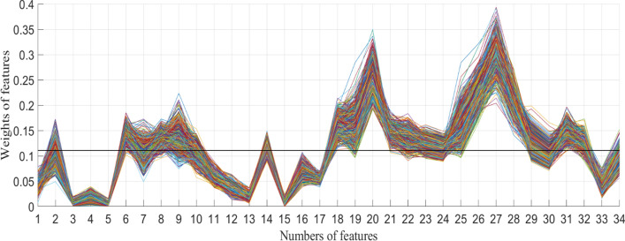
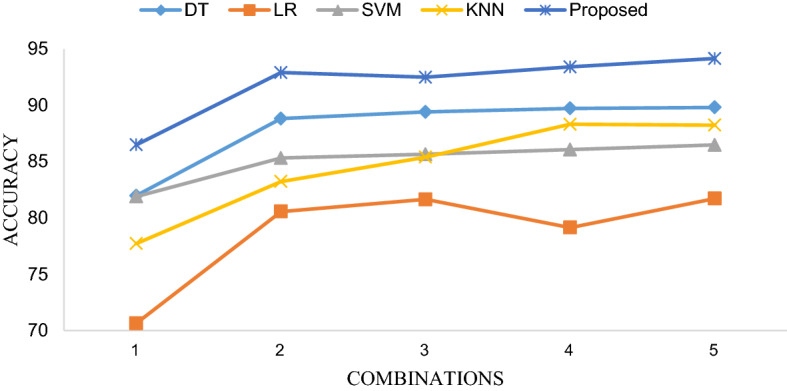
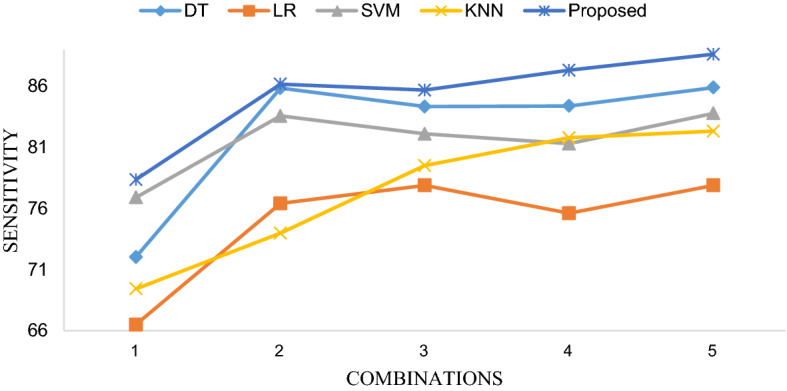
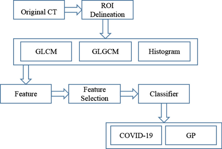
Methods: An integrated machine learning framework on chest CT images for differentiating COVID-19 from general pneumonia (GP) was developed and validated. Seventy-three confirmed COVID-19 cases were consecutively enrolled together with 27 confirmed general pneumonia patients from Ruian People's Hospital, from January 2020 to March 2020. To accurately classify COVID-19, region of interest (ROI) delineation was implemented based on ground-glass opacities (GGOs) before feature extraction. Then, 34 statistical texture features of COVID-19 and GP ROI images were extracted, including 13 gray-level co-occurrence matrix (GLCM) features, 15 gray-level-gradient co-occurrence matrix (GLGCM) features and 6 histogram features. High-dimensional features impact the classification performance. Thus, ReliefF algorithm was leveraged to select features. The relevance of each feature was the average weights calculated by ReliefF in n times. Features with relevance larger than the empirically set threshold T were selected. After feature selection, the optimal feature set along with 4 other selected feature combinations for comparison were applied to the ensemble of bagged tree (EBT) and four other machine learning classifiers including support vector machine (SVM), logistic regression (LR), decision tree (DT), and K-nearest neighbor with Minkowski distance equal weight (KNN) using tenfold cross-validation.
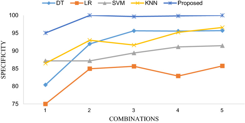
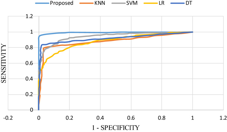
Results and conclusions: The classification accuracy (ACC), sensitivity (SEN), specificity (SPE) of our proposed method yield 94.16%, 88.62% and 100.00%, respectively. The area under the receiver operating characteristic curve (AUC) was 0.99. The experimental results indicate that the EBT algorithm with statistical textural features based on GGOs for differentiating COVID-19 from general pneumonia achieved high transferability, efficiency, specificity, sensitivity, and impressive accuracy, which is beneficial for inexperienced doctors to more accurately diagnose COVID-19 and essential for controlling the spread of the disease.
Keywords: Chest CT; General pneumonia; Machine learning; Novel coronavirus pneumonia.
Conflict of interest statement
The authors declare that they have no competing interests.
Figures
Similar articles
-
Differentiation of fat-poor angiomyolipoma from clear cell renal cell carcinoma in contrast-enhanced MDCT images using quantitative feature classification.Med Phys. 2017 Jul;44(7):3604-3614. doi: 10.1002/mp.12258. Epub 2017 Jun 9. Med Phys. 2017. PMID: 28376281
-
SVM-RLF-DNN: A DNN with reliefF and SVM for automatic identification of COVID from chest X-ray and CT images.Digit Health. 2024 May 27;10:20552076241257045. doi: 10.1177/20552076241257045. eCollection 2024 Jan-Dec. Digit Health. 2024. PMID: 38812845 Free PMC article.
-
CT-based radiomics for predicting the rapid progression of coronavirus disease 2019 (COVID-19) pneumonia lesions.Br J Radiol. 2021 Jun 1;94(1122):20201007. doi: 10.1259/bjr.20201007. Epub 2021 Apr 21. Br J Radiol. 2021. PMID: 33881930 Free PMC article.
-
Artificial intelligence model on chest imaging to diagnose COVID-19 and other pneumonias: A systematic review and meta-analysis.Eur J Radiol Open. 2022;9:100438. doi: 10.1016/j.ejro.2022.100438. Epub 2022 Aug 18. Eur J Radiol Open. 2022. PMID: 35996746 Free PMC article. Review.
-
Biphasic majority voting-based comparative COVID-19 diagnosis using chest X-ray images.Expert Syst Appl. 2023 Apr 15;216:119430. doi: 10.1016/j.eswa.2022.119430. Epub 2022 Dec 21. Expert Syst Appl. 2023. PMID: 36570382 Free PMC article. Review.
Cited by
-
Artificial Intelligence Approaches on X-ray-oriented Images Process for Early Detection of COVID-19.J Med Signals Sens. 2022 Jul 26;12(3):233-253. doi: 10.4103/jmss.jmss_111_21. eCollection 2022 Jul-Sep. J Med Signals Sens. 2022. PMID: 36120399 Free PMC article. Review.
-
"KAIZEN" method realizing implementation of deep-learning models for COVID-19 CT diagnosis in real world hospitals.Sci Rep. 2024 Jan 19;14(1):1672. doi: 10.1038/s41598-024-52135-y. Sci Rep. 2024. PMID: 38243054 Free PMC article.
-
Artificial intelligence and IoT based prediction of Covid-19 using chest X-ray images.Smart Health (Amst). 2022 Sep;25:100299. doi: 10.1016/j.smhl.2022.100299. Epub 2022 Jun 26. Smart Health (Amst). 2022. PMID: 35783463 Free PMC article.
-
Screening of COVID-19 Based on GLCM Features from CT Images Using Machine Learning Classifiers.SN Comput Sci. 2023;4(2):133. doi: 10.1007/s42979-022-01583-2. Epub 2022 Dec 29. SN Comput Sci. 2023. PMID: 36593973 Free PMC article.
-
CT radiomic models to distinguish COVID-19 pneumonia from other interstitial pneumonias.Radiol Med. 2021 Aug;126(8):1037-1043. doi: 10.1007/s11547-021-01370-8. Epub 2021 May 27. Radiol Med. 2021. PMID: 34043146 Free PMC article.
References
-
- Li D, Wang D, Dong J, Wang N, Huang H, Xu H, Xia C. False-negative results of real-time reverse-transcriptase polymerase chain reaction for severe acute respiratory syndrome coronavirus 2: role of deep-learning-based CT diagnosis and insights from two cases. Korean J Radiol. 2020;21(4):505–508. - PMC - PubMed
-
- Cheng Z, Lu Y, Cao Q, Qin L, Pan Z, Yan F, Clinical Yang W. Features and chest CT manifestations of coronavirus disease, (COVID-19) in a single-center study in Shanghai, China. AJR Am J Roentgenol. 2019;2020:1–6. - PubMed
-
- Li CX, Wu B, Luo F, Zhang N. Clinical Study and CT Findings of a Familial Cluster of Pneumonia with Coronavirus Disease 2019 (COVID-19) Sichuan Da Xue Xue Bao Yi Xue Ban. 2020;51(2):155–158. - PubMed
MeSH terms
Grants and funding
LinkOut - more resources
Full Text Sources
Medical
