. 2021 Jan;28(1):283-302.
doi: 10.1038/s41418-020-0600-6. Epub 2020 Aug 19.

CircRNA-vgll3 promotes osteogenic differentiation of adipose-derived mesenchymal stem cells via modulating miRNA-dependent integrin α5 expression

Affiliations

CircRNA-vgll3 promotes osteogenic differentiation of adipose-derived mesenchymal stem cells via modulating miRNA-dependent integrin α5 expression

Dandan Zhang et al. Cell Death Differ. 2021 Jan.

Abstract

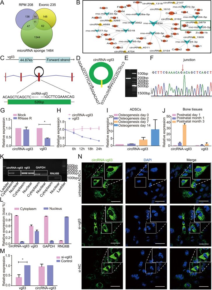
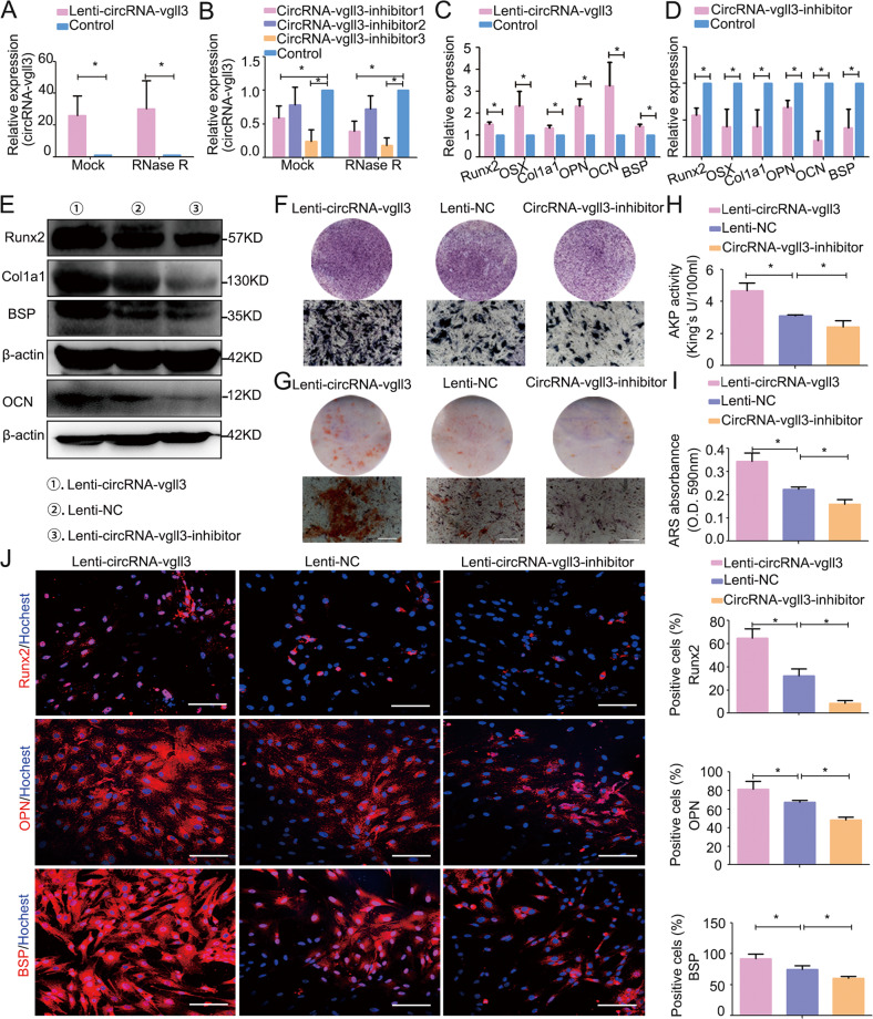
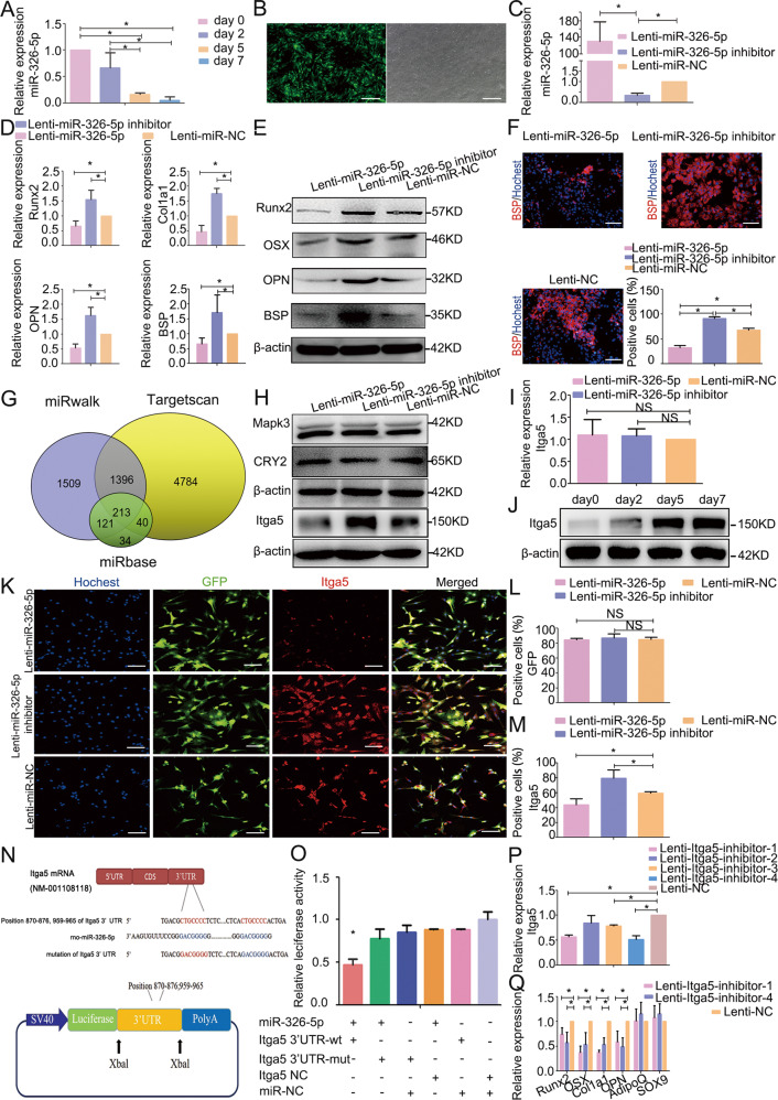
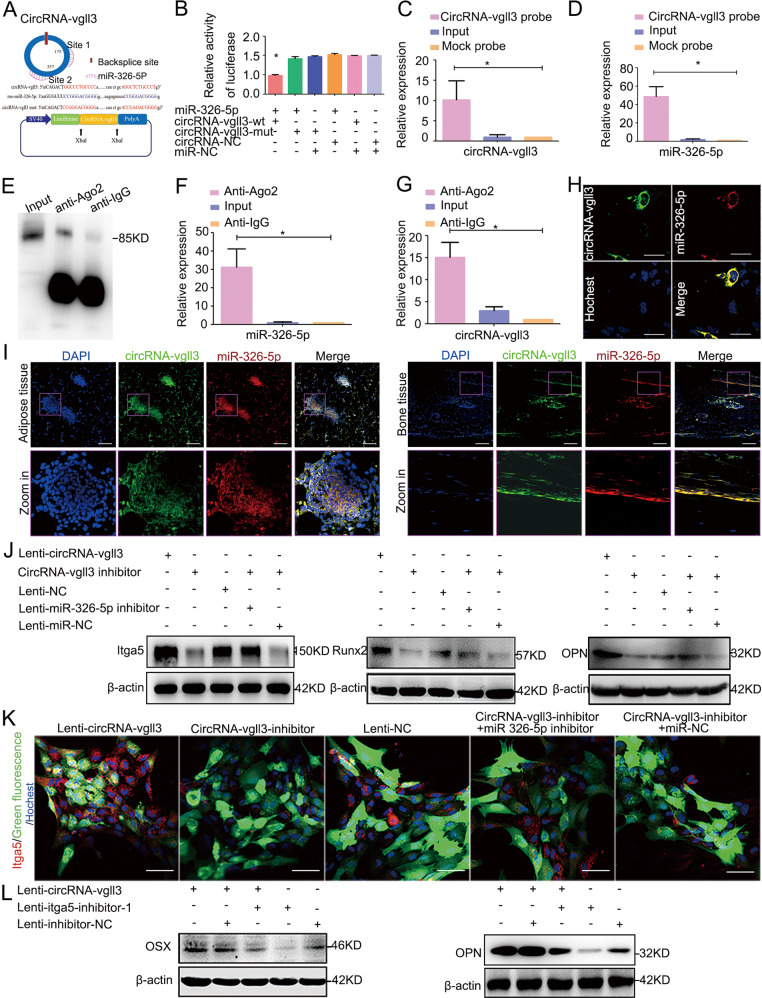
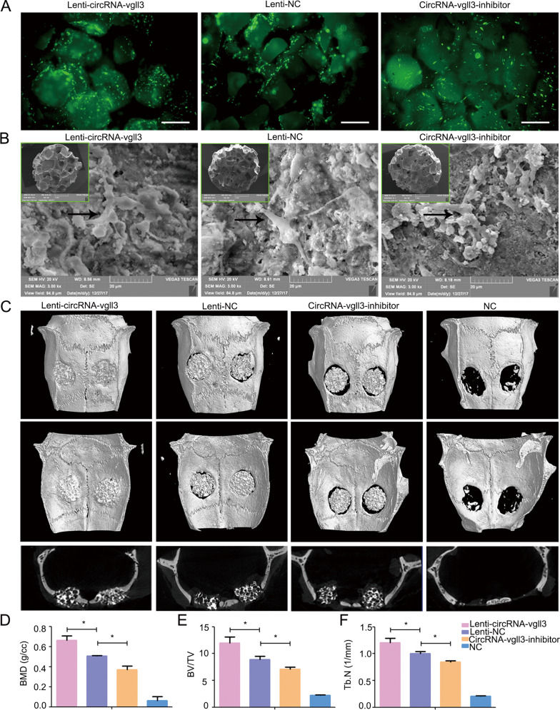
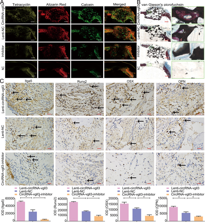
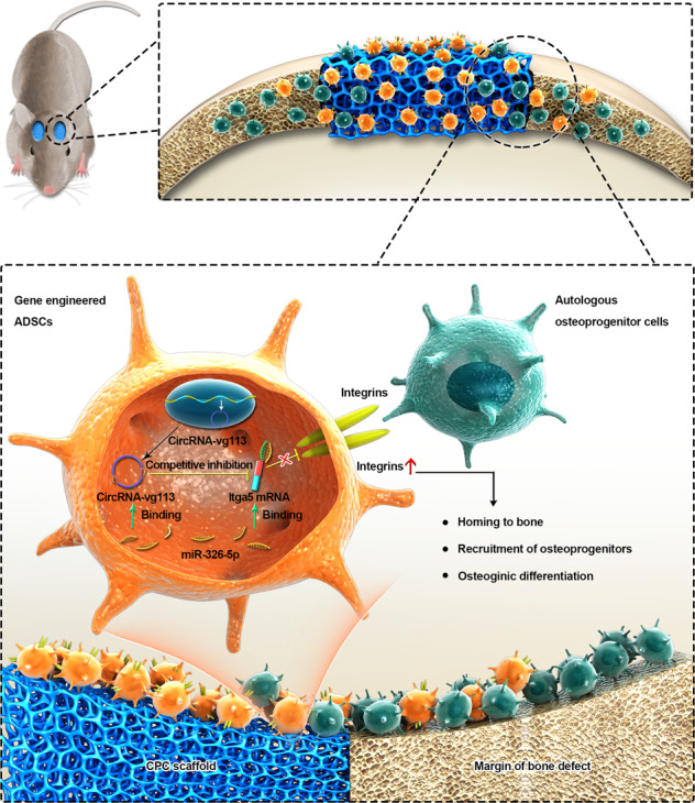
Adipose-derived mesenchymal stem cells (ADSCs) are promising candidate for regenerative medicine to repair non-healing bone defects due to their high and easy availability. However, the limited osteogenic differentiation potential greatly hinders the clinical application of ADSCs in bone repair. Accumulating evidences demonstrate that circular RNAs (circRNAs) are involved in stem/progenitor cell fate determination, but their specific role in stem/progenitor cell osteogenesis, remains mostly undescribed. Here, we show that circRNA-vgll3 originating from the vgll3 locus markedly enhances osteogenic differentiation of ADSCs; nevertheless, silencing of circRNA-vgll3 dramatically attenuates ADSC osteogenesis. Furthermore, we validate that circRNA-vgll3 functions in ADSC osteogenesis through a circRNA-vgll3/miR-326-5p/integrin α5 (Itga5) pathway. Itga5 promotes ADSC osteogenic differentiation and miR-326-5p suppresses Itga5 translation. CircRNA-vgll3 directly sequesters miR-326-5p in the cytoplasm and inhibits its activity to promote osteogenic differentiation. Moreover, the therapeutic potential of circRNA-vgll3-modified ADSCs with calcium phosphate cement (CPC) scaffolds was systematically evaluated in a critical-sized defect model in rats. Our results demonstrate that circRNA-vgll3 markedly enhances new bone formation with upregulated bone mineral density, bone volume/tissue volume, trabeculae number, and increased new bone generation. This study reveals the important role of circRNA-vgll3 during new bone biogenesis. Thus, circRNA-vgll3 engineered ADSCs may be effective potential therapeutic targets for bone regenerative medicine.


Conflict of interest statement

The authors declare that they have no conflict of interest.

Figures

Fig. 1
Fig. 1. Characterization of circRNA-vgll3 in ADSCs.
a Venn analysis of abundant (RPM > 0.1) exonic circRNAs with potential miRNA binding sites in ADSCs. b Bioinformatics prediction of 11 circRNAs, their predicted miRNAs, and miRNA targets. c CircRNA-vgll3 generated from the third exon of vgll3 gene locus. d-f PCR validation of circRNA-vgll3 using outward-facing primer and Sanger sequencing of the PCR product. g The relative gene expression of circRNA-vgll3 and vgll3 after RNase R treatment (n = 3, *P < 0.05 versus Mock group, statistical analysis was performed by Student’s t test). h The relative gene expression changes of circRNA-vgll3 and vgll3 after actinomycin D treatment for 6 h, 12 h,18 h, and 24 h, compared to that of 0 h (n = 3). i qPCR analysis of circRNA-vgll3 expression in BMP2-induced ADSCs and naive ADSCs. j qPCR analysis of circRNA-vgll3 expression in bone tissue development time points of postnatal day 1, month 1 and month 3. k-l PCR and qPCR analysis of cytoplasmic and nuclear RNA with circRNA-vgll3 primer, vgll3 primer, GAPDH primer (canonical marker of cytoplasmic fraction) and RNU6B primer (canonical marker of nuclear fraction) showed that circRNA-vgll3 mainly located in the cytoplasm and vgll3 located in the cytoplasm and nucleus. m qPCR analysis of the efficiency of the si-vgll3 in knocking down the mRNA levels of vgll3 (n = 3, *P < 0.05 versus Control group, statistical analysis was performed by Student’s t test). n FISH results depicted the cytoplasm location of circRNA-vgll3. Si-vgll3 treatment did not alter the labeling efficiency. Scale bars: 60 µm.
Fig. 2
Fig. 2. CircRNA-vgll3 positively regulates ADSC osteogenesis.
a Transfection efficiency was evaluated by qPCR and showed upregulation of circRNA-vgll3 expression in the Lenti-circRNA-vgll3-transfected group. The transfection efficiency was not influenced by RNase R treatment (n = 3, *P < 0.05 versus Control group, statistical analysis was performed by Student’s t test). b Three shRNAs for circRNA-vgll3 were cloned into vectors, and qPCR analysis showed that circRNA-vgll3 inhibitor 1 and 3 can effectively knockdown the expression of circRNA-vgll3. The knockdown efficiency was not influenced by RNase R treatment (n = 3, *P < 0.05 versus Control group, statistical analysis was performed by one-way ANOVA). cd mRNA expression levels of the markers Runx2, OSX, Col1a1, OPN, OCN, and BSP were elevated at osteogenesis day 7 by Lenti-circRNA-vgll3 whereas they were impaired by circRNA-vgll3 inhibitor when compared to Lenti-NC (n = 3, *P < 0.05 versus Control group, statistical analysis was performed by Student’s t test). e Western blot analysis detecting the levels of Runx2, Col1a1, BSP and OCN at osteogenesis day 7 showed that they were significantly promoted by Lenti-circRNA-vgll3 and greatly attenuated by transfection of the circRNA-vgll3 inhibitor in comparison to Lenti-NC. f ALP staining evaluated the effect of circRNA-vgll3 on ALP activity at osteogenesis day 7. Scale bars: 100 µm. g ARS staining evaluated the effect of circRNA-vgll3 on the ECM mineralization at osteogenesis day 14. Scale bars: 100 µm. hi Semiquantitative analysis of ALP activity and ARS activity (n = 3, *P < 0.05 versus Control group, statistical analysis was performed by one-way ANOVA). j Cellular immunofluorescence showing the cellular expression levels of Runx2, OPN and BSP at osteogenesis day 7 (n = 3, *P < 0.05 versus Control group, statistical analysis was performed by one-way ANOVA, Scale bars: 100 µm).
Fig. 3
Fig. 3. MiR-326-5p regulates ADSC osteogenesis by targeting Itga5.
a MiR-326-5p expression levels during ADSC osteogenic differentiation were assessed by qPCR and showed a gradual trend of downregulation (n = 3, *P < 0.05, statistical analysis was performed by one-way ANOVA). b Transfection efficiency of miR-326-5p as evidenced by fluorescence pictures. Scale bars: 200 µm. c Transfection efficiency of Lenti-miR-326-5p and Lenti-miR-326-5p inhibitor was evaluated by qPCR (n = 3, *P < 0.05 versus Lenti-miR-NC group, statistical analysis was performed by one-way ANOVA). d The mRNA expression of osteogenic marker genes Runx2, Col1a1, OPN and BSP was markedly inhibited in the Lenti-miR-326-5p-transfected cells whereas it was promoted by the Lenti-miR-326-5p inhibitor compared to Lenti-NC at osteogenesis day 7 (n = 3, *P < 0.05 versus Lenti-miR-NC group, statistical analysis was performed by one-way ANOVA). (e). Western blotting for the protein expression of Runx2, OSX, OPN and BSP at osteogenesis day 7. f Immunocytochemistry showed BSP was markedly inhibited in the Lenti-miR-326-5p-transfected cells whereas it was promoted by the Lenti-miR-326-5p inhibitor compared to Lenti-NC. (n = 3, *P < 0.05, statistical analysis was performed by one-way ANOVA, Scale bars: 100 µm). g The target gene prediction databases miRanda, miRwalk and Targetscan indicate that there are totally 213 target genes in intersection for miR-326-5p. h The protein levels of CRY2 and Mapk3 were not influenced by overexpression or knockdown of miR-326-5p in ADSCs. The protein level of Itga5 was reduced in Lenti-miR-326-5p-transfected ADSCs whereas it was promoted in Lenti-miR-326-5p inhibitor-transfected ADSCs at osteogenesis day 7. i qPCR results of Itga5 mRNA expression in Lenti-miR-326-5p-transfected ADSCs at osteogenesis day 7. j Western blot analysis at days 0, 2, 5, and 7 indicated that the protein expression of Itga5 was gradually increased. km. Immunocytochemistry analysis of Itga5 expression levels in Lenti-miR-326-5p-transduced ADSCs. Scale bars: 100 µm. n Schematic diagram depicting the constructed firefly luciferase reporter system and the predicted binding sites of miR-326-5p in the Itga5 3′UTR. o Luciferase reporter assay showed that co-transfection of miR-326-5p with the constructed Itga5 3'UTR-wt plasmid obviously decreased luciferase activity (*P < 0.05, statistical analysis was performed by one-way ANOVA). p Four Lenti-itga5-inhibitors were transduced into ADSCs, and Lenti-itga5-inhibitor-1 and Lenti-itga5-inhibitor-4 effectively knocked down the expression of Itga5 (n = 3, *P < 0.05 versus Lenti-NC group, statistical analysis was performed by one-way ANOVA). q Knockdown of Itga5 by Lenti-itga5-inhibitor-1 and Lenti-itga5-inhibitor-4 decreased the mRNA levels of Runx2, OSX, Col1a1 and OPN, while the expression of AdipoQ and SOX9 showed no obvious changes with Itga5 inhibition in ADSCs at osteogenesis day 7 (n = 3, *P < 0.05 versus Lenti-NC group, statistical analysis was performed by one-way ANOVA).
Fig. 4
Fig. 4. CircRNA-vgll3 upregulates the level of Itga5 through miR-326-5p.
a The predicted miR-326-5p binding sites on circRNA-vgll3. b Luciferase reporter assay showed that co-transfection of the miR-326-5p plasmid with circRNA-vgll3-wt plasmid significantly impaired the luciferase activity (*P < 0.05 versus NC group, statistical analysis was performed by one-way ANOVA). cd RNA pull-down analysis showed distinctive enrichment of circRNA-vgll3 and miR-326-5p in the biotin-labeled circRNA-specific probe group (n = 3, *P < 0.05 versus Mock probe group, statistical analysis was performed by one-way ANOVA). eg RNA RIP was implemented on ADSCs using antibodies against Ago2 to evaluate whether circRNA-vgll3 was related to RISC. Western blot analysis of the efficiency of Ago2 enrichment by anti-Ago2 (e); qPCR measuring the RNA levels in immunoprecipitants showed that the miR-326-5p level in the anti-Ago2 group was higher than that in the anti-IgG cells (f), accompanied by a higher circRNA-vgll3 level in the anti-Ago2 group than that in the anti-IgG group (g) (n = 3, *P < 0.05 versus anti-IgG group, statistical analysis was performed by one-way ANOVA). h FISH assay showed both miR-326-5p and circRNA-vgll3 localized in the cytoplasm of ADSCs. Scale bars: 60 µm. i FISH assay showed that both miR-326-5p and circRNA-vgll3 localized in the cytoplasm of adipose and bone tissues. Scale bars: 200 µm. j ADSCs were co-transfected with the circRNA-vgll3 inhibitor and the miR-326-5p inhibitor, and the expression levels of Itga5, Runx2 and OPN were evaluated by western blotting at osteogenesis day 7. The levels of Itga5, Runx2, and OPN were significantly decreased by transfection with circRNA-vgll3 inhibitor; however, the miR-326-5p inhibitor obviously rescued the low levels of Itga5, Runx2, and OPN caused by the circRNA-vgll3 inhibitor. k Immunocytochemistry detection of Itga5 after co-transfection of circRNA-vgll3 inhibitor and the miR-326-5p inhibitor in ADSCs. Scale bars: 50 µm. l Co-transfection of Lenti-circRNA-vgll3 and Itga5 inhibitor in ADSCs impaired the upregulated expression of OSX and OPN caused by circRNA-vgll3 overexpression at osteogenesis day 7.
Fig. 5
Fig. 5. CircRNA-vgll3 regulates bone formation in vivo.
a Fluorescence images of ADSCs on CPC scaffolds. Scale bars: 200 µm. b SEM images revealed that the transfected ADSCs were tightly adherent to the surface and pores of CPCs with a large amount of ECM fibril networks. c Micro-CT showed that new bone regeneration was promoted in the Lenti-circRNA-vgll3 group, whereas it was impaired by circRNA-vgll3 inhibitor 8 weeks post implantation. df Micro-CT quantitative analysis showed that the BMD, BV/TV and Tb. N of the overexpression group were markedly enhanced compared to the Lenti-NC group, whereas they were inhibited in the circRNA-vgll3 inhibitor skulls than those in the Lenti-NC skulls. *P < 0.05, statistical analysis was performed by one-way ANOVA.
Fig. 6
Fig. 6. Fluorochrome-labeling and histological analysis of in vivo bone regeneration.
a The area of Tetracycline-labeled, Alizarin Red-labeled and Calcein-labeled bone was elevated by Lenti-circRNA-vgll3 group, whereas the circRNA-vgll3 inhibitor reduced this area. Scale bars: 1000 µm. b van Gieson’s picrofuchsin staining showed that Lenti-circRNA-vgll3 increased the new bone percentage in the scaffold tissue whereas the circRNA-vgll3 inhibitor impaired it. Scale bars: 1000 µm. c Immunohistochemical analysis of Itga5, Runx2, OSX and OPN in the defect area at 8 weeks post-operation. Scale bars: 25 µm. *P < 0.05, statistical analysis was performed by one-way ANOVA.
Fig. 7
Fig. 7. A model for circRNA-vgll3-modified ADSCs in bone regeneration.
CircRNA-vgll3-modified ADSCs composition with CPC scaffolds promote the repair of critical-sized bone defects. CircRNA-vgll3 directly sequesters miR-326-5p in the cytoplasm and inhibits its activity, which in turn upregulates the protein levels of Itga5. The upregulated Itga5 protein levels help mediate the homing of ADSCs to the bone, the recruitment of osteoprogenitor cells, and the promotion of osteogenic differentiation in ADSCs.

References

    1. Uccelli A, Moretta L, Pistoia V. Mesenchymal stem cells in health and disease. Nat Rev Immunol. 2008;8:726–36. doi: 10.1038/nri2395. - DOI - PubMed
    1. Bunpetch V, Zhang ZY, Zhang X, Han S, Zongyou P, Wu H, et al. Strategies for MSC expansion and MSC-based microtissue for bone regeneration. Biomaterials. 2019;196:67–79. doi: 10.1016/j.biomaterials.2017.11.023. - DOI - PubMed
    1. Wankhade UD, Shen M, Kolhe R, Fulzele S. Advances in adipose-derived stem cells isolation, characterization, and application in regenerative tissue engineering. Stem Cells Int. 2016;2016:3206807. doi: 10.1155/2016/3206807. - DOI - PMC - PubMed
    1. Tabatabaei Qomi R, Sheykhhasan M. Adipose-derived stromal cell in regenerative medicine: a review. World J Stem Cells. 2017;9:107–17. doi: 10.4252/wjsc.v9.i8.107. - DOI - PMC - PubMed
    1. Lotfy A, Salama M, Zahran F, Jones E, Badawy A, Sobh M. Characterization of mesenchymal stem cells derived from rat bone marrow and adipose tissue: a comparative study. Int J Stem Cells. 2014;7:135–42. doi: 10.15283/ijsc.2014.7.2.135. - DOI - PMC - PubMed

Publication types