. 2020 Aug;584(7822):630-634.
doi: 10.1038/s41586-020-2624-y. Epub 2020 Aug 19.

An intramembrane chaperone complex facilitates membrane protein biogenesis

Affiliations

An intramembrane chaperone complex facilitates membrane protein biogenesis

Patrick J Chitwood et al. Nature. 2020 Aug.

Abstract

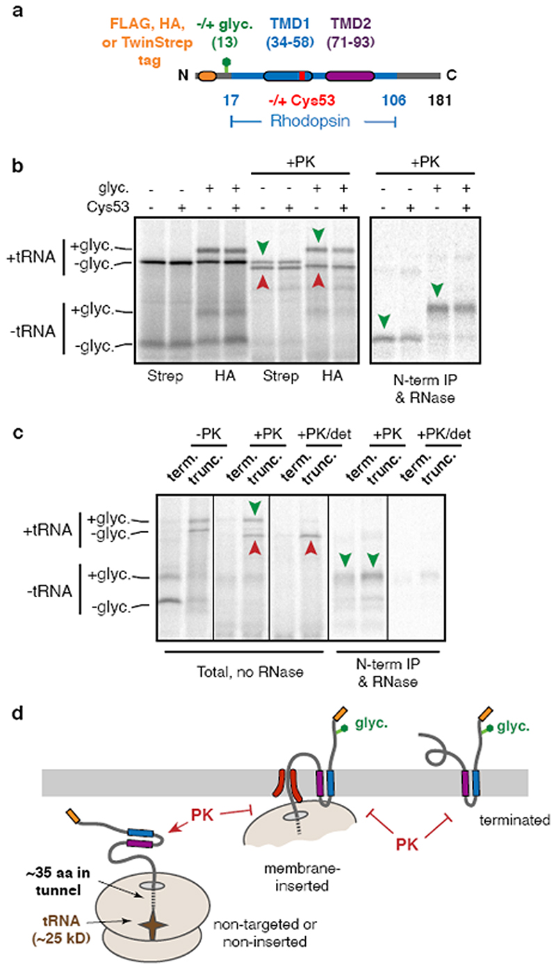
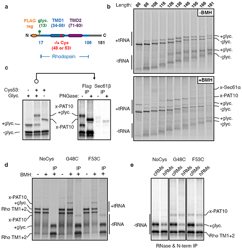
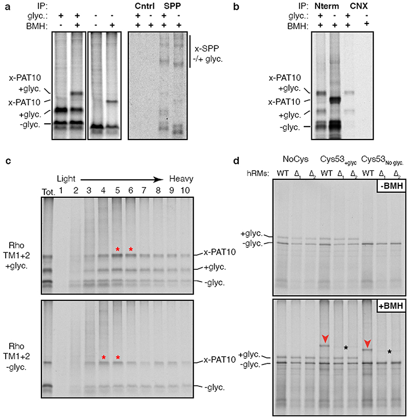
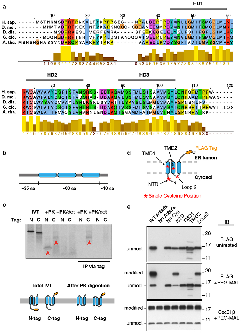
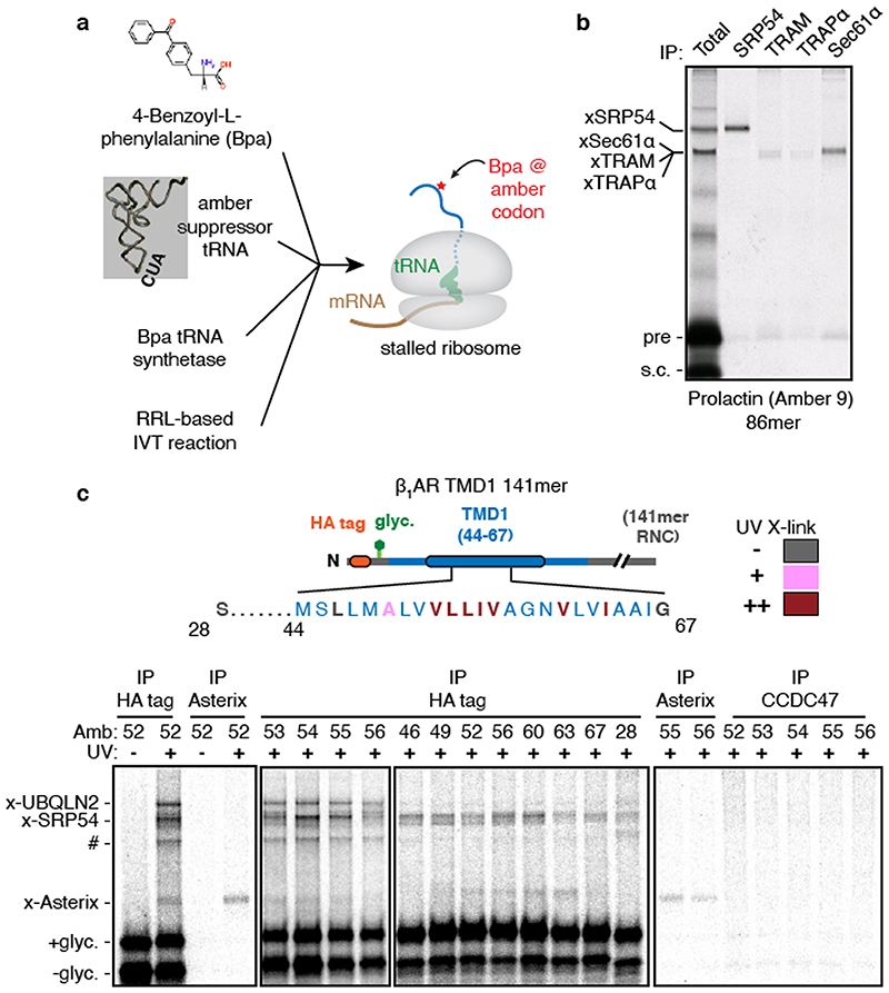
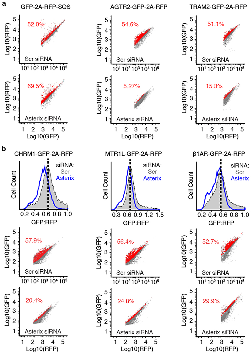
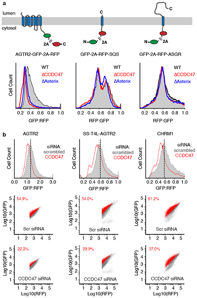
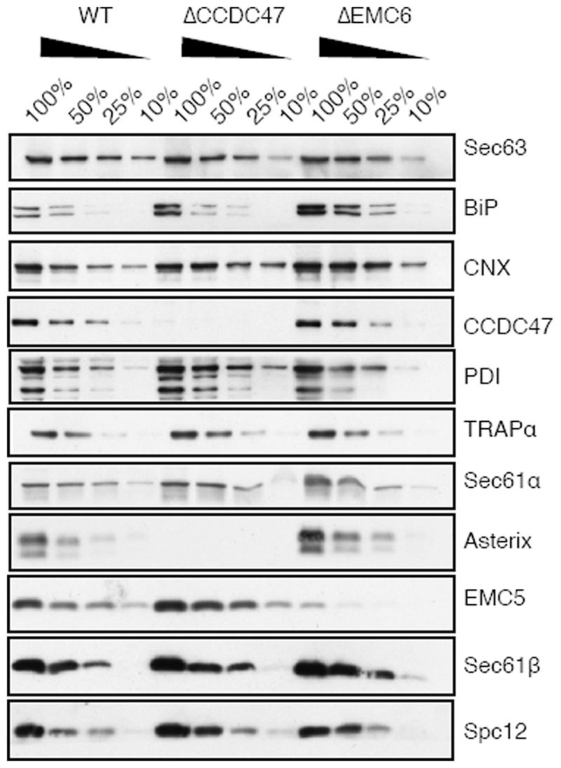
Integral membrane proteins are encoded by approximately 25% of all protein-coding genes1. In eukaryotes, the majority of membrane proteins are inserted, modified and folded at the endoplasmic reticulum (ER)2. Research over the past several decades has determined how membrane proteins are targeted to the ER and how individual transmembrane domains (TMDs) are inserted into the lipid bilayer3. By contrast, very little is known about how multi-spanning membrane proteins with several TMDs are assembled within the membrane. During the assembly of TMDs, interactions between polar or charged amino acids typically stabilize the final folded configuration4-8. TMDs with hydrophilic amino acids are likely to be chaperoned during the co-translational biogenesis of membrane proteins; however, ER-resident intramembrane chaperones are poorly defined. Here we identify the PAT complex, an abundant obligate heterodimer of the widely conserved ER-resident membrane proteins CCDC47 and Asterix. The PAT complex engages nascent TMDs that contain unshielded hydrophilic side chains within the lipid bilayer, and it disengages concomitant with substrate folding. Cells that lack either subunit of the PAT complex show reduced biogenesis of numerous multi-spanning membrane proteins. Thus, the PAT complex is an intramembrane chaperone that protects TMDs during assembly to minimize misfolding of multi-spanning membrane proteins and maintain cellular protein homeostasis.


Conflict of interest statement

Competing interests: The authors declare no competing interests.

Figures

Extended Data Fig. 1
Extended Data Fig. 1. Characterisation of Rho TM1+2 insertion.
(a) Diagram of Rho TM1+2 constructs used throughout this study. Variations on this construct include different N-terminal epitope tags, the presence or absence of a glycosylation site near the N-terminus, the presence, absence, or position of a cysteine within TMD1, various mutations within TMD1, and the presence or absence of TMD2. All of the constructs were tested either by protease protection or glycosylation to verify that no appreciable differences were observed in their insertion efficiencies. Note that although the exact amino acid numbering varies depending on the N-terminal tag, the numbering system corresponding to the FLAG-tagged version is used throughout. Thus, the 146mer refers to a truncation at the 146th codon in the numbering scheme indicated in Fig. 1a and in this diagram, even in constructs containing a different tag. (b) Representative example of insertion assays on two different tagged versions of Rho TM1+2. The TwinStrep tagged version (Strep) lacking a glycosylation site was compared to an HA tagged version containing a glycosylation site. Identical constructs containing either the wild-type Rho TM1 sequence or a point mutant (F53C) were tested in parallel to confirm no insertion defects result from insertion of a cysteine in TM1 (used for BMH-mediated crosslinking in later experiments). In this experiment,S-methionine labelled ribosome nascent chain complexes (RNCs) of 181 amino acids were produced by in vitro translation using rabbit reticulocyte lysate (RRL) in the presence of ER-derived rough microsomes (RMs) after which the microsomes were isolated and resuspended. Aliquots of the reactions were left untreated or digested with proteinase K (PK) and analysed directly by SDS-PAGE and visualised by autoradiography (left panel). Green arrow heads represent the fully inserted and PK-protected population and red arrowheads denote the non-inserted and proteolytically cleaved products. The cleaved product contains the region of polypeptide protected by the ribosomal tunnel and the attached tRNA. Aliquots of the PK-digested sample were treated with EDTA and RNase to release the polypeptide from the ribosome and tRNA and immunoprecipitated (IP) via the N-terminal tag. Only the fully inserted products are recovered by IP (green arrowheads). (c) Comparison of the topology of truncated RNCs and terminated Rho TM1+2. In this experiment, the FLAG-tagged Rho TM1+2 containing a glycosylation site with (term.) or without (trunc.) a stop codon was translated in the presence of RM after which the RMs were isolated by centrifugation. Aliquots of the isolated RMs were analysed directly (-PK), after PK digestion (+PK), or after PK digestion in the presence of detergent (+PK/det). Where indicated, the +PK and +PK/det samples were released from the attached tRNA and immunoprecipitated via the N-terminal tag. Note comparable glycosylation near the N-terminus and complete protection from PK for both the truncated and terminated products. (d) Diagram representing the interpretation of the experiments in panels b and c. The relatively short cytosolic loop between TMD1 and TMD2 is not accessible to PK digestion either as an RNC or a terminated product. For gel source data, see Supplementary Figure 1.
Extended Data Fig. 2
Extended Data Fig. 2. Additional characterisation of crosslinking to PAT10.
(a) Diagram of constructs either lacking or containing a cysteine in place of Gly48 or Phe53 within TMD1 of a Rho TM1+2 cassette. (b) S-labelled FLAG Rho TM1+2 ribosome nascent chains (RNCs) of varying lengths (as described in Figure 1a) containing a Cys at position 53 were generated by in vitro translation in the presence of RMs. Membranes were isolated by centrifugation through a sucrose cushion and resuspended in physiological salt buffer (PSB). An equivalent amount of each translation reaction was taken before (-BMH) and after (+BMH) the addition BMH for analysis by SDS-PAGE. The tRNA-linked nascent chains and free nascent chains are indicated. The free nascent chains arise from partial hydrolysis of the tRNA during electrophoresis under moderately basic conditions. Glycosylation is first observed at the 96mer length, which is 38 amino acids downstream of the end of TMD1. This matches the length of the ribosome tunnel, and indicates that membrane insertion and glycosylation occurs only after TMD1 is fully exposed outside the ribosome. Crosslinks to Sec61α are most prominent for the 106mer. Crosslinks to PAT10 are most prominent from the 126mer onwards, after the Sec61α crosslinks diminish. Note that all of the crosslinked adducts are seen to the tRNA-attached nascent chain, verifying that they are co-translational. The Sec61α crosslink and others are not as visible when total translation products are analysed, which is why we typically immunoprecipitate the sample via the nascent chain (e.g., in Fig. 1a). This reduces the background, allowing otherwise obscured crosslinks to be visualised clearly. Furthermore, we usually digest the samples with RNase after the experiment but before SDS-PAGE to remove the tRNA, thereby avoiding the heterogeneity that results from partial tRNA hydrolysis during sample handling and SDS-PAGE. All of the indicated crosslinking adducts observed were completely dependent on the presence of BMH. (c) The indicated “Input” crosslinking sample from Fig. 2b (reproduced here on the left) was subjected to immunoprecipitation (IP) using anti-FLAG or anti-Sec61p antibodies under denaturing conditions. The IP samples were either left untreated or digested with PNGase F to remove N-linked glycans. Equivalent amounts were loaded in each lane. Note that Sec61p is not an appreciable crosslinking partner of these RNCs, and to the extent a crosslink is observed, it migrates slightly faster than the PAT10 crosslink. (d) The indicated Rho TM1+2 variants were translated in vitro in the presence of RMs and treated with BMH as indicated. In this experiment, the crosslinking was done directly on total translation reactions, not after isolation of the microsome fraction. Instead, translation reactions were diluted 5-fold with buffer to dilute the reduced glutathione and minimise quenching of BMH. An aliquot of each reaction was analysed directly by SDS-PAGE. Cross-linking efficiency is reduced compared to other experiments because membranes were not isolated by centrifugation through a sucrose cushion to remove reduced glutathione from translation extract. One aliquot of the BMH-treated translation reactions were treated with RNase A and EDTA, denatured, and IPed via the N-terminal FLAG tag (IP). (e) As in panel d except Rho TM1+2 variants were generated in the presence of RMs derived from either canine pancreas (cRM) or HEK293 cells (hRM). In this experiment, the microsomes were isolated by centrifugation through a sucrose cushion prior to BMH crosslinking (note higher cross linking efficiency). While the PAT10 crosslink is seen in both cRM and hRM, cross-linking efficiency of the inserted (glycosylated) product is significantly lower in cRMs, which is one reason we used HEK293- derived RMs for most of the experiments in this study. For gel source data, see Supplementary Figure 1.
Extended Data Fig. 3
Extended Data Fig. 3. Analysis of PAT complex candidates.
(a) Radiolabelled 146mer RNCs of Rho TM1+2 constructs containing or lacking a glycosylation site were generated by in vitro translation in the presence of RMs. Membranes were isolated by centrifugation through a sucrose cushion, subjected to BMH crosslinking where indicated, treated with RNase A, then analysed by SDS-PAGE. One aliquot of the BMH treated reactions were solubilised under native conditions and IPed with either an antibody raised against the HA epitope tag (Cntrl) for a specificity control, or an antibody against signal peptide peptidase (SPP). Direct crosslinks to SPP are observed as two distinct adducts seen on very long exposures (compare to Fig. 1d, which is a shorter exposure), but native IPs do not enriched for a PAT10 engaged substrate. (b) Radiolabelled 146mer RNCs of FLAG tagged Rho TM1+2 containing (+) or lacking (-) a glycosylation site (glyc.) were generated by in vitro translation with RM, crosslinked with BMH, then immunoprecipitated under native conditions via the N-terminal FLAG tag on the substrate (Nterm) or with an antibody recognising the C-terminus of calnexin (CNX). Only the glycosylated substrate is recovered with CNX, consistent with its binding via the glycan. (c) Aliquots of the crosslinked and natively solubilised samples from panel b were run on a 5-25% sucrose gradient before analysis by SDS-PAGE and autoradiography. Red asterisks denote peak fractions containing the PAT complex as detected by the PAT10 crosslinking product. The PAT complex crosslinked to unglycosylated Rho TM1+2 migrates slightly smaller on the gradient than glycosylated Rho TM1+2, likely the result of CNX (∼90 kD) no longer associated with the nascent chain. (d) The insertion and BMH mediated cross-linking for 146mer RNCs of the parent Rho TM1+2 construct or versions lacking a cysteine (NoCys) or lacking a glycosylation site (Cys53No glyc). The radiolabelled RNCs were produced by in vitro translation in the presence of RMs isolated from wild type (WT) or two different CCDC47 KO cell lines (A1 and A2) generated from two different guide RNAs. Aliquots of the reaction before (-BMH) and after (+BMH) addition BMH were analysed by SDS-PAGE. No appreciable difference in insertion efficiency was observed in KO microsomes for Rho TM1+2 as monitored by glycosylation efficiency. Red arrowheads indicate the PAT10 crosslink which is lost upon CCDC47 KO. The faint crosslinked adduct observed in the KO samples (black asterisks) migrates slightly faster on the gel and likely represents weak Rho TM1+2 crosslinks to Sec61p (see Extended Data Fig. 2c). For gel source data, see Supplementary Figure 1.
Extended Data Fig. 4
Extended Data Fig. 4. Conservation and topology of Asterix.
(a) Alignment of Asterix homologs for five divergent species with a bar chart representing conservation scores of each amino acid. Indicated are three hydrophobic domains (HD1 to HD3), each of ∼15 amino acids, that are candidate TMDs. (b) Representation of human Asterix amino acid sequence and the relative lengths of hydrophilic (grey bar) and hydrophobic (blue) regions. (c) Two matched human Asterix constructs containing either an N- or C-terminal FLAG tag were generated by in vitro translation in the presence of RMs. One aliquot of the reaction was set aside for analysis by SDS-PAGE of total translation products (IVT). The remainder of the reaction was treated with proteinase K (+PK) without or with detergent (det). These protease- digested samples were either analysed directly or after immunoprecipitation (IP) via the FLAG tag. Red arrowheads indicate fragments protected from PK in the absence, but not presence of detergent. The PK-protected fragment from the C-terminally tagged Asterix was recovered by IP, suggesting that the C-terminus is located within the ER lumen and the N- terminus is located in the cytosol. The relative size difference between the N-and C- terminally tagged constructs observed after PK digestion can be attributed to digestion or protection of the FLAG tag. Below the gel is a cartoon depiction of one possible topology based on the results and the protease protected fragments that remain after digestion with PK. The other possible topology is a single-spanning orientation with HD2 and HD3 in the lumen. (d) Schematic of human Asterix with a C-terminal FLAG tag in its predicted 3-TMD topology based on the protease digestion results in panel c. To test this prediction, a cysteine- free version of Asterix (No Cys) was modified with single cysteines at the position indicated by the red-asterisks. If the topology prediction is correct, only the N-terminal domain (NTD) cysteine and the Loop 2 cysteine should be accessible to sulfhydryl modifying reagents added to the cytosolic side of them membrane. If the protein only spans the membrane once with the N-terminus facing the cytosol, then the Loop 2 cysteine should not be modified. As shown in panel a, wild type Asterix naturally has four cysteines, only one of which should be exposed to the cytosol because it is in the NTD. (e) Asterix KO HEK-293 cells were transiently transfected with the indicated Asterix-FLAG constructs, semi-permeabilised in 0.01% digitonin, washed to remove digitonin, and treated with PEG-Maleimide (average molecular weight 5 kDa) in order to modify any cytosolically exposed cysteine residues. WT Asterix contains 4 native cysteine residues, one in the N-terminus preceding TMD1 and 3 others within the putative TMD regions. Modification was only observed for the NTD cysteine and the cysteine in Loop 2, supporting a 3-TMD topology as depicted in panel d. The single cysteine present in the cytosolic domain of Sec61p was used as a positive control demonstrating equal modification efficiency in all samples, and the No Cys construct verifies sulfhydryl-dependent modification. Protection of the cysteines in TMD1 and TMD2 from modification verifies that membrane integrity was maintained in the experiment. For gel source data, see Supplementary Figure 1.
Extended Data Fig. 5
Extended Data Fig. 5. Characterisation of site-specific photo-crosslinking.
(a) Schematic of the strategy for site-specific incorporation of the photo-crosslinking amino acid Benzoyl- phenylalanine (Bpa) during in vitro translation (IVT). Bpa, a synthetic amber-suppressor tRNA, and recombinant Bpa tRNA synthetase are added to an IVT reaction. The nascent protein that is produced incorporates Bpa at an amber codon. UV irradiation results in Bpa activation and crosslinking to adjacent proteins. (b) The photo-crosslinking strategy was tested using a well-validated translocation intermediate: the 86mer of the secretory protein prolactin. The amber codon was installed at position 9, within the hydrophobic core of the signal sequence. At this length, the majority of the nascent chain is precursor (pre), with a small amount that is signal-cleaved (s.c.). The primary crosslinks to SRP54 and components of the translocation site (Sec61α, TRAM, and TRAPa) were verified by immunoprecipitation. (c) Site-specific photo-crosslinking of a 141mer RNC containing the UV-activated photo-crosslinking amino acid benzoyl-phenylalanine (Bpa) at the indicated amber positions (Amb). The diagram above the autoradiographs shows a schematic of the construct with the appropriate amino acid numbering. Amino acids in red show the strongest crosslinks to Asterix, pink show reduced crosslinks, and grey no detectable crosslinks. Total translation products recovered by IP via an HA tag on the nascent chain are shown adjacent to parallel IPs of selected samples using the indicated antibodies. Although not all IPs are shown, each position was tested for crosslinking to Asterix and CCDC47. RNCs that failed to engage SRP crosslink to UBQLN2, a quality control factor that binds exposed TMDs. Note that this crosslink diminishes markedly when increased RMs are used in the reaction (lanes 916, compared to lanes 5-8), presumably because the RMs contribute SRP, which is otherwise limiting in the reaction. A subset of RNCs fail to release from SRP and crosslink to SRP54. The crosslink indicated by the hashtag (#) is likely to be a mixture of similarly migrating crosslinks. Because this crosslink diminishes substantially with increased RMs (similar to the UBQLN2 crosslink), it is likely to be SGTA, another TMD-binding factor in the cytosol of this size. A small proportion of this crosslink could be the similarly sized Sec61α or TRAM. Of the membrane-inserted RNCs, the main crosslink is to Asterix, seen prominently for residues 52 to 63. At this length, the TMD has moved away from Sec61α, so crosslinks to this factor are minimal. No crosslinking to CCDC47 were ever observed. Note that by testing five sequential positions in the centre of the TMD, all sides of the helix have been sampled.
Extended Data Fig. 6
Extended Data Fig. 6. Effect of Asterix depletion on multi-spanning membrane proteins.
(a) The raw data for three of the histograms of the GFP:RFP ratio (or RFP:GFP ratio in the case of GFP-2A-RFP-SQS) shown in Fig. 3. The mode of the control histogram (dotted black line in Fig. 3) was used to determine the statistical mode of GFP:RFP (or RFP:GFP) ratio. This mode was used as a gate to colour the dot plots shown below the histograms such that all cells above the gate were coloured red and those below the gate were coloured grey. The percent of cells above the gate for each plot is indicated. (b) Flow- cytometry analysis of the indicated GPCR reporters using the dual-colour assay system exactly as in Fig. 3. Cell lines containing the reporter stably integrated at a single FRT site located downstream of a doxycycline-inducible reporter were used for these assays. This allows assay of cells using a single transfection (which proved to be less toxic than sequential transfections with both the siRNA and the reporter), and provided control of the length of time of reporter expression. Each reporter cell line was treated with scrambled (Scr) or Asterix-targeting siRNAs, then at the time of effective knockdown (verified in separate experiments using immunoblotting), the reporter was induced for ∼6-8 hours. Induction only after knockdown allows us to monitor the reporter that was produced in the absence of Asterix rather than a heterogeneous mixture of reporter expressed during the knockdown. The histograms of the GFP:RFP ratio in the scrambled- versus Asterix-siRNA cells are shown in grey and blue, respectively, in the upper plot for each construct. The two dot plots below the histogram are the corresponding raw data plotted as described in panel a. Each reporter shows a distribution of lower GFP:RFP ratio, with some reporters being more impacted than others. This is not seen with the tail-anchored protein SQS using the same assay format.
Extended Data Fig. 7
Extended Data Fig. 7. Effect of CCDC47 depletion on membrane protein biogenesis.
(a) The diagrams depict dual-colour fluorescent reporters for protein stability as an indirect measure of successful biogenesis. The membrane protein of interest is tagged with one fluorescent protein (FP), which is separated from a second FP by the viral 2A peptide sequence. When the 2A sequence is translated, peptide bond formation is skipped without perturbing elongation by the ribosome. Thus, translation results in two separate proteins made in a 1:1 stoichiometry that are separated at the 2A sequence. If biogenesis of the membrane protein is impaired, it will be degraded along with its tagged FP, resulting in an altered ratio of the two FPs. Thus, treatment conditions that impair biogenesis of the membrane protein will be reflected as a relative change in the ratio of FPs. The three reporters encoding angiotensin type-2 receptor II (AGTR2), squalene synthase (SQS) and Asialglycoprotein receptor (ASGR) were transiently transfected into wild-type (WT), CCDC47 KO (ΔCCDC47) or Asterix KO (AAsterix) HEK293 cells and analysed by dual- colour flow cytometry. Histograms represent the distribution of FP ratio in WT (grey), ΔCCDC47 (red) and AAsterix (blue) cells. A biogenesis defect is only seen for the multi- spanning membrane protein AGTR2, but not for the tail-anchored protein SQS or the signal- anchored single pass protein ASGR. (b) Assays similar to those in Fig. 3, but for cell lines treated with scrambled versus CCDC47 siRNAs as indicated. We find that the phenotypes for Asterix and CCDC47 knockdowns are very similar for all reporters (three are shown here), with CCDC47 consistently being somewhat more modest. The reason for this seems to be that CCDC47 knockdown is slower and less efficient than Asterix knockdown. Note that similar phenotypes are seen for AGTR2 and SS-T4L-AGTR2, a version that contains an N- terminal signal sequence and T4 lysozyme preceding TMD1. In earlier studies, we found that initiating translocation with a signal sequence completely bypasses the requirement for EMC- mediated TMD1 insertion. The fact that SS-T4L-AGTR2 remains sensitive to PAT complex depletion (as judged by either Asterix or CCDC47 knockdowns) argues that the PAT complex acts independently of EMC.
Extended Data Fig. 8
Extended Data Fig. 8. Expression of ER biogenesis factors in ΔCCDC47 and AEMC6 cells.
ER rough microsomes were isolated from WT, ΔCCDC47 and AEMC6 HEK293 cells and normalised to an absorbance of 75 at 280nm. Serial dilutions of each sample were analysed by SDS-PAGE and immunoblotting for the indicated antigens. Note that BiP levels are elevated in both knockout cell lines consistent with an activated UPR caused by altered ER homeostasis. For gel source data, see Supplementary Figure 1.
Extended Data Fig. 9
Extended Data Fig. 9. TMD1 insertion does not require the PAT complex or TMD2.
(a) Rho TM1+2 RNCs of varying nascent chain lengths (indicated at top of gels) were translated in vitro in the presence of RM prepared from ΔCCDC47 or AAsterix HEK293 cells as indicated. Membranes were isolated by centrifugation through a sucrose cushion and treated with the chemical cross linking reagent BMH. The samples were denatured in 1% SDS and immunoprecipitated via the substrates’s N-terminal FLAG tag. Notice that the glycosylation of substrate is very similar in efficiency and timing as that seen in RM prepared from wild type HEK293 cells (see Fig. 1a for comparison). Furthermore, the appearance and disappearance of the SRP54 and Sec61α cross linking adducts are not appreciably altered from the results seen in RM prepared from wild type cells. Thus, the early steps of Rhodopsin biogenesis are not impaired appreciably in the absence of Asterix or CCDC47. As expected, the crosslink to Asterix/PAT10 is not seen (verified by anti-Asterix immunoprecipitation; not shown). Crosslinking products seen at the approximate size of the Asterix crosslink are therefore other protein(s). (b) The Rho TM1 construct in which TMD2 is replaced with a hydrophilic linker sequence (see diagram in Fig. 4a) was analysed for crosslinking as in Fig. 1a. Note that the absence of TMD2 does not affect the crosslinking between TMD1 and Asterix. By contrast, mutation of the most polar residue in TMD1 (N52) markedly reduces Asterix crosslinking and reduces TMD1 proximity to Sec61α (see Fig. 4a).
Extended Data Fig. 10
Extended Data Fig. 10. Analysis of Asterix interaction with TMD1 by photo- crosslinking.
(a) Experimental strategy for comparing Asterix interaction with a membrane protein intermediate versus full length product. In this experiment, the photo-crosslinking amino acid Bpa (yellow star) is incorporated into position 52 within TMD1 of β1AR by in vitro translation. The intermediate is represented by the TM1-3 product containing the first three TMDs of β1AR. The full length (FL) β1AR contains all seven TMDs followed by a long flexible linker. TM1-3 is stalled 35 amino acids downstream of TMD3 (with TMD4 inside the ribosomal tunnel), allowing TMD3 to be outside the ribosome. β1AR FL is stalled 152 amino acids downstream of TMD7, providing a sufficiently long tether for all seven TMDs to have emerged, inserted into the membrane, and assembled together. The translation products are then irradiated with UV light to activate the Bpa and any crosslinking to Asterix is subsequently detected by denaturing IP via Asterix. (b) Results from a photo-crosslinking experiment as depicted in panel a. The microsomes from the IVT reaction were isolated, resuspended, irradiated with UV light (or left untreated), and denatured. The samples were then divided in two and immunoprecipitated via the nascent chain or via Asterix. Six-fold more of the Asterix IPs were loaded on the gel relative to the nascent chain IPs. As expected, the Bpa in TMD1 crosslinks to Asterix in the TM1-3 intermediate. Note that the crosslinked band in the 6x Asterix IP is the same intensity as the glycosylated band in the 1x nascent chain IP. Although elongation to the full length product was somewhat inefficient, clear glycosylated and non-glycosylated products are observed in the nascent chain IPs. No band is seen in the 6x Asterix IP sample that is of comparable intensity to the glycosylated band in the 1x nascent chain IP. This argues that the proximity of TMD1 to Asterix has diminished substantially in the full length nascent chain relative to the TM1-3 intermediate. Of note, a heterogeneous set of crosslinks (marked by red stars) are seen at a lower part of the gel in the 6x Asterix IP. These correspond to the sizes expected for Asterix crosslinks (i.e., shifted by ∼10 kD) to the major incomplete translation products (marked by blue stars). These crosslinks provide an internal control and further supports the conclusion that incomplete products engage Asterix, while a complete 7-TMD product does not.
Fig. 1
Fig. 1. A protein complex containing CCDC47 engages nascent membrane proteins.
(a) Cysteine-based crosslinking ofS-labelled ribosome nascent chain complexes (RNCs) of the indicated length representing intermediates during targeting and insertion of mammalian rhodopsin. The construct is shown in the upper diagram and a schematic of the results in the lower diagram. RNCs were produced by in vitro translation containing ER-derived rough microsomes (RMs) from HEK293 cells. The upper gel shows the translation products and all of their crosslinks as visualised by autoradiography of immunoprecipitations (IPs) via the nascent chain’s N-terminal FLAG tag. The lower autoradiograph shows the IP products using antibodies against Sec61α. The non-glycosylated (-glyc.) and glycosylated (+glyc) translation products and the crosslinks to PAT10, Sec61α, and SRP54 are indicated. (b) Sucrose gradient separation of theS-labelled membrane-targeted 146mer RNC after BMH crosslinking, native solubilisation, and release from the ribosome by RNase digestion. Endogenous haemoglobin (∼60 kD) from the translation extract was visualised by Coomassie staining of the same gel. (c) BMH-crosslinked 146mer RNCs containing a FLAG or Strep tag were released from the ribosome by RNase digestion, subjected to native FLAG IPs, and analysed by quantitative mass spectrometry. Proteins enriched 2-fold or more in the FLAG-tagged RNCs are indicated. (d) 146mer RNCs containing or lacking either a glycosylation site (glyc.) or cysteine at position 53 (Cys53) were crosslinked with BMH and analysed directly (input) or after native IP using antibodies against CCDC47 or signal peptide peptidase (SPP). Nascent chains were released from the ribosome with RNase A before IP. (e) RMs prepared from wild type (WT) or CCDC47 knock-out (ΔCCDC47) HEK293 cells were immunoblotted for CCDC47 and Sec63. (f) 146mer RNCs targeted to RMs from WT or ΔCCDC47 (A) cells were treated with BMH, released from the ribosome with RNase, and immunoprecipitated under denaturing (denat.) or native conditions with the antibodies indicated. For gel source data, see Supplementary Figure 1.
Fig. 2
Fig. 2. Asterix is the substrate-binding subunit of the PAT complex.
(a) Affinity purification of CCDC47 from natively solubilised RMs using two unrelated CCDC47 antibodies (#1 and #2). The elution samples represent ∼200-fold more than the input and flow-through (FT) samples. (b) S-radiolabelled 146mer RNCs of Rho TM1+2 (see Fig. 1a) containing or lacking Cys53 or a glycosylation site were targeted to RMs, treated with BMH, digested with RNase, and analysed directly (input) or after denaturing IP with anti-Asterix antibodies. (c) Either Asterix or CCDC47 was depleted from HEK293 cells using siRNAs and compared to scrambled siRNA treatment (control). The indicated antigens were detected by immunoblotting. (d) Affinity purification via CCDC47 as in panel a, but with a negative control using anti-HA antibodies. Asterix and CCDC47 were detected by immunoblot. The elution samples represent 4-fold more than the input and FT samples. (e) Cartoon depicting topology of the PAT complex as deduced from predictions and direct analysis (see Extended Data Fig. 4). (f) Site-specific photo-crosslinking of a 141mer RNC containing the UV- activated photo-crosslinking amino acid benzoyl-phenylalanine (Bpa) at position 52 within TMD1. Total translation products recovered by IP via an HA tag on the nascent chain are shown adjacent to parallel IPs using the indicated antibodies. RNCs that failed to engage SRP crosslink to UBQLN2, a quality control factor that binds exposed TMDs. A subset of RNCs fail to release from SRP and crosslink to SRP54. Of the membrane-inserted RNCs, the main crosslink is to Asterix. At this length, the TMD has moved away from Sec61α, so crosslinks to this factor are minimal. (g) Summary of Asterix crosslinks observed (or not) from different positions in or near TMD1 of β1AR (see Extended Data Fig. 5). No CCDC47 crosslinks were seen from any of these positions. For gel source data, see Supplementary Figure 1.
Fig. 3
Fig. 3. The PAT complex facilitates biogenesis of multi-spanning membrane proteins.
Stable cell lines containing the indicated inducible reporters were treated with scrambled or Asterix-targeting siRNAs, reporter expression was induced for ∼6 hours, and the cells were analysed by flow cytometry. A cartoon depicting the topology, number of TMDs, and fluorescent proteins for each of the membrane protein reporters is show to the left of its respective flow cytometry data. The viral P2A peptide sequence results in two proteins from a single translation reaction as indicated. The plots show histograms of fluorescent protein ratios in control cells (grey) and Asterix-knockdown cells (blue). The dashed black line indicates the mode for the control population.
Fig. 4
Fig. 4. The PAT complex engages TMDs via exposed polar residues.
(a) The Rho TM1 construct (diagram) containing the N52L mutation was analysed for crosslinking as in Fig. 1a. Compared to the identical construct without the N52L mutation (Extended Data Fig. 9b), crosslinks to Asterix are markedly diminished and strong crosslinks to Sec61α are only seen for the 116mer. (b) Crosslinking reactions of 146mer RNCs of Rho TM1 containing the indicated mutations. (c) Translation reactions as in panel B (but without crosslinking) were either analysed directly (input) or subjected to native IP using anti-CCDC47 antibodies. Nascent chains were released from the ribosome with RNase A before IP. The glycosylated substrate recovered with CCDC47 was visualised by autoradiography. Note that the efficiencies of crosslinking to Asterix in panel b corelate to the efficiencies of recovery with CCDC47. (d) Terminated (Term.) or truncated (RNC) Rho TM1+2 was inserted into RMs and treated with BMH where indicated. Before SDS-PAGE, some of the samples were digested with RNase A as indicated to remove the tRNA. (e) Full length β1AR (FL) or constructs truncated after each TMD were translated in the presence of RMs and analysed either individually (left panel) or pooled before analysis. All constructs contained a stop codon and are terminated. The membrane-targeted population from the pooled reaction was isolated by sedimentation and divided in two aliquots. One aliquot was set aside (Total Pool) and the other was used for a native IP with anti-CCDC47 antibody. Both samples were then subjected to denaturing pulldown via the C-terminal His tag to ensure that only completed translation products were visualised. The graph below the gel represents scanning densitometry of the last two lanes. Note that substantially less full length β1AR is recovered with CCDC47 relative to each of the truncation products. For gel source data, see Supplementary Figure 1.

Comment in

References

    1. UniProt Consortium T. UniProt: the universal protein knowledgebase. Nucleic Acids Res. 2018;46:2699–2699. - PMC - PubMed
    1. von Heijne G. The membrane protein universe: what’s out there and why bother? J. Intern. Med. 2007;261:543–57. - PubMed
    1. Shao S, Hegde RS. Membrane protein insertion at the endoplasmic reticulum. Annu. Rev. Cell Dev. Biol. 2011;27:25–56. - PMC - PubMed
    1. Lu P, et al. Accurate computational design of multipass transmembrane proteins. Science. 2018;359:1042–1046. - PMC - PubMed
    1. Harrington SE, Ben-Tal N. Structural determinants of transmembrane helical proteins. Structure. 2009;17:1092–103. - PubMed

Publication types

MeSH terms