. 2020 Oct;586(7829):452-456.
doi: 10.1038/s41586-020-2636-7. Epub 2020 Aug 19.

Structural basis for dimerization quality control

Affiliations

Structural basis for dimerization quality control

Elijah L Mena et al. Nature. 2020 Oct.

Abstract

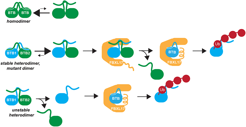
Most quality control pathways target misfolded proteins to prevent toxic aggregation and neurodegeneration1. Dimerization quality control further improves proteostasis by eliminating complexes of aberrant composition2, but how it detects incorrect subunits remains unknown. Here we provide structural insight into target selection by SCF-FBXL17, a dimerization-quality-control E3 ligase that ubiquitylates and helps to degrade inactive heterodimers of BTB proteins while sparing functional homodimers. We find that SCF-FBXL17 disrupts aberrant BTB dimers that fail to stabilize an intermolecular β-sheet around a highly divergent β-strand of the BTB domain. Complex dissociation allows SCF-FBXL17 to wrap around a single BTB domain, resulting in robust ubiquitylation. SCF-FBXL17 therefore probes both shape and complementarity of BTB domains, a mechanism that is well suited to establish quality control of complex composition for recurrent interaction modules.


Conflict of interest statement

Competing interests

M.R. and J.K. are founders and consultants of Nurix, a biotechnology company working in the ubiquitin field.

Figures

Extended Data Figure 1:
Extended Data Figure 1:. Variant BTB domains of KEAP1 are recognized by SCFFBXL17.
a. Mutations in KEAP1’s BTB domain result in efficient recognition by SCFFBXL17. The same mutations as in Fig. 1a were introduced into the KEAP1S172A variant that had previously been used for crystallization. 35S-labeled double mutants, but not the KEAP1S172A single mutant, were retained by immobilized MBPFBXL17, as detected by gel electrophoresis and autoradiography. This experiment was performed once. b. SCFFBXL17 strongly prefers mutant over wildtype BTB domains. Increasing concentrations of MBPFBXL17 were immobilized on amylose beads and incubated with 100nM wildtype or mutant KEAP1. Depletion of KEAP1 from the supernatant was measured by quantitative LiCor imaging of Coomassie-stained SDS-PAGE gels. The affinity of FBXL17 to wildtype KEAP1 was too low to be determined reliable by this method. Three independent experiments were performed with similar results. c. Mutant BTB domains form homodimers in vitro. Recombinant BTB domains of KEAP1, KEAP1F64A, and KEAPV98A (MW ~15kDa) were analyzed by size exclusion chromatography detecting A280. Expected position of BTB dimer versus monomer, as well as of control proteins with known MW are shown on top. Three independent experiments were performed with similar results. d. BTB domains of wildtype KEAP1 and mutant KEAP1F64A form homodimers in solution, as determined by SEC-MALS. This experiment was performed twice. e. Mutant BTB domains unfold via an intermediate species. Wild-type or mutant BTB domains of KEAP1 (0.04 mg/ml) were incubated with various concentrations of urea, equilibrated overnight, and their resulting secondary structure was monitored by ellipticity at 222 nm using circular dichroism. The experiment was performed once for the mutants and twice for the wild-type KEAP1. f. The intermediate seen in the unfolding of KEAP1V98A likely reflects local conformational changes, rather than monomerization. Urea-dependent unfolding curves for the BTB domain of KEAP1V98A were repeated at 10-fold higher BTB concentration (low: 0.04mg/ml; high: 0.4 mg/ml); only the second transition shifted to higher urea concentrations, identifying it as a dimer-unfolded transition. This experiment was performed once. g. Mutation of Phe64 or Val98 to Ala in the BTB domain of KEAP1 reduces contacts between helices of two interacting subunits of the KEAP1 dimer.
Extended Data Figure 2:
Extended Data Figure 2:. Cryo-EM data collection and processing.
a. Representative micrograph (graphene oxide coated grid, imaged using a Talos Arctica and a Gatan K3 camera) showing CUL1-SKP1-FBXL17-KEAP1V98A particles. b. Resolution estimation using the FSC = 0.143 criterion indicates an overall resolution of 8.5 Å for the cryo-EM reconstruction. c. Data processing scheme. Datasets were initially processed independently and then combined for the final refinement. EM volumes are shown in grey and orientation distributions are given for intermediate refinement steps. The final reconstruction is shown with and without sharpening applied, and additionally colored by local resolution (determined using RELION3). d, e. Initial model generation. We originally obtained an initial reference by generating a low-resolution volume with the overall shape of the complex observed in 2D class averages (d) and later also verified this solution using CRYOSPARC2 ab initio model generation (e; see methods for details).
Extended Data Figure 3:
Extended Data Figure 3:. SCFFBXL17 binds the BTB domain, but its active site is next to the Kelch repeats of KEAP1.
a. Elution profile of the SCFFBXL17-KEAP1V99A complex by size exclusion chromatography detecting A280. Control proteins with known MW are shown on top. This experiment was performed three times. b. Cryo-EM density map of the SCFFBXL17-KEAP1V98A complex. Dark gray: CUL11-450; light gray: SKP1; orange: FBXL17; blue: KEAP1. c. Despite an overlap in binding sites, CUL3 does not compete with SCFFBXL17 for substrate ubiquitylation. 35S-labeled wildtype or mutant KEAP1 were ubiquitylated by SCFFBXL17 in reticulocyte lysate either in the presence or absence of a CUL3 variant shown to bind BTB proteins. Ubiquitylated KEAP1 was detected by gel electrophoresis and autoradiography. Three independent experiments were performed with similar results. d. SCFFBXL17 ubiquitylates full-length BTB proteins with Kelch repeats better than isolated BTB domains. 35S-labeled full-length KEAP1F64A, the BTB domain of KEAP1F64A, full-length KLHL12V50A, or the BTB domain of KLHL12V50A were incubated in reticulocyte lysate with recombinant FBXL17, and ubiquitylation was detected by gel electrophoresis and autoradiography. Two independent experiments were performed with similar results. e. Full-length BTB proteins or isolated BTB domains bind similarly well to FBXL17. 35S-labeled full-length BTB proteins or isolated BTB domains, as indicated on the right, were incubated with immobilized MBPFBXL17 and bound proteins were detected by gel electrophoresis and autoradiography. Two independent experiments were performed with similar results.
Extended Data Figure 4:
Extended Data Figure 4:. Structural features of the SKP1/FBXL17-BTB complex.
a. Elution profile of the SKP1/FBXL17-BTB(KEAP1F64A) complex by size exclusion chromatography detecting A280. Control proteins with known MW are shown on top. This experiment was performed three times. b. FBXL17 binds to SKP1 via its F-box domain, in a manner highly similar to the LRR-domain containing F-box proteins SKP2 and FBXL3 ,. The structures of SKP1-FBXL17, SKP1-SKP2, and SKP1-FBXL3 were aligned via SKP1. FBXL17 is shown in orange, SKP2 in magenta, and FBXL3 in yellow. c. FBXL17 uses conserved residues in its F-box to bind SKP1. The highlighted residues in FBXL17 (orange) that bind SKP1 (gray) were adopted from ref. . d. The substrate binding LRR domain of FBXL17 is longer and more curved than the LRR domains of SKP2 or FBXL3. Complexes were aligned via SKP1 (FBXL17, orange; SKP2, magenta; FBXL3, yellow). e. Structural models of BTB-FBXL17 complexes, using BTB domains that are similar in shape, but distinct in sequence. All complexes between FBXL17 and these confirmed substrates can be formed without steric clashes. f. SKP1 and Elongin C, which also adopt BTB folds, cannot be bound to FBXL17, due to steric clashes shown in the insets.
Extended Data Figure 5:
Extended Data Figure 5:. Validation of SKP1/FBXL17-BTB structure through FBXL17 mutations.
a. Single mutations of FBXL17 rarely affect co-translational recognition of KEAP1 in cells. FBXL17FLAG mutants were affinity-purified from 293T cells that also expressed MYCSKP1, HAKEAP1, and dominant negative CUL1 to prevent degradation of the BTB protein. Bound proteins were detected by gel electrophoresis and Western blotting. Red, mutations that abolish binding to FBXL17; orange, mutations that weaken binding to FBXL17; green, wild-type FBXL17; black: mutations that had no effect on KEAP1 binding. This experiment was performed once. b. Single mutations of FBXL17 rarely interfere with the proteasomal degradation of KEAP1. 293T cells were transfected with HAKEAP1 and either wild-type or mutant FBXL17FLAG, as denoted on the right, MYCSKP1, and dominant-negative CUL1 (dnCUL1), as indicated. The abundance of KEAP1 was monitored by gel electrophoresis and αHA-Western blotting. This experiment was performed once. c. The CTH is required, but not sufficient, for BTB recognition by SCFFBXL17. Immobilized recombinant MBP, MBPFBXL17, MBPFBXL17ΔCTH, or CTHMBP were incubated with 35S-labeled fused dimers of the BTB domains of KLHL12 (green) and KEAP1 (orange). Bound proteins were detected by gel electrophoresis and autoradiography. This experiment was performed once. d. The CTH is required for in vitro ubiquitylation of mutant KEAP1. Recombinant FBXL17-SKP1 or FBXL17ΔCTH-SKP1 were added to reticulocyte lysate after the synthesis of either wild-type or mutant KEAP1. Reticulocyte lysate contains all other components required for in vitro ubiquitylation through SCFFBXL17. Unmodified and ubiquitylated KEAP1 were detected by gel electrophoresis and autoradiography. This experiment was performed once.
Extended Data Figure 6:
Extended Data Figure 6:. Validation of SKP1/FBXL17-BTB structure through KEAP1 mutations.
a. Single mutations in KEAP1 do not inhibit the co-translational SCFFBXL17-dependent degradation of the BTB protein. 293T cells were transfected with either wild-type (left three lanes) or mutant HAKEAP1 (right two lanes; mutations denoted on the right), as well as MYCSKP1, FBXL17FLAG and dominant negative CUL1 (dnCUL1), as indicated on top. KEAP1 levels were monitored by gel electrophoresis and αHA Western blotting. This experiment was performed once. b. Single mutation of residues in KEAP1 at the interface with FBXL17 do not inhibit co-translational binding of the BTB protein to SCFFBXL17. 293T cells were transfected with wild-type or mutant HAKEAP1, MYCSKP1, FBXL17FLAG, and dominant-negative CUL1 (dnCUL1). FBXL17FLAG was affinity-purified and bound proteins were detected by gel electrophoresis and Western blotting. This experiment was performed once. c. Ala109 (red stick) in KEAP1 (blue) is positioned further from FBXL17 compared to the corresponding A60 residue in KLHL12 (green). KEAP1 and KLHL12 BTB domains were overlain bound to FBXL17 (KEAP1, actual structure; KLHL12, model).
Extended Data Figure 7:
Extended Data Figure 7:. Sequence alignment of BTB domains.
Residues involved in BTB dimerization are marked by a blue dot; residues at the interface between the BTB domain and FBXL17 are marked by an orange dot; residues at the interface between the BTB domain and CUL3 are marked by a magenta dot. Sites of mutations used for X-ray crystallography or electron microscopy are marked by a red star.
Extended Data Figure 8:
Extended Data Figure 8:. Binding and destabilization of BTB dimers by SCFFBXL17.
a. A FRET-based assay to monitor BTB dimer formation. Blue curve: The BTB domain of KEAP1F64A was labeled with Alexa 555, then denatured and refolded. Red curve: The BTB domain of KEAP1F64A was labeled with Alexa 647, then denatured and refolded. Green curve: Two separate BTB domain pools of KEAP1F64A were labeled with either Alexa 555 or Alexa 647, mixed in equimolar concentrations, denatured, and then refolded. ~50% of dimers are labeled with distinct fluorophores in each BTB subunit, giving rise to donor fluorescence quenching and acceptor emission as indication of FRET. Three independent experiments were performed with similar results. b. KEAP1F64A dimers dissociate very slowly and inefficiently. BTB domains of KEAP1F64A were labeled with either Alexa 555 or Alexa 647, respectively. The labeled BTB domains were then mixed, incubated overnight, and analyzed for FRET that results from stochastic rebinding of BTB monomers, leading to formation of BTB dimers containing one subunit labeled with Alexa 555 and the other subunit labeled with Alexa 647. However, in comparison to complex reformation by refolding (see above), little FRET was detected. This experiment was performed twice. c. FBXL17 can modulate BTB complex composition in vitro. The KLHL12 locus was tagged with a 3xFLAG epitope by CRISPR/Cas9-mediated genome editing. Endogenous KLHL123xFLAG complexes were affinity-purified from 293T cells and incubated with recombinant MBP (control), FBXL17, or inactive FBXL17ΔCTH. Proteins that remained bound to KLHL123xFLAG were determined by mass spectrometry. d. Overexpression of FBXL17ΔFBOX, which can bind but not ubiquitylate BTB proteins, prevents BTB heterodimerization. The endogenous KLHL123xFLAG was affinity-purified either in the presence or absence of FBXL17ΔFbox, and bound proteins were determined by mass spectrometry. e. FBXL17 can bind BTB dimers. FLAGKLHL12 was affinity-purified from 293T cells also expressing MYCKLHL12 and FBXL17HA. FLAGKLHL12 complexes were eluted with FLAG-peptide and FBXL17HA-containing complexes were then purified over αHA-agarose. Bound MYCKLHL12, indicative of FBXL17 associating with KLHL12 dimers, was then detected by Western blotting. This experiment was performed once. f. Binding of FBXL17 to BTB dimers requires its CTH to be disengaged from its binding site at the BTB dimer interface. A structural model of a KEAP1 BTB dimer bound to FBXL17 was generated using the KEAP1F64A dimer and the FBXL17-KEAP1F64A complex structures. Clashes are predominantly at the CTH of FBXL17. g. Residues of FBXL17, which in the structural model of a FBXL17-BTB dimer complex are in proximity to the leaving BTB subunit, contribute to stable substrate recognition. Indicated FBXL17 residues at the interface with the leaving BTB subunit (above) were mutated in the sensitized background of the FBXL17C680D variant and analyzed for binding to endogenous BTB proteins by affinity purification and Western blotting. This experiment was performed once.
Extended Data Figure 9:
Extended Data Figure 9:. The amino-terminal β-strand is important for BTB complex formation and recognition.
a. Chimeric KLHL12 with β-strand and adjacent dimer interface residues of KEAP1, but not wild-type KLHL12, forms heterodimers with KEAP1 in vivo. 293T cells were transfected with KLHL12FLAG (wild-type or chimera) and KLHL12HA or HAKEAP1, as indicated. KLHL12FLAG variants were immunoprecipitated and bound proteins detected by gel electrophoresis and Western blotting. This experiment was performed once. b. BTB heterodimers are inactive in signaling. 293T cells were transfected with FLAG-tagged wild-type KLHL12, chimeric KLHL12, or wild-type KEAP1. The FLAG-tagged BTB proteins were affinity-purified and bound endogenous targets of KLHL12 (SEC31, PEF1, ALG2) or KEAP1 (NRF2) were detected by Western blotting. This experiment was performed once. c. A chimeric KLHL12 that contains helix and β-strand residues of KEAP1 efficiently heterodimerizes with KEAP1, yet fails to bind substrates of either KLHL12 or KEAP1. KLHL12FLAG, chimeric KLHL12FLAG or KEAP1FLAG were affinity-purified from 293T cells and bound proteins were determined by CompPASS mass spectrometry.
Extended Data Figure 10:
Extended Data Figure 10:. The amino-terminal β-strand of BTB domains serves as a molecular barcode for functional dimerization.
Sequence alignment of amino-terminal β-strands across human BTB domains shows divergence, indicative of rapid evolution of this protein sequence.
Figure 1:
Figure 1:. SCFFBXL17 binds monomeric BTB domains.
a. 35S-labeled KEAP1 variants are recognized by immobilized FBXL17. Binding of mutant KEAP1 to FBXL17 was performed five times. b. Mutant (blue) and wildtype (green) KEAP1 BTB domains adopt the same dimer fold, as shown by X-ray crystallography at 2.2-2.5 Å resolution. c. 8.5 Å resolution cryo-EM structure of a complex between CUL1 (residues 1-410; medium gray); SKP1 (light gray); FBXL17 (orange); and KEAP1V99A (blue). X-ray coordinates of the FBXL17-SKP1-BTB complex (Fig. 2) and CUL1-RBX1 were fitted into the cryo-EM density. d. FBXL17 and CUL3 recognize overlapping surfaces on the BTB domain of KEAP1. CUL3 (magenta) was superposed onto the KEAP1 BTB domain based on PDB ID 5NLB.
Figure 2:
Figure 2:. Crystal structure of substrate-bound FBXL17 reveals specificity determinants of DQC.
a. Side view of the 3.2 Å X-ray structure of a complex between SKP1 (gray), FBXL17 (orange), and the BTB domain of KEAP1F64A (residues 48-180; blue). b. Top view of the SKP1-FBXL17-BTB complex showing how FBXL17 encircles the BTB domain through its LRRs and the C-terminal helix (CTH). c. Side view of the SKP1-FBXL17-BTB complex. d. The CTH binds a conserved area of the BTB domains (blue: high conservation; red: low conservation). e. The CTH crosses the dimerization interface of the BTB domain in a position typically occupied by another subunit in the BTB dimer (green).
Figure 3:
Figure 3:. Multiple surfaces of FBXL17 contribute to substrate binding.
a. Detailed view of the interface between FBXL17 (orange) and the BTB domain of KEAP1F64A (blue). b. Combined mutation of FBXL17 residues in LRRs and CTH prevents recognition of HAKEAP1, as shown by FBXL17FLAG affinity-purification and quantitative αHA-LiCor Western blotting. c. Combined mutations in FBXL17 interfere with proteasomal degradation of KEAP1, as monitored by quantitative Western blotting. d. Mutations in FBXL17 prevent recognition of endogenous BTB proteins, as determined by affinity-purification and mass spectrometry. The heat map shows total spectral counts normalized to FBXL17. e. Mutation of residues in HAKEAP1 inhibits binding to SCFFBXL17, as seen upon FBXL17FLAG affinity-purification and quantitative αHA-Western blotting. f. Combined mutations in KEAP1 inhibit SCFFBXL17-dependent degradation, as monitored by quantitative Western blotting.
Figure 4:
Figure 4:. A domain-swapped β-sheet in BTB domains controls access to SCFFBXL17.
a. KEAP1F64A BTB dimers labeled with distinct fluorophores dissociate, as shown by loss of donor fluorescence quenching, upon incubation with FBXL17. Inactive FBXL17ΔCTH, excess unlabeled BTB domains (KEAP1F64A; KEAP1V98A), GroEL, or MBP did not have strong effects. Dissociation by FBXL17 was measured three times. b. Overnight incubation of FRET-labeled KEAP1F64A BTB dimers with FBXL17, FBXL17ΔCTH, or excess unlabeled KEAP1F64A BTB domain. Dissociation was measured 2-3 times. c. The amino-terminal β-strand of the KEAP1 BTB domain forms an intermolecular sheet in dimers, but adopts an intramolecular conformation in the BTB monomer bound to SCFFBXL17. d. Binding of 35S-labeled KEAP1 variants with mutations in the domain-swapped β-sheet to MBPFBXL17. This experiment was performed once. e. 35S-labeled unfused BTB domains of KLHL12V50A, fused BTB domains of KLHL12V50A or fused BTB domains that were cut within the linker were bound to MBPFBXL17. Two independent experiments were performed with similar results. f. Structural model of a KLHL12-KEAP1 heterodimer shows clashes at the dimerization helix region and amino-terminal β-strand. g. 35S-labeled KLHL12-KEAP1 BTB heterodimers, mutant heterodimers, and KEAP1 BTB homodimers were bound MBPFBXL17. Two independent experiments were performed with similar results. h. 35S-labeled homodimeric KEAP1 (green), mutants with helix and β-strand residues of KLHL12 placed into the first subunit of a KEAP1 homodimer (blue), or KLHL12-KEAP1 heterodimers were incubated with MBPFBXL17 and analyzed for binding by gel electrophoresis and autoradiography. Two independent experiments were performed with similar results. i. The amino-terminal β-strand and its interaction residues in the partner BTB domain evolve rapidly (high conservation: blue; no conservation: red).
Figure 5:
Figure 5:. Model of the DQC mechanism.
BTB homodimers have identical amino-terminal β-strand mostly in the domain swapped position. This prevents SCFFBXL17 from engaging and ubiquitylating the homodimer. BTB heterodimers or mutant BTB dimers have poorly compatible helices and β-strands. Their amino-terminal β-strand will be mostly displaced, which allows for capture of these aberrant dimers by SCFFBXL17. SCFFBXL17 could further destabilize these dimers or rely on spontaneous dimer dissociation to associate with a monomeric BTB subunit for ubiquitylation and degradation.

References

    1. Balchin D, Hayer-Hartl M & Hartl FU In vivo aspects of protein folding and quality control. Science 353, aac4354, doi:10.1126/science.aac4354 (2016). - DOI - PubMed
    1. Mena EL et al. Dimerization quality control ensures neuronal development and survival. Science 362, doi:10.1126/science.aap8236 (2018). - DOI - PubMed
    1. Gordley RM, Bugaj LJ & Lim WA Modular engineering of cellular signaling proteins and networks. Curr Opin Struct Biol 39, 106–114, doi:10.1016/j.sbi.2016.06.012 (2016). - DOI - PMC - PubMed
    1. Ji AX & Prive GG Crystal structure of KLHL3 in complex with Cullin3. PLoS One 8, e60445, doi:10.1371/journal.pone.0060445 (2013). - DOI - PMC - PubMed
    1. Zhuang M et al. Structures of SPOP-substrate complexes: insights into molecular architectures of BTB-Cul3 ubiquitin ligases. Molecular cell 36, 39–50, doi:10.1016/j.molcel.2009.09.022 (2009). - DOI - PMC - PubMed

Supplemental References

    1. Duda DM et al. Structural insights into NEDD8 activation of cullin-RING ligases: conformational control of conjugation. Cell 134, 995–1006, doi:S0092-8674(08)00942-2 [pii] 10.1016/j.cell.2008.07.022 (2008). - DOI - PMC - PubMed
    1. Kabsch W Xds. Acta Crystallogr D Biol Crystallogr 66, 125–132, doi:10.1107/S0907444909047337 (2010). - DOI - PMC - PubMed
    1. Evans PR & Murshudov GN How good are my data and what is the resolution? Acta Crystallogr D Biol Crystallogr 69, 1204–1214, doi:10.1107/S0907444913000061 (2013). - DOI - PMC - PubMed
    1. Winn MD et al. Overview of the CCP4 suite and current developments. Acta Crystallogr D Biol Crystallogr 67, 235–242, doi:10.1107/S0907444910045749 (2011). - DOI - PMC - PubMed
    1. Adams PD et al. PHENIX: a comprehensive Python-based system for macromolecular structure solution. Acta Crystallogr D Biol Crystallogr 66, 213–221, doi:10.1107/S0907444909052925 (2010). - DOI - PMC - PubMed

Publication types