. 2020 Nov;177(22):5114-5130.
doi: 10.1111/bph.15233. Epub 2020 Sep 21.

Identification of novel therapeutic targets for blocking acantholysis in pemphigus

Affiliations

Identification of novel therapeutic targets for blocking acantholysis in pemphigus

Imke A K Burmester et al. Br J Pharmacol. 2020 Nov.

Abstract

Background and purpose: Pemphigus is caused by autoantibodies against desmoglein (Dsg) 1, Dsg3, and/or non-Dsg antigens. Pemphigus vulgaris (PV) is the most common manifestation of pemphigus, with painful erosions on mucous membranes. In most cases, blistering also occurs on the skin, leading to areas of extensive denudation. Despite improvements in pemphigus treatment, time to achieve remission is long, severe adverse events are frequent and 20% of patients do not respond adequately. Current clinical developments focus exclusively on modulating B cell function or autoantibody half-life. However, topical modulation of PV autoantibody-induced blistering is an attractive target because it could promptly relieve symptoms.

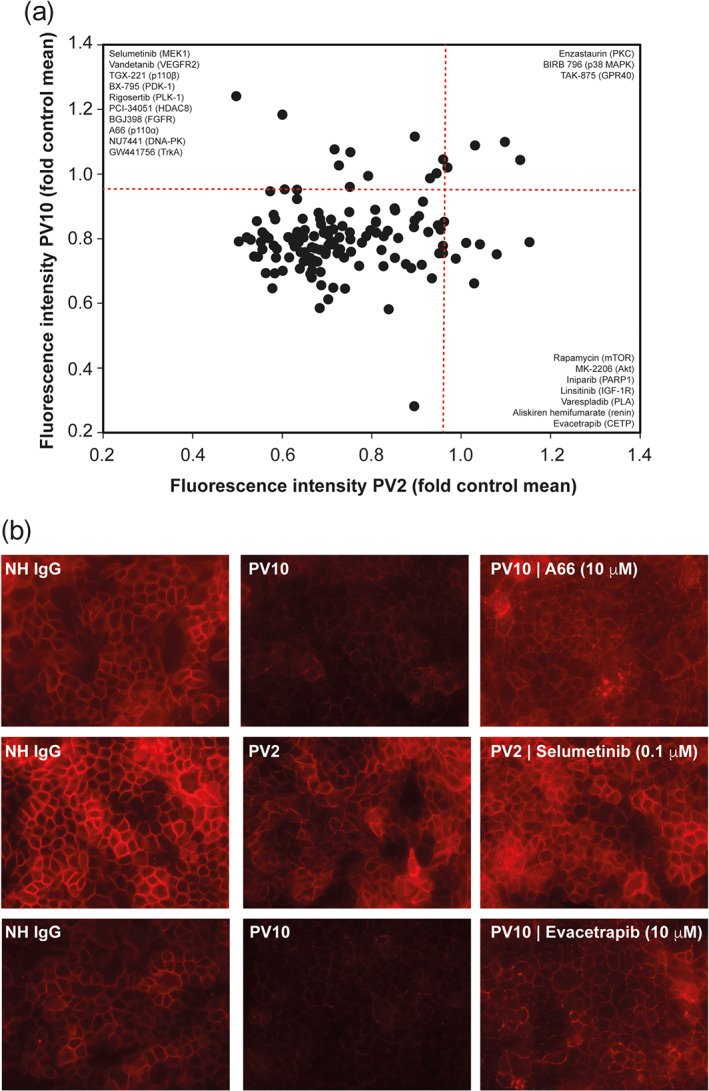
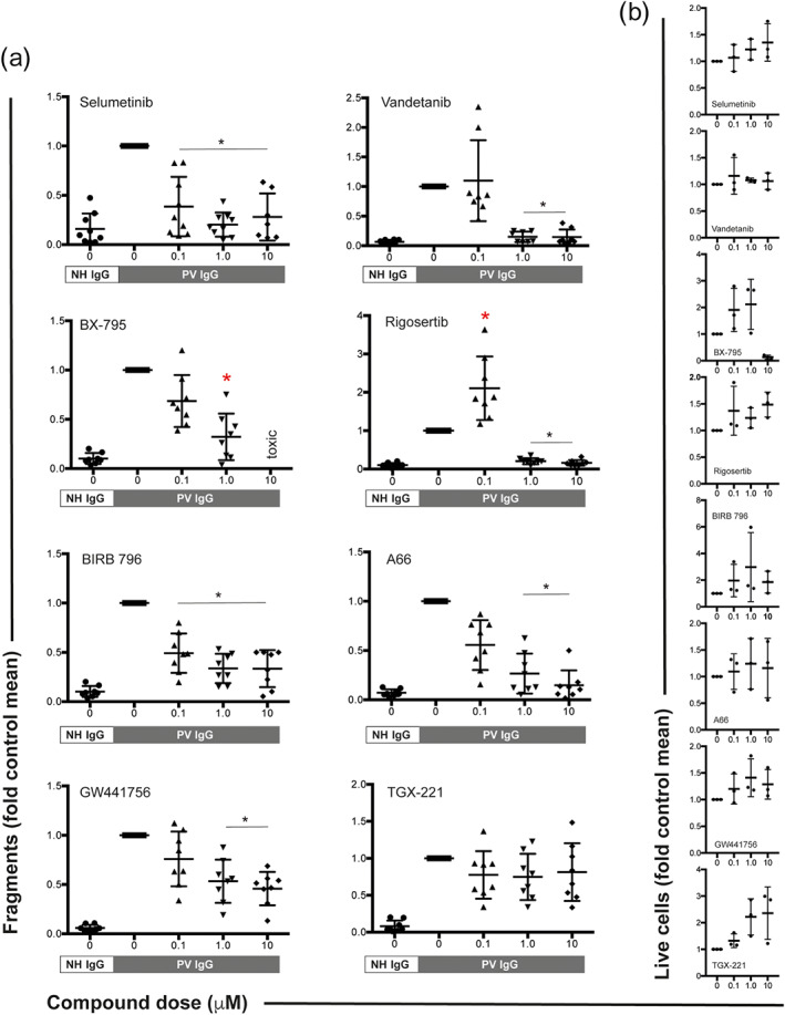
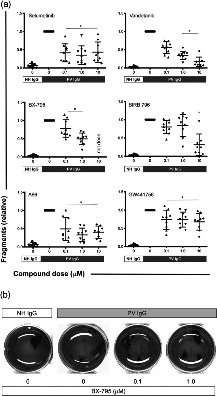
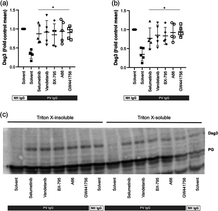
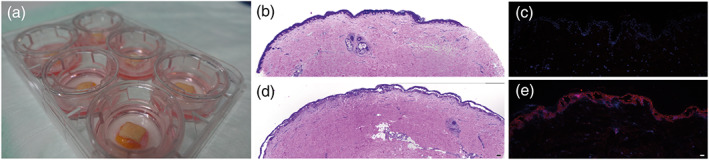
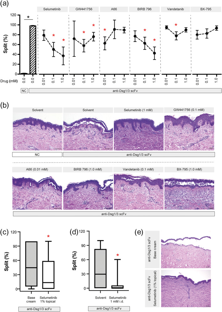
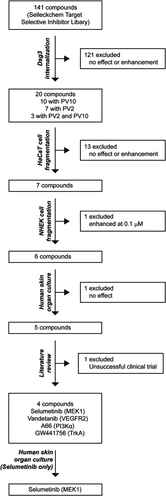
Experimental approach: To address this issue, we performed an unbiased screening in a complex biological system using 141 low MW inhibitors from a chemical library. Specifically, we evaluated PV IgG-induced Dsg3 internalization in HaCaT keratinocytes. Validation of the 20 identified compounds was performed using keratinocyte fragmentation assays, as well as a human skin organ culture (HSOC) model.

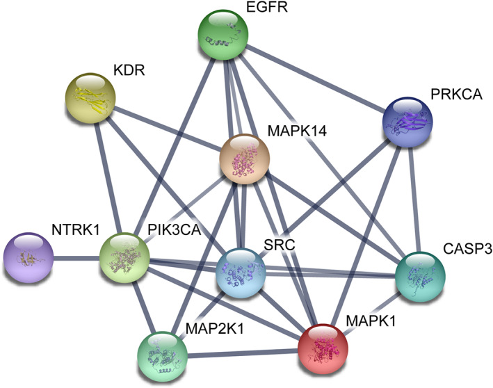
Key results: Overall, this approach led to the identification of four molecules involved in PV IgG-induced skin pathology: MEK1, TrkA, PI3Kα, and VEGFR2.

Conclusion and implications: This unbiased screening revealed novel mechanisms by which PV autoantibodies induce blistering in keratinocytes and identified new treatment targets for this severe and potentially life-threatening skin disease.

Keywords: autoimmunity; cell signaling; model system; pemphigus; skin.


Conflict of interest statement

R.J.L. has received research funding from Miltenyi Biotec, Biogen, Biotest, Almirall, True North Therapeutics, UCB Pharma, ArgenX, TxCell, Topadur, Incyte, and Admirx and fees for consulting or speaking from ArgenX, Immunogenetics, Novartis, and Lilly. D.Z. has received support through research and development grants as well as for consulting or lecturing from Biotest, Fresenius, Miltenyi Biotec, Roche Pharma, Biogen, AbbVie, UCB, Janssen, Euroimmun, Dompe, Novartis, and ArgenX. C.M.H. is advisor to ArgenX and viDa Therapeutics. All other authors declare no conflicts of interest.

Figures

FIGURE 1
FIGURE 1
Modulation of PV IgG‐induced Dsg3 internalization in HaCaT cells by target‐selective, low MW inhibitors. (a) HaCaT keratinocytes were incubated with one of the two pemphigus vulgaris (PV) immunadsorption materials, and Dsg3 expression on the cellular surface was determined by immunohistochemistry. A compound was considered to block PV IgG induction if cFI/(pcFI + ΔFI/2) was 0.95 or higher. The red line indicates the predefined cut‐off. All data were normalized to NH IgG. Collectively, 10 compounds inhibited PV10 IgG‐induced Dsg3 internalization (upper left corner), and seven compounds were identified with PV2 and three compounds inhibited Dsg3 internalization for both patients' IgG. (b) Representative images from the experiments shown in panel (a)
FIGURE 2
FIGURE 2
Inhibitors of MEK1, VEGFR2, PDK1, PLK1, p38 MAPK, PI3Kα, and TrkA impair PV IgG‐induced cell fragmentation in HaCaT keratinocytes. (a) HaCaT cell sheaths were exposed to defined mechanical stress in the presence of pemphigus vulgaris (PV) IgG. Normal human (NH) IgG served as a negative control. Inhibitors of MEK1 (selumetinib), VEGFR2 (vandetanib), PDK1 (BX‐795), PLK1 (rigosertib), p38 MAPK (BIRB 796), PI3Kα (A66), and TrkA (GW441756) impaired the PV IgG‐induced fragmentation of the cell sheaths. The remaining 13 compounds (Figure 1) had no effect on cell fragmentation. The PI3Kβ inhibitor TGX‐221 is shown as one example of a compound that had no effect on PV IgG‐induced cell fragmentation. All data were normalized to the fragmentation induced by PV IgG. Data are shown as individual values, with means ± SD from seven to nine independent replicates per group. *P<0.05, significantly different, as indicated, from PV IgG; Kruskal–Wallis H test followed by Dunn's post hoc test. (b) All compounds, with the exception of BX‐795, had no toxic effects at the indicated doses. Data are shown as individual values, with medians ± SD from three replicates per group and is an exploratory screening of the potential toxicity
FIGURE 3
FIGURE 3
Inhibitors of MEK1, VEGFR2, PDK1, p38 MAPK, PI3Kα, and TrkA impair PV IgG‐induced cell fragmentation in NHEK keratinocytes. (a) NHEK cell sheaths from three donors were exposed to defined mechanical stress in the presence of PV IgG from three different patients. Normal human (NH) IgG served as a negative control. Inhibitors of MEK1 (selumetinib), VEGFR2 (vandetanib), PDK1 (BX‐795), p38 MAPK (BIRB 796), PI3Kα (A66) and TrkA (GW441756) impaired the PV IgG‐induced fragmentation of the cell sheaths. All data were normalized to the fragmentation induced by PV IgG. Data are shown as individual values, with means ± SD from eight to nine independent replicates per group. *P<0.05, significantly different, as indicated, from PV IgG; Kruskal–Wallis H test followed by Dunn's post hoc test. (b) Representative images from one experiment are shown in panel (a). The brightness of all images was increased to the same extent using GIMP (www.gimp.org) to better contrast the sheaths from the medium
FIGURE 4
FIGURE 4
Inhibitors of MEK1, VEGFR2, PDK1, PI3Kα, and TrkA impair PV IgG‐induced internalization of the cytoskeletal‐anchored as well as from non‐cytoskeletal‐anchored Dsg3 pool in HaCaT keratinocytes. PV IgG was added to HaCaT keratinocytes for 24 h and Dsg3 internalization from the cytoskeletal‐anchored and the non‐cytoskeletal‐anchored Dsg3 was assayed using western blotting of Triton‐X lysed cells. (a–b) Inhibitors of MEK1 (selumetinib), VEGFR2 (vandetanib), PDK1 (BX‐795), PI3Kα (A66), and TrkA (GW441756) almost completely inhibited PV IgG‐induced Dsg3 internalization from both Dsg3 pools. Graphs show the relative Dsg3 staining in (a) the Triton X‐insoluble and (b) soluble fraction of HaCaT cells. Data are shown as individual values, with medians ± SD from five replicates per group. *P<0.05, significantly different, as indicated, from PV IgG+Solvent; ANOVA with Dunnett's post test. (c) Representative western blot showing the Triton X‐insoluble and soluble fraction of HaCaT cells 24 h after the addition of PV IgG. Abbreviations used: Dsg3, desmogelein 3; PK, plakoglobin
FIGURE 5
FIGURE 5
Anti‐Dsg1/3 scFv induces acantholysis in a full thickness human skin organ culture model. (a) Images of six skin specimens in culture. (b) H&E‐stained section from a skin section incubated with normal human (NH) IgG shows no intraepidermal blistering. (c) Anti‐Dsg1/3 scFv‐ (red) and DAPI (blue)‐stained sections from a skin section incubated with NH IgG show no autoantibody binding to keratinocytes. (d) H&E‐stained section from a skin section incubated with pemphigus vulgaris (PV) IgG, with extensive intraepidermal blistering. (e) Anti‐Dsg1/3 scFv‐ (red) and DAPI (blue)‐stained sections from a skin section incubated with anti‐Dsg1/3 scFv show the presence of autoantibodies bound to keratinocytes, as well as intraepidermal blistering. Scale bars correspond to 100 μm
FIGURE 6
FIGURE 6
Inhibitors of MEK1, TrkA, PI3Kα, p38 MAPK, and VEGFR2 impair PV IgG‐induced intraepidermal split formation in the human skin organ culture model. (a) The anti‐Dsg1/3 scFv was injected into human skin samples, in the absence or presence of the indicated inhibitors. Data are shown as individual values, with medians ± SD from 8–19 sections per group from three organ cultures. *P<0.05, significantly different from anti‐Dsg1/3 scFv alone; Kruskal–Wallis H test followed by Dunn's post hoc test. (b) Representative H&E‐stained images from the experiments are shown in panel (a). For better contrast, white correction was applied using GIMP (www.gimp.org). Scale bars correspond to 100 μm. (c) The same experimental set‐up as for panel (a), with the exception that selumetinib was emulsified in base cream (1%), which was applied on top of the organ culture. For control, skin specimens were treated with base cream alone. Data are shown as box (first and third quartiles) and whisker (range) plots with medians, and are based on 39–40 sections from four to five organ cultures. * P < 0.05, significantly different from base cream alone; rank‐sum test. (d) The same experimental set‐up as for panel (a), performed in parallel to the experiments from panel (c) as a positive treatment control. Data are shown as box ( first and third quartiles) and whisker ( ) plots with medians and based on 37–44 sections from five to six organ cultures; * P < 0.05, significantly different from solvent alone; rank‐sum test. (e) Representative H&E‐stained images from the experiments are shown in panel (c). For better contrast, white correction was applied using GIMP (www.gimp.org). Scale bars correspond to 100 μm
FIGURE 7
FIGURE 7
Summary of screening and validation. A total of 141 compounds from the Selleckchem Target Selective Inhibitor Library (Table S1) were used for the initial screening. Of these, 20 compounds were defined as potential inhibitors of PV IgG‐induced pathology in keratinocytes and were used in the first validation experiment. Next, in the HaCaT cell dissociation assay, seven compounds were selected for further validation. In the NHEK cell dissociation assay, 6/6 compounds displayed inhibitory effects on PV IgG‐induced pathology. Ultimately, these remaining compounds were tested in the human skin organ culture model, and 5/6 of the compounds impaired the induction of intra‐epidermal blistering induced by a monoclonal bispecific anti‐Dsg1/3 scFv. Based on the literature (Sayar et al., 2014), we excluded p38 MAPK from the list of potential therapeutic targets. In summary, the screening and subsequent validation identified four novel therapeutic targets for the modulation of PV IgG‐induced intraepidermal blistering, namely, MEK1, TrkA, PI3Kα, and VEGFR2
FIGURE 8
FIGURE 8
Interactions among the identified therapeutic targets for intraepidermal blistering induced by anti‐Dsg1/3 autoantibodies. The interaction map was drawn using STRING (string‐db.org; accessed 26 November 2018). The interactions are displayed in the confidence setting, where line thickness indicates the strength of data support at high confidence (0.7). The colour of nodes was selected at random. The following genes served as input: EGFR (EGFR), PRKCA (PKC), SRC (Src), MAP2K1 (MEK), KDR (VEGFR2), MAPK14 (p38 MAPK), MAPK1 (ERK), PIK3CA (PI3Kα), NTRK1 (TrkA), and CASP3 (caspase‐3)

References

    1. Alexander, S. P. H. , Christopoulos, A. , Davenport, A. P. , Kelly, E. , Mathie, A. , Peters, J. A. , … CGTP Collaborators . (2019). THE CONCISE GUIDE TO PHARMACOLOGY 2019/20: G protein‐coupled receptors. British Journal of Pharmacology, 176, S21–S141. 10.1111/bph.14748 - DOI - PMC - PubMed
    1. Alexander, S. P. H. , Fabbro, D. , Kelly, E. , Mathie, A. , Peters, J. A. , Veale, E. L. , … CGTP Collaborators . (2019a). THE CONCISE GUIDE TO PHARMACOLOGY 2019/20: Catalytic receptors. British Journal of Pharmacology, 176, S247–S296. 10.1111/bph.14751 - DOI - PMC - PubMed
    1. Alexander, S. P. H. , Fabbro, D. , Kelly, E. , Mathie, A. , Peters, J. A. , Veale, E. L. , … CGTP Collaborators . (2019b). THE CONCISE GUIDE TO PHARMACOLOGY 2019/20: Enzymes. British Journal of Pharmacology, 176, S297–S396. 10.1111/bph.14752 - DOI - PMC - PubMed
    1. Alexander, S. P. H. , Kelly, E. , Mathie, A. , Peters, J. A. , Veale, E. L. , Armstrong, J. F. , … Southan, C. (2019). THE CONCISE GUIDE TO PHARMACOLOGY 2019/20: Introduction and other protein targets. British Journal of Pharmacology, 176(Suppl 1), S1–S20. - PMC - PubMed
    1. Alexander, S. P. H. , Roberts, R. E. , Broughton, B. R. S. , Sobey, C. G. , George, C. H. , Stanford, S. C. , … Ahluwalia, A. (2018). Goals and practicalities of immunoblotting and immunohistochemistry: A guide for submission to the British Journal of Pharmacology . British Journal of Pharmacology, 175(3), 407–411. 10.1111/bph.14112 - DOI - PMC - PubMed

Publication types