

Case Reports
doi: 10.1002/mds.28277.
Epub 2020 Sep 24.
Coronavirus Disease 2019 and Parkinsonism: A Non-post-encephalitic Case
Affiliations
- PMID: 32815213
- PMCID: PMC7461093
- DOI: 10.1002/mds.28277
Item in Clipboard
Case Reports
Coronavirus Disease 2019 and Parkinsonism: A Non-post-encephalitic Case
Mov Disord.
2020 Oct.
No abstract available
Keywords: CNS infection; COVID-19; SARS-CoV-2; parkinsonism; postinfectious parkinsonism.
Figures
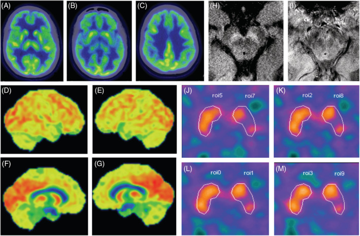
Comprehensive neuroimaging ancillary evaluation: fluorodeoxyglucose–positron emission tomography, neuromelanin and nigrosome‐1 magnetic resonance imaging, and 99mTc‐TRODAT‐1 (Technetium‐99m labeled tropane derivative) single‐photon emission computed tomography). (A–C) Fluorodeoxyglucose–positron emission tomography scan axial slices (through the thalamus, striatum, and corona radiata) show cerebral glucose metabolism within normal limits in the basal ganglia and cortical regions. (D–G) Three‐dimensional stereotactic surface projection (3d‐SSP) projection of the cortical radiotracer uptake to a lateral and medial surface perspective of both hemispheres shows normal glucose metabolism. (H) Normal neuromelanin content is seen in an axial magnetic resonance imaging slice of the midbrain through the substantia nigra. (I) Nigrossome‐1 and its related “swallow‐tail” sign are readily identifiable in both cerebral peduncles. (J–M) Nigrostriatal denervation at the left mid‐putamen (semiquantification of dopamine transporter binding—0.60), contralateral to the most significant clinically affected side. The specific dopamine transporter binding potential in the basal ganglia is calculated as the difference between dorsal putamen activity (PUT) and the reference region, that is, the occipital activity (OA), divided by the OA: (PUT−OA)/OA. [Color figure can be viewed at wileyonlinelibrary.com ]
References
Publication types
MeSH terms
LinkOut - more resources
Full Text Sources
Miscellaneous
