

Arrangement of Ceramides in the Skin: Sphingosine Chains Localize at a Single Position in Stratum Corneum Lipid Matrix Models
- PMID: 32816488
- PMCID: PMC7498151
- DOI: 10.1021/acs.langmuir.0c01992
Arrangement of Ceramides in the Skin: Sphingosine Chains Localize at a Single Position in Stratum Corneum Lipid Matrix Models
Abstract
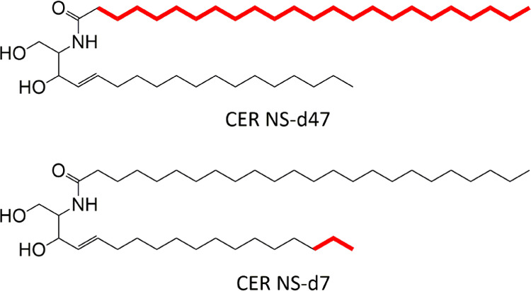
Understanding the structure of the stratum corneum (SC) is essential to understand the skin barrier process. The long periodicity phase (LPP) is a unique trilayer lamellar structure located in the SC. Adjustments in the composition of the lipid matrix, as in many skin abnormalities, can have severe effects on the lipid organization and barrier function. Although the location of individual lipid subclasses has been identified, the lipid conformation at these locations remains uncertain. Contrast variation experiments via small-angle neutron diffraction were used to investigate the conformation of ceramide (CER) N-(tetracosanoyl)-sphingosine (NS) within both simplistic and porcine mimicking LPP models. To identify the lipid conformation of the twin chain CER NS, the chains were individually deuterated, and their scattering length profiles were calculated to identify their locations in the LPP unit cell. In the repeating trilayer unit of the LPP, the acyl chain of CER NS was located in the central and outer layers, while the sphingosine chain was located exclusively in the middle of the outer layers. Thus, for the CER NS with the acyl chain in the central layer, this demonstrates an extended conformation. Electron density distribution profiles identified that the lipid structure remains consistent regardless of the lipid's lateral packing phase, this may be partially due to the anchoring of the extended CER NS. The presented results provide a more detailed insight on the internal arrangement of the LPP lipids and how they are expected to be arranged in healthy skin.
Conflict of interest statement
The authors declare no competing financial interest.
Figures
Similar articles
-
Stratum corneum lipid matrix: Location of acyl ceramide and cholesterol in the unit cell of the long periodicity phase.Biochim Biophys Acta. 2016 Aug;1858(8):1926-34. doi: 10.1016/j.bbamem.2016.05.006. Epub 2016 May 8. Biochim Biophys Acta. 2016. PMID: 27169629
-
Phytosphingosine ceramide mainly localizes in the central layer of the unique lamellar phase of skin lipid model systems.J Lipid Res. 2022 Sep;63(9):100258. doi: 10.1016/j.jlr.2022.100258. Epub 2022 Aug 2. J Lipid Res. 2022. PMID: 35931203 Free PMC article.
-
The molecular arrangement of ceramides in the unit cell of the long periodicity phase of stratum corneum models shows a high adaptability to different ceramide head group structures.Biochim Biophys Acta Biomembr. 2024 Jun;1866(5):184324. doi: 10.1016/j.bbamem.2024.184324. Epub 2024 Apr 29. Biochim Biophys Acta Biomembr. 2024. PMID: 38688405
-
Properties of ceramides and their impact on the stratum corneum structure: a review. Part 1: ceramides.Skin Pharmacol Physiol. 2007;20(5):220-9. doi: 10.1159/000104420. Epub 2007 Jun 22. Skin Pharmacol Physiol. 2007. PMID: 17587886 Review.
-
Ceramides and skin function.Am J Clin Dermatol. 2003;4(2):107-29. doi: 10.2165/00128071-200304020-00004. Am J Clin Dermatol. 2003. PMID: 12553851 Review.
Cited by
-
The Importance of Free Fatty Chain Length on the Lipid Organization in the Long Periodicity Phase.Int J Mol Sci. 2021 Apr 1;22(7):3679. doi: 10.3390/ijms22073679. Int J Mol Sci. 2021. PMID: 33916267 Free PMC article.
-
In Silico Prediction of Stratum Corneum Partition Coefficients via COSMOmic and Molecular Dynamics Simulations.J Phys Chem B. 2023 Mar 30;127(12):2719-2728. doi: 10.1021/acs.jpcb.2c08566. Epub 2023 Mar 17. J Phys Chem B. 2023. PMID: 36930176 Free PMC article.
-
Evaluation of Constrained and Restrained Molecular Dynamics Simulation Methods for Predicting Skin Lipid Permeability.ACS Omega. 2021 Dec 15;6(51):35363-35374. doi: 10.1021/acsomega.1c04684. eCollection 2021 Dec 28. ACS Omega. 2021. PMID: 34984268 Free PMC article.
-
Using molecular simulation to understand the skin barrier.Prog Lipid Res. 2022 Nov;88:101184. doi: 10.1016/j.plipres.2022.101184. Epub 2022 Aug 19. Prog Lipid Res. 2022. PMID: 35988796 Free PMC article. Review.
-
The skin barrier: An extraordinary interface with an exceptional lipid organization.Prog Lipid Res. 2023 Nov;92:101252. doi: 10.1016/j.plipres.2023.101252. Epub 2023 Sep 4. Prog Lipid Res. 2023. PMID: 37666282 Free PMC article. Review.
References
-
- Boddé H. E.; Kruithof M. A. M.; Brussee J.; Koerten H. K. Visualisation of normal and enhanced HgCl2 transport through human skin in vitro. Int. J. Pharm. 1989, 53, 13–24. 10.1016/0378-5173(89)90356-6. - DOI
MeSH terms
Substances
LinkOut - more resources
Full Text Sources
Miscellaneous
