. 2020 Oct 1;124(4):1045-1055.
doi: 10.1152/jn.00231.2020. Epub 2020 Aug 20.

Intermuscular coherence between homologous muscles during dynamic and static movement periods of bipedal squatting

Affiliations

Intermuscular coherence between homologous muscles during dynamic and static movement periods of bipedal squatting

Rouven Kenville et al. J Neurophysiol. .

Abstract

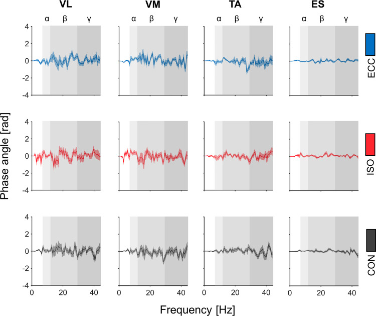
Coordination of functionally coupled muscles is a key aspect of movement execution. Demands on coordinative control increase with the number of involved muscles and joints, as well as with differing movement periods within a given motor sequence. While previous research has provided evidence concerning inter- and intramuscular synchrony in isolated movements, compound movements remain largely unexplored. With this study, we aimed to uncover neural mechanisms of bilateral coordination through intermuscular coherence (IMC) analyses between principal homologous muscles during bipedal squatting (BpS) at multiple frequency bands (alpha, beta, and gamma). For this purpose, participants performed bipedal squats without additional load, which were divided into three distinct movement periods (eccentric, isometric, and concentric). Surface electromyography (EMG) was recorded from four homologous muscle pairs representing prime movers during bipedal squatting. We provide novel evidence that IMC magnitudes differ between movement periods in beta and gamma bands, as well as between homologous muscle pairs across all frequency bands. IMC was greater in the muscle pairs involved in postural and bipedal stability compared with those involved in muscular force during BpS. Furthermore, beta and gamma IMC magnitudes were highest during eccentric movement periods, whereas we did not find movement-related modulations for alpha IMC magnitudes. This finding thus indicates increased integration of afferent information during eccentric movement periods. Collectively, our results shed light on intermuscular synchronization during bipedal squatting, as we provide evidence that central nervous processing of bilateral intermuscular functioning is achieved through task-dependent modulations of common neural input to homologous muscles.NEW & NOTEWORTHY It is largely unexplored how the central nervous system achieves coordination of homologous muscles of the upper and lower body within a compound whole body movement, and to what extent this neural drive is modulated between different movement periods and muscles. Using intermuscular coherence analysis, we show that homologous muscle functions are mediated through common oscillatory input that extends over alpha, beta, and gamma frequencies with different synchronization patterns at different movement periods.

Keywords: bipedal squat; compound movement; intermuscular coherence; neural oscillations.


Conflict of interest statement

No conflicts of interest, financial or otherwise, are declared by the authors.

Figures

Fig. 1.
Fig. 1.
Normalized power spectral density (PSD) of electromyography (EMG) envelopes per muscle and period. PSD of EMG envelopes are illustrated for all muscles during each movement period. Power spectra were averaged across muscles, epochs, and participants and normalized to total power. Mean values of normalized PSD are displayed per movement period: eccentric movement period (ECC; blue), isometric movement period (ISO; red), and concentric movement period (CON; gray), while individual values are displayed as gray lines. Each row represents distinct muscles which are highlighted in orange. Alpha, beta, and gamma frequency bands are indicated through rectangles colored in different gradations of gray. ESl and ESr, left and right erector spinae; TAl and TAr, left and right tibialis anterior; VLl and VLr, left and right vastus lateralis; VMl and VMr, left and right vastus medialis.
Fig. 2.
Fig. 2.
Overview of intermuscular coherence (IMC) spectra I. A: schematic setup of electromyography (EMG) sensors. All EMG recording sites are highlighted through blacked-out muscles with respective labels situated beside them. Please note that the upper half is drawn from dorsal perspective and the lower half is drawn from ventral perspective. Orange circles indicate EMG sensor positions. B: grand-averaged IMC for all opposing muscle pairs (highlighted in orange). Each row of IMC spectra represents different movement periods: eccentric movement period (ECC; blue), isometric movement period (ISO; red), and concentric movement period (CON; gray). Dashed green lines indicate confidence limit (CL) above which observed coherence values are considered significant. Alpha, beta, and gamma frequency bands are indicated through rectangles colored in different gradations of gray. ESl and ESr, left and right erector spinae; TAl and TAr, left and right tibialis anterior; VLl and VLr, left and right vastus lateralis; VMl and VMr, left and right vastus medialis.
Fig. 3.
Fig. 3.
Averaged alpha IMCarea per muscle and movement period. Averaged log-transformed IMCarea are illustrated per muscle and period for alpha frequency band. *Significant differences between IMCarea of different muscles within movement periods. Respective P values are reported in the results section. Here, blue symbols indicate IMCarea for eccentric movement period (ECC), whereas red symbols indicate IMCarea for isometric movement period (ISO) and black symbols represent IMCarea for concentric movement period (CON). Here, circles indicate IMCarea for vastus lateralis (VL), triangles for vastus medialis (VM), diamonds for tibialis anterior (TA), and squares for erector spinae (ES). IMCarea, summed intermuscular coherence estimates.
Fig. 4.
Fig. 4.
Averaged IMCarea per muscle. Averaged log-transformed IMCarea are illustrated per muscle for alpha (A), beta (B), and gamma (C) frequency bands. *Significant differences between IMCarea of different muscles. Respective P values are reported in the results section. Here, circles indicate IMCarea vastus lateralis (VL), triangles for vastus medialis (VM), diamonds for tibialis anterior (TA), and squares for erector spinae (ES). IMCarea, summed intermuscular coherence estimates.
Fig. 5.
Fig. 5.
Averaged IMCarea and mean EMG amplitudes (RMS) per movement period. A: averaged log-transformed IMCarea are illustrated per period for alpha, beta, and gamma frequency bands. B: averaged, normalized root mean square (RMSmean) values are depicted per period. *Significant differences between IMCarea of different movement periods. Respective P values are reported in the results section. Here, blue symbols indicate IMCarea for eccentric movement period (ECC), whereas red symbols indicate IMCarea for isometric movement period (ISO) and black symbols represent IMCarea for concentric movement period (CON). EMG, electromyography; IMCarea, summed intermuscular coherence estimates.
Fig. 6.
Fig. 6.
Overview of phase angle spectra. Each column depicts averaged phase angle spectra and corresponding standard errors of the mean for all opposing muscle pairs. Each row represents phase angle spectra of different movement periods: eccentric movement period (ECC; blue), isometric movement period (ISO; red), and concentric movement period (CON; gray). Alpha, beta, and gamma frequency bands are indicated through rectangles colored in different gradations of gray.

Similar articles

Cited by

References

    1. Aune TK, Aune MA, Ettema G, Vereijken B. Comparison of bilateral force deficit in proximal and distal joints in upper extremities. Hum Mov Sci 32: 436–444, 2013. doi:10.1016/j.humov.2013.01.005. - DOI - PubMed
    1. Baker SN. Oscillatory interactions between sensorimotor cortex and the periphery. Curr Opin Neurobiol 17: 649–655, 2007. doi:10.1016/j.conb.2008.01.007. - DOI - PMC - PubMed
    1. Baker SN, Kilner JM, Pinches EM, Lemon RN. The role of synchrony and oscillations in the motor output. Exp Brain Res 128: 109–117, 1999. doi:10.1007/s002210050825. - DOI - PubMed
    1. Baker SN, Pinches EM, Lemon RN. Synchronization in monkey motor cortex during a precision grip task. II. effect of oscillatory activity on corticospinal output. J Neurophysiol 89: 1941–1953, 2003. doi:10.1152/jn.00832.2002. - DOI - PubMed
    1. Bayraktaroglu Z, von Carlowitz-Ghori K, Curio G, Nikulin VV. It is not all about phase: amplitude dynamics in corticomuscular interactions. Neuroimage 64: 496–504, 2013. doi:10.1016/j.neuroimage.2012.08.069. - DOI - PubMed

Publication types