

Case Reports
doi: 10.3174/ajnr.A6762.
Epub 2020 Aug 13.
COVID-19-Associated PRES-like Encephalopathy with Perivascular Gadolinium Enhancement
Affiliations
- PMID: 32816769
- PMCID: PMC7963244
- DOI: 10.3174/ajnr.A6762
Item in Clipboard
Case Reports
COVID-19-Associated PRES-like Encephalopathy with Perivascular Gadolinium Enhancement
AJNR Am J Neuroradiol.
2020 Dec.
Abstract
We describe the case of a 63-year-old woman who developed a coronavirus disease 2019-associated acute encephalopathy with perivascular gadolinium enhancement.
© 2020 by American Journal of Neuroradiology.
Figures
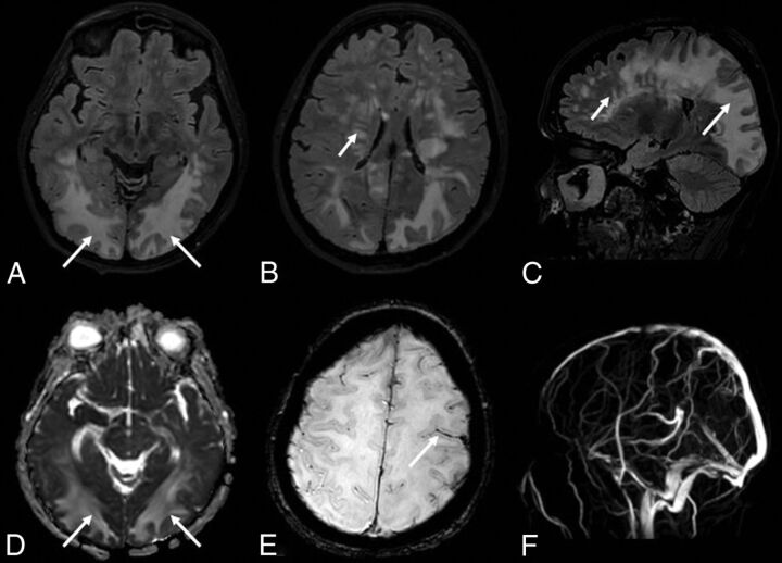
The FLAIR sequence shows multiple lesions of the white matter of both hemispheres (A–C), more striking in the posterior regions with a tumefactive appearance (long arrows) and with a multifocal perivascular pattern in the deep white matter regions (short arrows). The lesions (arrows) show high values on the apparent diffusion coefficient map (D), suggesting increased water content in the parenchyma. The SWI sequence (E) shows a subarachnoid blood effusion along the left precentral sulcus (arrow). MR venography (F) excludes intracranial thrombosis.
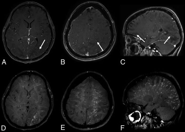
The contrast-enhanced 3D conventional T1-weighted sequence (A–C) shows multiple spotlike gadolinium enhancement (arrows) in the posterior white matter and left hemisphere. The postcontrast 3D-T1-weighted black-blood sequence (D–F) shows a stratiform perivascular gadolinium enhancement in the white matter of both cerebral hemispheres. Neither wall enhancement of the large intracranial arterial vessels nor leptomeningeal enhancement are observed on postcontrast sequences.