Case Reports
. 2020 Aug 19;13(8):e236218.
doi: 10.1136/bcr-2020-236218.

Acute perimyocarditis with cardiac tamponade in COVID-19 infection without respiratory disease

Affiliations
Case Reports

Acute perimyocarditis with cardiac tamponade in COVID-19 infection without respiratory disease

Havard Dalen et al. BMJ Case Rep. .

Abstract

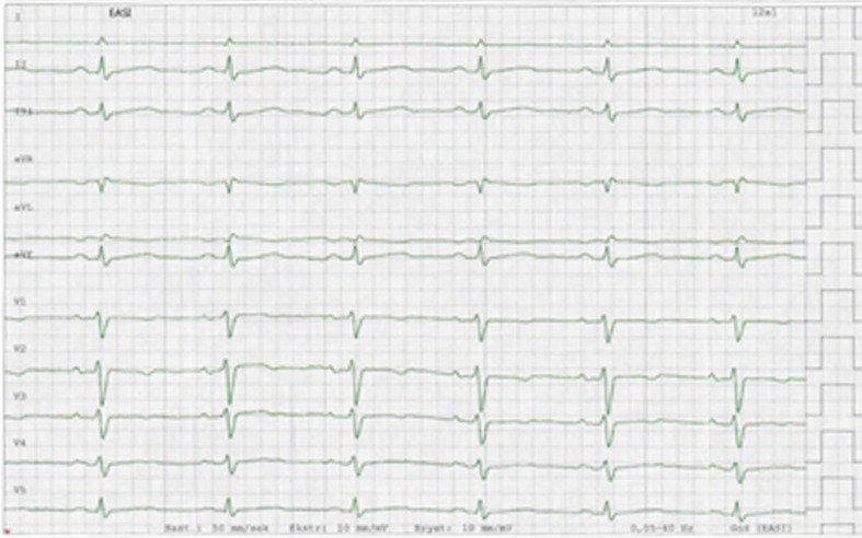
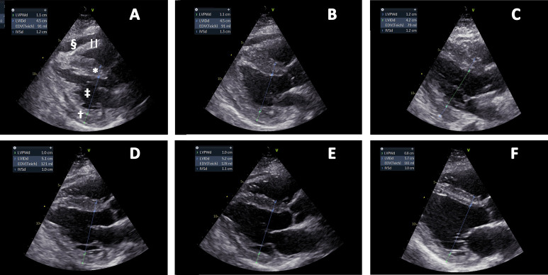
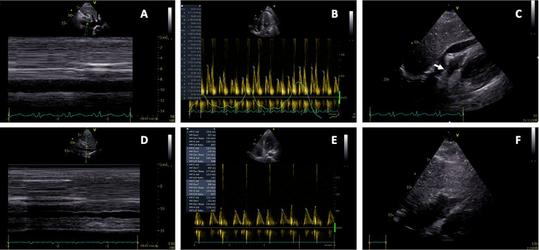
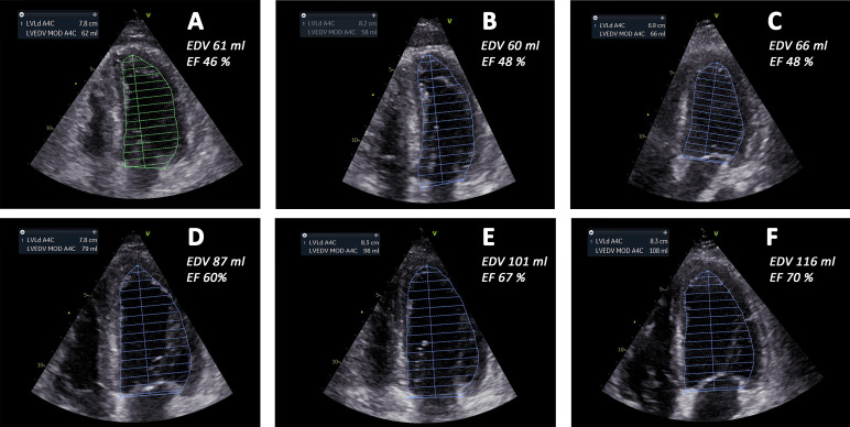
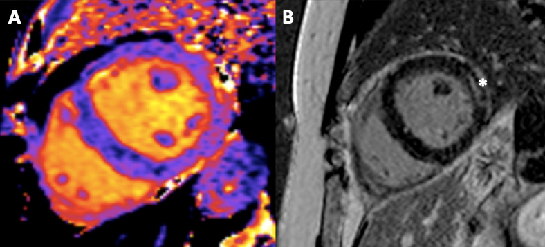
The COVID-19 pandemic with its severe respiratory disease has caused overflow to hospitals and intensive care units. Elevated troponins and natriuretic peptides are related to cardiac injury and poor prognosis. We present a young woman with COVID-19 infection with haemodynamic instability caused by acute perimyocarditis and cardiac tamponade. Troponin T was modestly elevated. Focused cardiac ultrasound made the diagnosis. Echocardiography revealed transient thickening of the myocardial walls. After pericardial drainage and supportive care, she improved significantly within 1 week without targeted therapy. The case illustrates the importance of cardiac diagnostic imaging in patients with COVID-19 and elevated cardiac biomarkers.

Keywords: infectious diseases; pericardial disease.


Conflict of interest statement

Competing interests: None declared.

Figures

Figure 1
Figure 1
EASI ECG representative for the course of disease. The ECG was recorded on day 14. Lead V6 is lacking in the stored version. There is insignificant ST-elevation in inferior leads and T-wave inversion in precordial leads. Furthermore, low voltage findings are present with peak-to-peak QRS amplitude less than 5 mm in the standard leads and 10 mm in the precordial leads (V5 and V6). Similar findings were seen in the printed 12-lead ECGs recorded at admittance to hospital and during the hospital stay.
Figure 2
Figure 2
Parasternal long-axis views day 8–day 16. (A) Day 8, (B) day 9, (C) day 10, (D) day 11, (E) day 12 and (F) day 16, respectively. Interventricular septum* and left ventricular posterior wall thicknesses (IVSd and LVPWd) are reduced from (A) to (F), accompanied by increased end-diastolic left ventricular internal dimension (LVIDd). Pericardial effusion§ is shown in (A) and (B) and increased right ventricular wall||thickness is best visualised in (A). Annotations are shown in (A) only.
Figure 3
Figure 3
Echocardiography immediately before and after pericardial drainages due to tamponade. (A–C) These show pericardial tamponade: (A) subcostal view of a dilated inferior caval vein, (B) mitral inflow with exaggerated respiratory variability (>25%) and (C) subcostal view with pericardial fluid, and a compressed right ventricle and right atrium (arrow). (D–F) These show postdrainage: (D) subcostal view of inferior caval vein with normal respiratory variability, (E) mitral inflow with normal respiratory variability and (F) subcostal view without pericardial fluid and no compression of right ventricle and right atrium.
Figure 4
Figure 4
End-diastolic four-chamber views from day 8 to day 16. (A) Day 8, (B) day 9, (C) day 10, (D) day 11, (E) day 12 and (F) day 16, respectively. (A–F) These show left ventricular end-diastolic volume (LVEDV) traces from four-chamber views. In the top left boxes, the volume estimate is shown. End-diastolic volumes increased from day 8 to day 16, accompanied by increased ejection fraction (EF) and increased stroke volume. End-diastolic volume (EDV) and EF of the left ventricle were calculated by Simpson’s equation in four-chamber and two-chamber views and the appropriate numbers are indicated in the panels. A4C, four-chamber; LVLd, left ventricular length; MOD, method of disc summation.
Figure 5
Figure 5
Cardiac MRI performed at day 15. (A) Shows a native short-axis T1-map and (B) shows late gadolinium enhancement* in the left ventricular anterolateral wall. The findings are consistent with a subacute perimyocarditis.

References

    1. Chen T, Wu D, Chen H, et al. . Clinical characteristics of 113 deceased patients with coronavirus disease 2019: retrospective study. BMJ 2020;368:m1091. 10.1136/bmj.m1091 - DOI - PMC - PubMed
    1. Zhou F, Yu T, Du R, et al. . Clinical course and risk factors for mortality of adult inpatients with COVID-19 in Wuhan, China: a retrospective cohort study. Lancet 2020;395:1054–62. 10.1016/S0140-6736(20)30566-3 - DOI - PMC - PubMed
    1. Guo T, Fan Y, Chen M, et al. . Cardiovascular implications of fatal outcomes of patients with coronavirus disease 2019 (COVID-19). JAMA Cardiol 2020;5:811 10.1001/jamacardio.2020.1017 - DOI - PMC - PubMed
    1. Chapman AR, Bularga A, Mills NL. High-Sensitivity cardiac troponin can be an ally in the fight against COVID-19. Circulation 2020;141:1733–5. 10.1161/CIRCULATIONAHA.120.047008 - DOI - PubMed
    1. Alhogbani T. Acute myocarditis associated with novel middle East respiratory syndrome coronavirus. Ann Saudi Med 2016;36:78–80. 10.5144/0256-4947.2016.78 - DOI - PMC - PubMed

Publication types

MeSH terms