

Extracellular Vesicle-Derived miR-124 Resolves Radiation-Induced Brain Injury
- PMID: 32816912
- PMCID: PMC7541572
- DOI: 10.1158/0008-5472.CAN-20-1599
Extracellular Vesicle-Derived miR-124 Resolves Radiation-Induced Brain Injury
Abstract
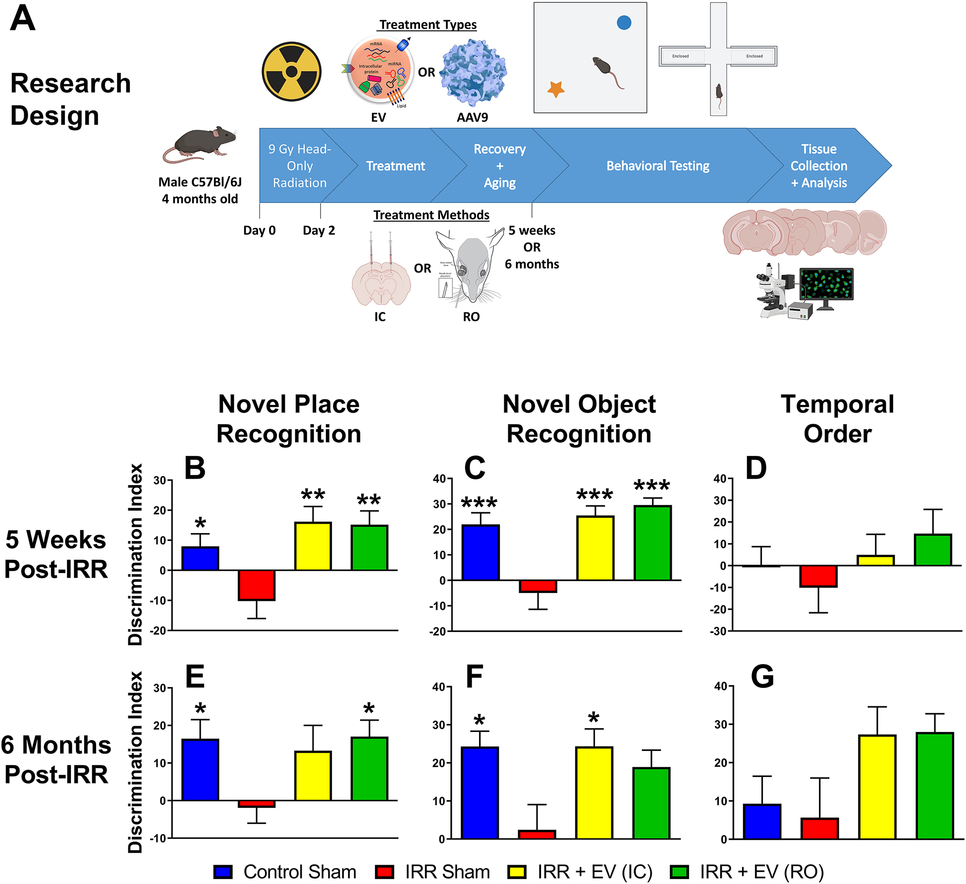
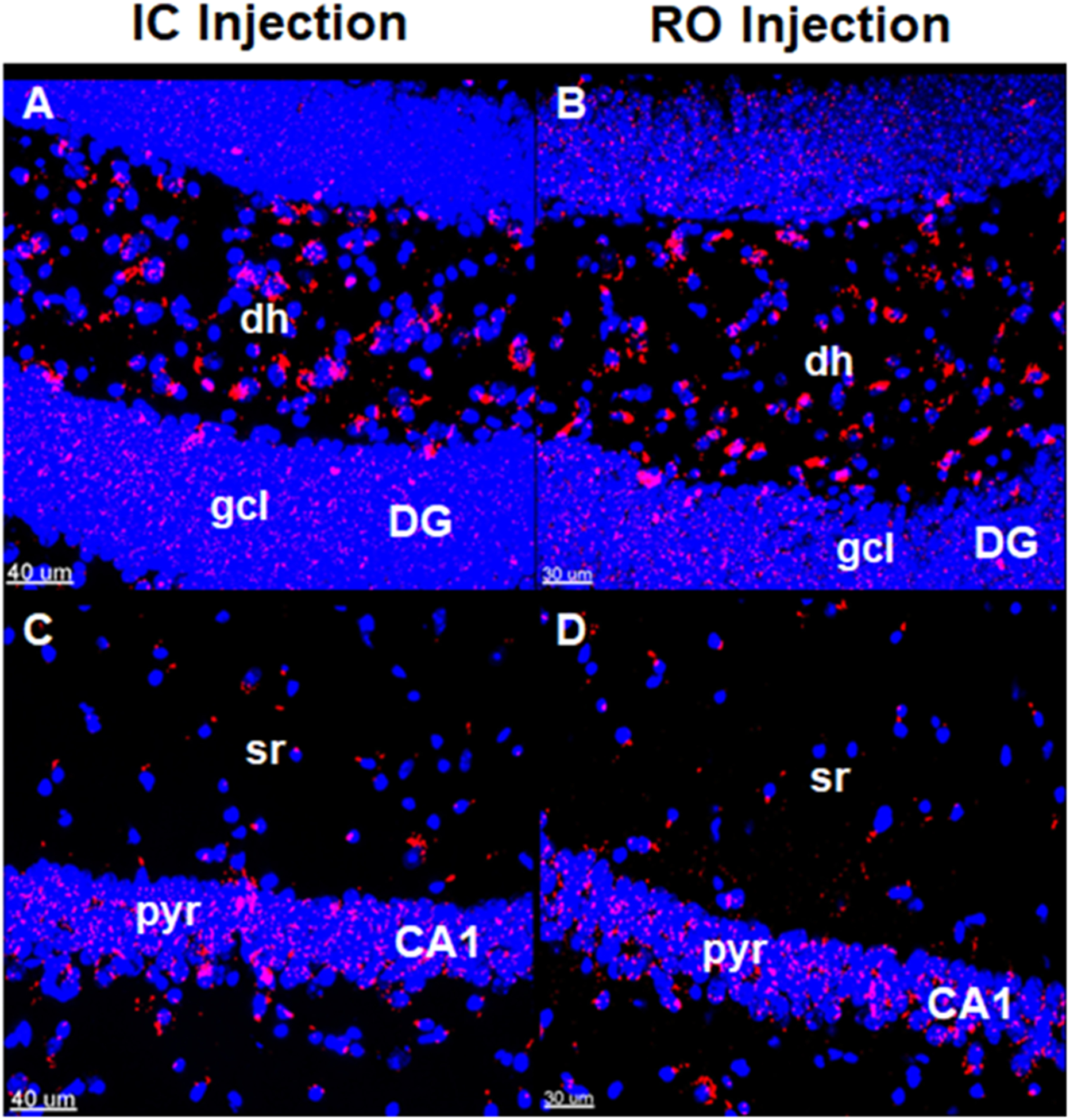
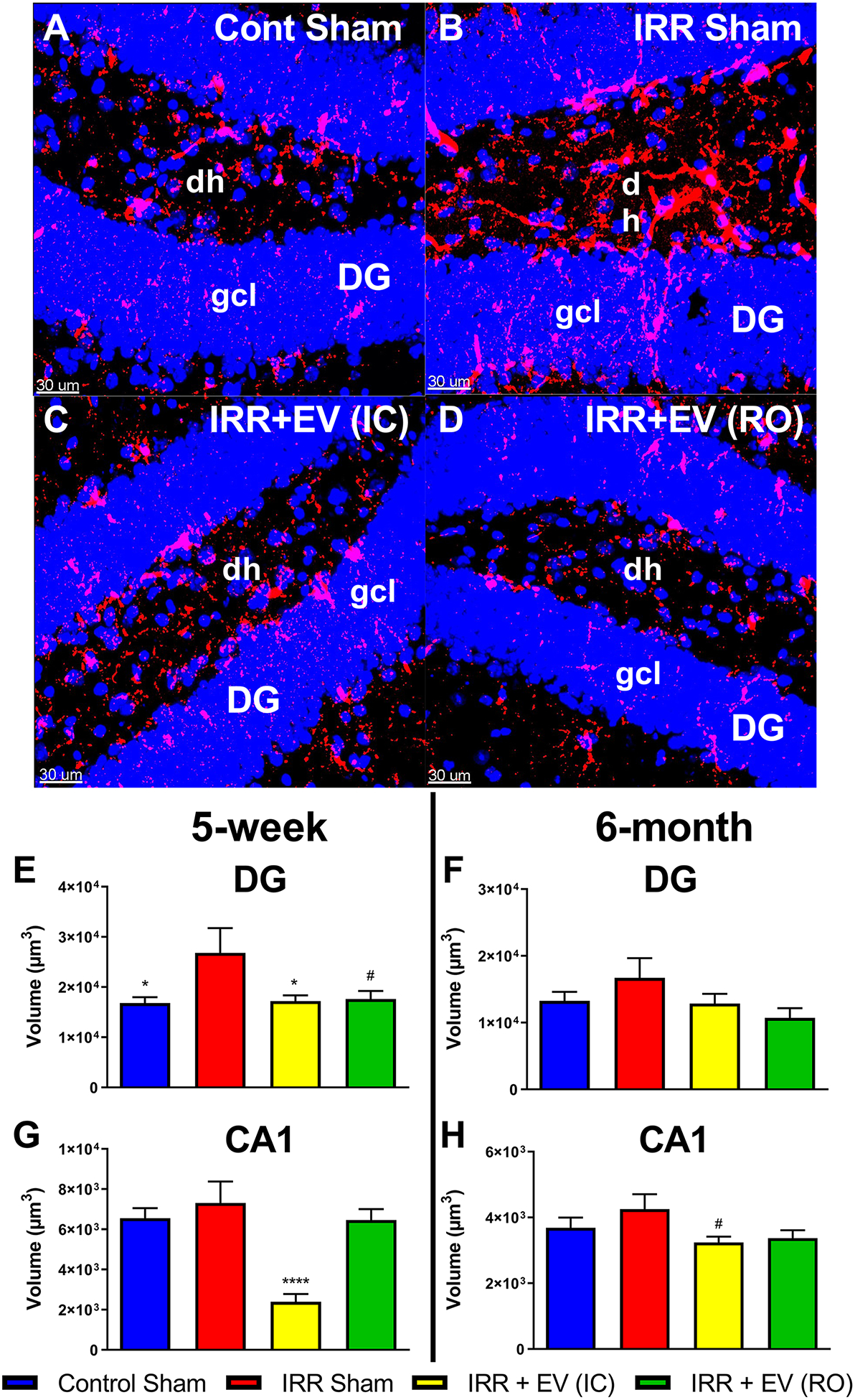
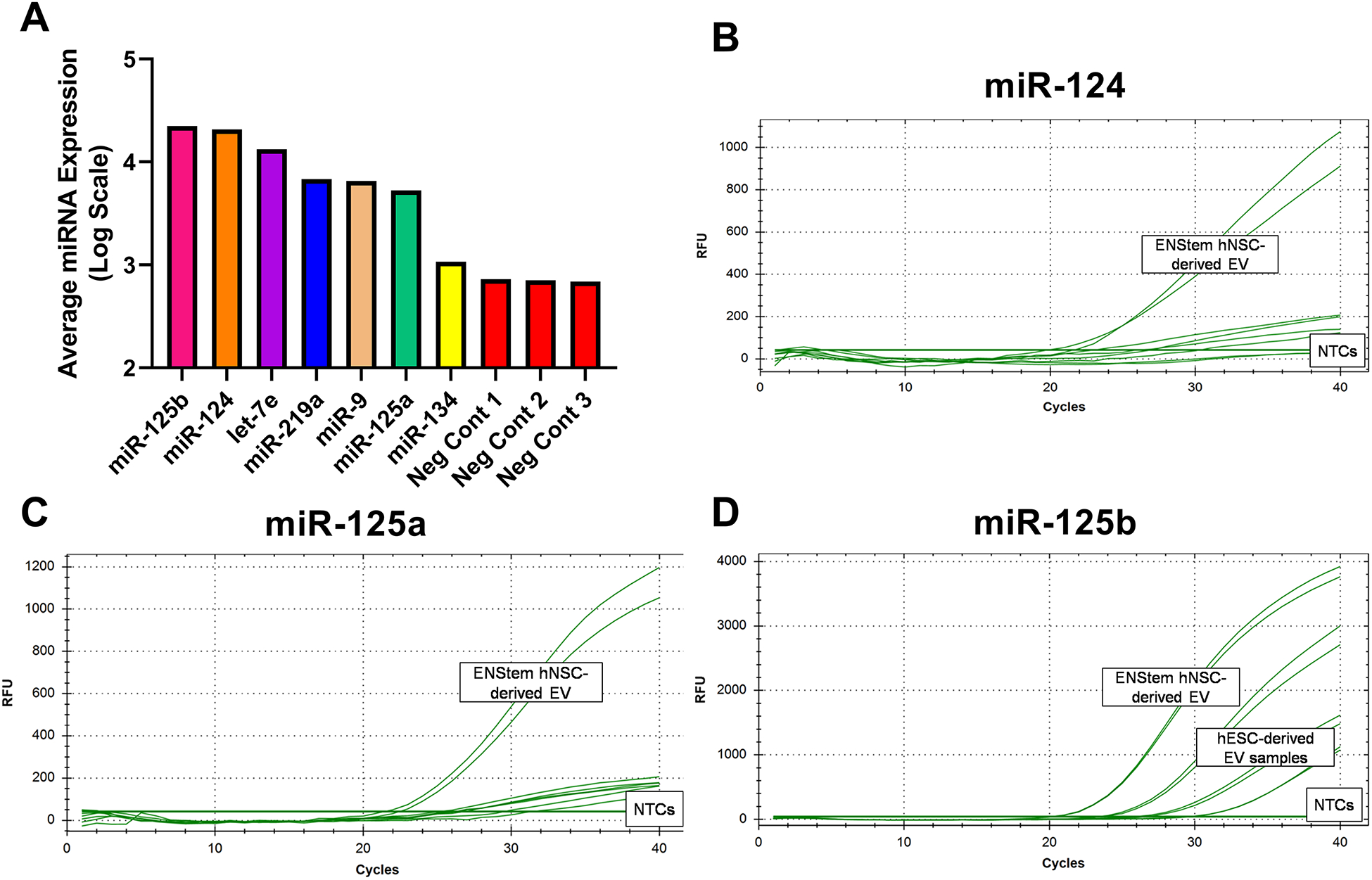
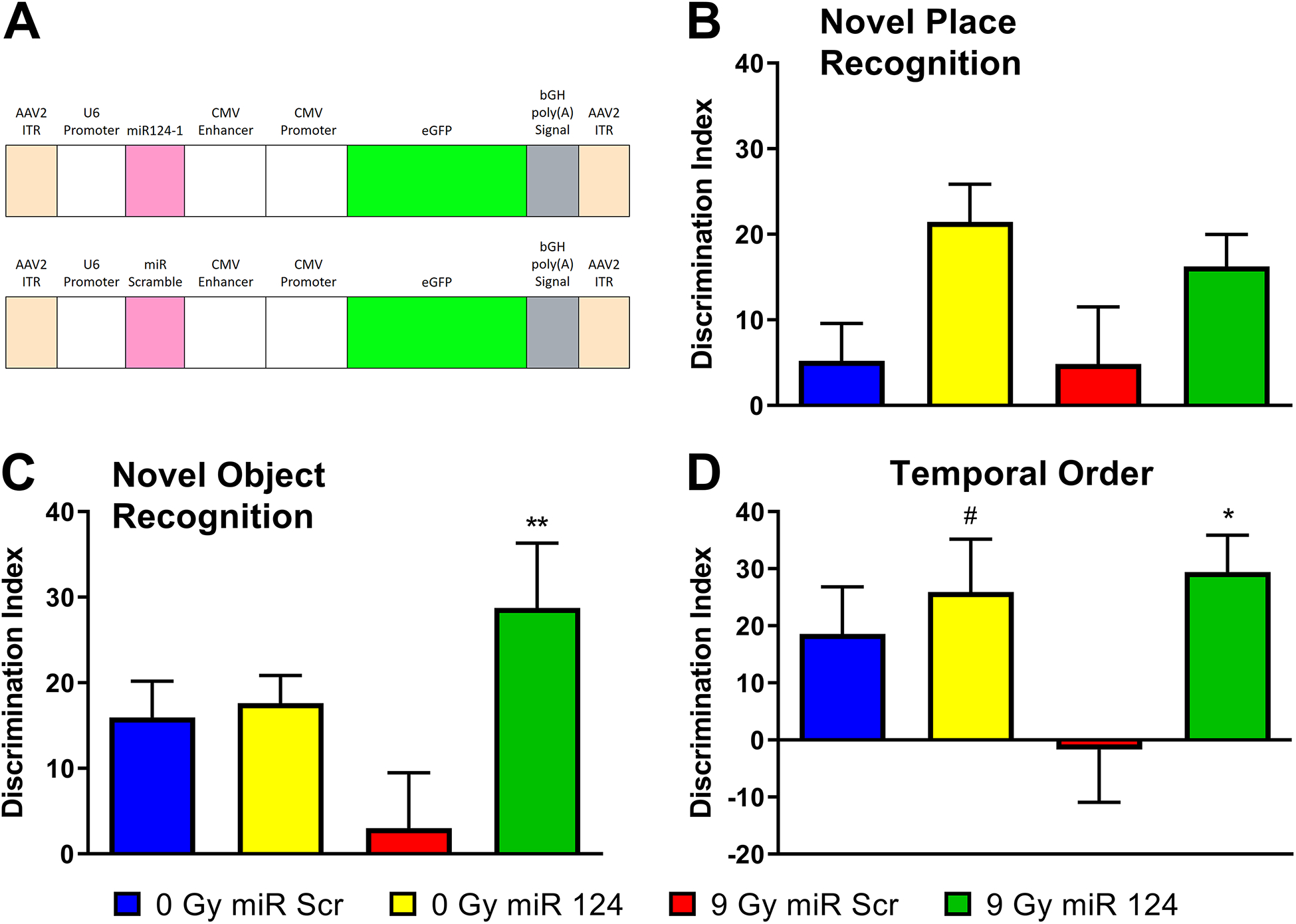
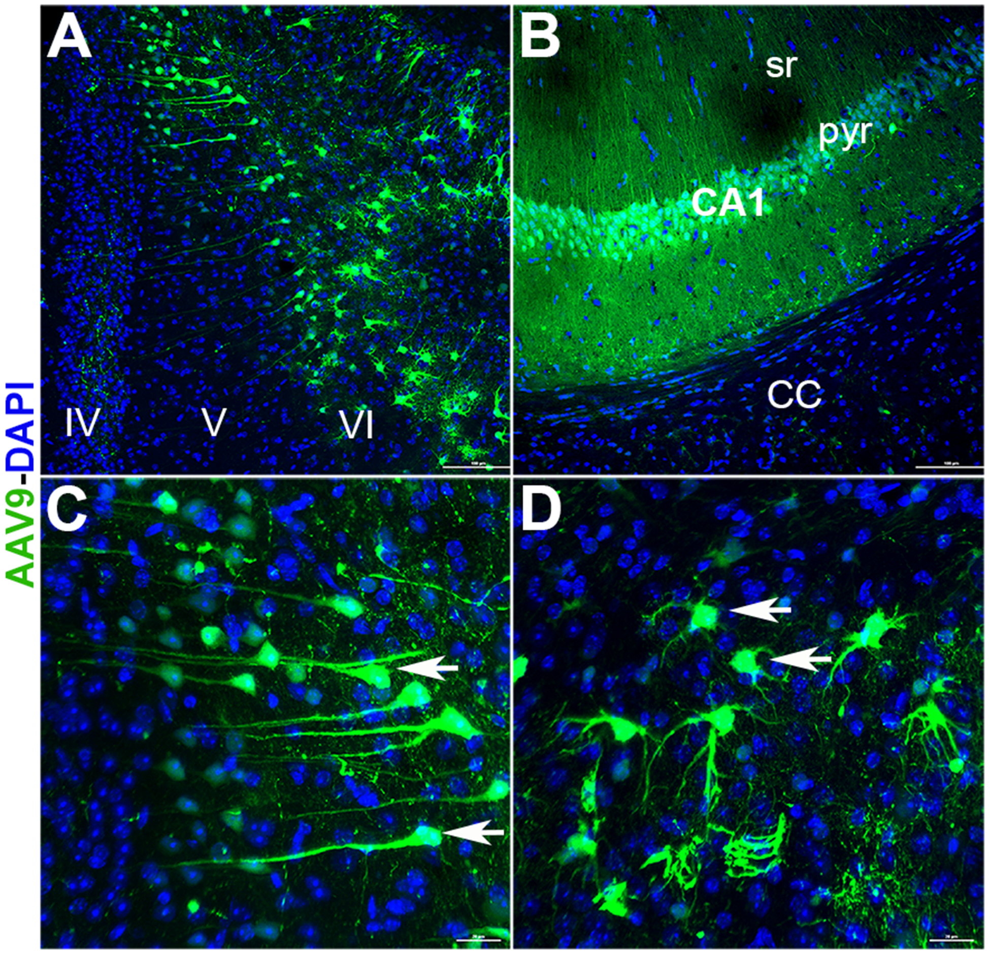
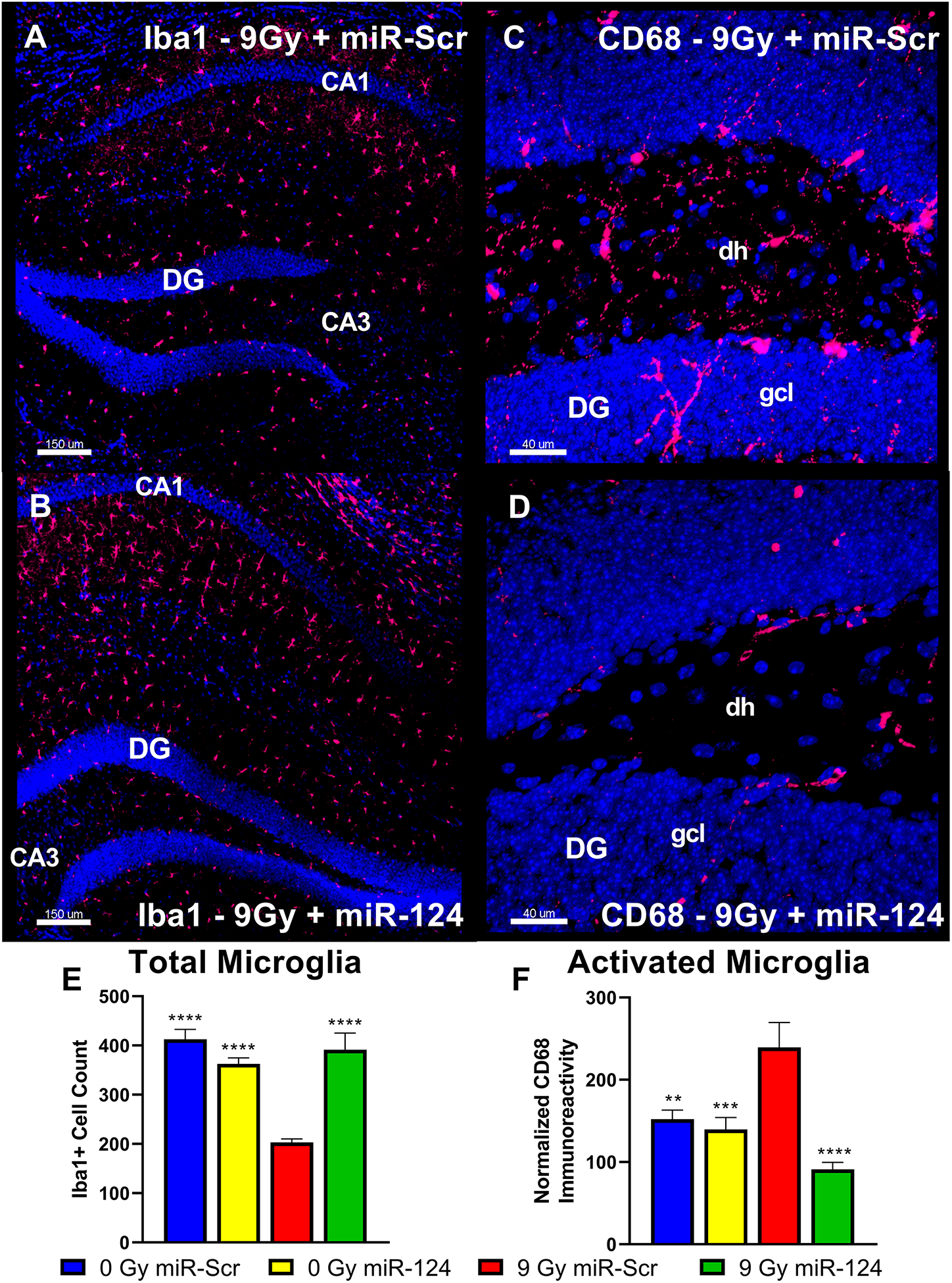
Radiation-induced cognitive dysfunction (RICD) is a progressive and debilitating health issue facing patients following cranial radiotherapy to control central nervous system cancers. There has been some success treating RICD in rodents using human neural stem cell (hNSC) transplantation, but the procedure is invasive, requires immunosuppression, and could cause other complications such as teratoma formation. Extracellular vesicles (EV) are nanoscale membrane-bound structures that contain biological contents including mRNA, miRNA, proteins, and lipids that can be readily isolated from conditioned culture media. It has been previously shown that hNSC-derived EV resolves RICD following cranial irradiation using an immunocompromised rodent model. Here, we use immunocompetent wild-type mice to show that hNSC-derived EV treatment administered either intravenously via retro-orbital vein injection or via intracranial transplantation can ameliorate cognitive deficits following 9 Gy head-only irradiation. Cognitive function assessed on the novel place recognition, novel object recognition, and temporal order tasks was not only improved at early (5 weeks) but also at delayed (6 months) postirradiation times with just a single EV treatment. Improved behavioral outcomes were also associated with reduced neuroinflammation as measured by a reduction in activated microglia. To identify the mechanism of action, analysis of EV cargo implicated miRNA (miR-124) as a potential candidate in the mitigation of RICD. Furthermore, viral vector-mediated overexpression of miR-124 in the irradiated brain ameliorated RICD and reduced microglial activation. Our findings demonstrate for the first time that systemic administration of hNSC-derived EV abrogates RICD and neuroinflammation in cranially irradiated wild-type rodents through a mechanism involving miR-124. SIGNIFICANCE: Radiation-induced neurocognitive decrements in immunocompetent mice can be resolved by systemic delivery of hNSC-derived EVs involving a mechanism dependent on expression of miR-124.
©2020 American Association for Cancer Research.
Conflict of interest statement
The authors declare no potential conflicts of interest.
Figures
Similar articles
-
Long-term cognitive effects of human stem cell transplantation in the irradiated brain.Int J Radiat Biol. 2014 Sep;90(9):816-20. doi: 10.3109/09553002.2014.927934. Epub 2014 Jun 25. Int J Radiat Biol. 2014. PMID: 24882389 Free PMC article.
-
Selective inhibition of microglia-mediated neuroinflammation mitigates radiation-induced cognitive impairment.Radiat Res. 2013 May;179(5):549-56. doi: 10.1667/RR3026.1. Epub 2013 Apr 5. Radiat Res. 2013. PMID: 23560629 Free PMC article.
-
Functional equivalence of stem cell and stem cell-derived extracellular vesicle transplantation to repair the irradiated brain.Stem Cells Transl Med. 2020 Jan;9(1):93-105. doi: 10.1002/sctm.18-0227. Epub 2019 Sep 30. Stem Cells Transl Med. 2020. PMID: 31568685 Free PMC article.
-
miRNA-based therapeutic potential of stem cell-derived extracellular vesicles: a safe cell-free treatment to ameliorate radiation-induced brain injury.Int J Radiat Biol. 2019 Apr;95(4):427-435. doi: 10.1080/09553002.2018.1522012. Epub 2018 Sep 25. Int J Radiat Biol. 2019. PMID: 30252569 Free PMC article. Review.
-
Extracellular Vesicles Derived From Neural Stem Cells, Astrocytes, and Microglia as Therapeutics for Easing TBI-Induced Brain Dysfunction.Stem Cells Transl Med. 2023 Mar 17;12(3):140-153. doi: 10.1093/stcltm/szad004. Stem Cells Transl Med. 2023. PMID: 36847078 Free PMC article. Review.
Cited by
-
Microglia as Therapeutic Target for Radiation-Induced Brain Injury.Int J Mol Sci. 2022 Jul 27;23(15):8286. doi: 10.3390/ijms23158286. Int J Mol Sci. 2022. PMID: 35955439 Free PMC article. Review.
-
Glia-Selective Deletion of Complement C1q Prevents Radiation-Induced Cognitive Deficits and Neuroinflammation.Cancer Res. 2021 Apr 1;81(7):1732-1744. doi: 10.1158/0008-5472.CAN-20-2565. Epub 2020 Dec 15. Cancer Res. 2021. PMID: 33323383 Free PMC article.
-
Extracellular Vesicles: The Invisible Heroes and Villains of COVID-19 Central Neuropathology.Adv Sci (Weinh). 2024 Mar;11(10):e2305554. doi: 10.1002/advs.202305554. Epub 2023 Dec 24. Adv Sci (Weinh). 2024. PMID: 38143270 Free PMC article. Review.
-
Unraveling the Emerging Niche Role of Extracellular Vesicles (EVs) in Traumatic Brain Injury (TBI).CNS Neurol Disord Drug Targets. 2024;23(11):1357-1370. doi: 10.2174/0118715273288155240201065041. CNS Neurol Disord Drug Targets. 2024. PMID: 38351688 Review.
-
The Use of Neural Stem Cells-Derived Exosomes to Prevent Late Radiation-Induced Cognitive Impairments in Mice.Bull Exp Biol Med. 2023 Feb;174(4):571-577. doi: 10.1007/s10517-023-05749-7. Epub 2023 Mar 10. Bull Exp Biol Med. 2023. PMID: 36894818
References
-
- Stupp R, Hegi ME, Mason WP, van den Bent MJ, Taphoorn MJ, Janzer RC, et al. Effects of radiotherapy with concomitant and adjuvant temozolomide versus radiotherapy alone on survival in glioblastoma in a randomised phase III study: 5-year analysis of the EORTC-NCIC trial. Lancet Oncol 2009;10:459–66 - PubMed
-
- Meyers CA. Neurocognitive dysfunction in cancer patients. Oncology (Williston Park) 2000;14:75–9; discussion 9, 81–2, 5 - PubMed
-
- Roman DD, Sperduto PW. Neuropsychological effects of cranial radiation: current knowledge and future directions. Int J Radiat Oncol Biol Phys 1995;31:983–98 - PubMed
-
- Abayomi OK. Pathogenesis of irradiation-induced cognitive dysfunction. Acta Oncol 1996;35:659–63 - PubMed
