

doi: 10.1183/13993003.01948-2020.
Print 2020 Sep.
Elevated ACE-2 expression in the olfactory neuroepithelium: implications for anosmia and upper respiratory SARS-CoV-2 entry and replication
Affiliations
- PMID: 32817004
- PMCID: PMC7439429
- DOI: 10.1183/13993003.01948-2020
Item in Clipboard
Elevated ACE-2 expression in the olfactory neuroepithelium: implications for anosmia and upper respiratory SARS-CoV-2 entry and replication
Eur Respir J.
.
Abstract
ACE2 protein is expressed at high levels in the human olfactory epithelium relative to upper airway epithelial cells. This may explain COVID-19-associated olfactory dysfunction, and suggests a SARS-CoV-2 reservoir site and potential intranasal therapy.
Conflict of interest statement
Conflict of interest: M. Chen has nothing to disclose. Conflict of interest: W. Shen has nothing to disclose. Conflict of interest: N.R. Rowan has nothing to disclose. Conflict of interest: H. Kulaga has nothing to disclose. Conflict of interest: A. Hillel has nothing to disclose. Conflict of interest: M. Ramanathan Jr has nothing to disclose. Conflict of interest: A.P. Lane has nothing to disclose.
Figures
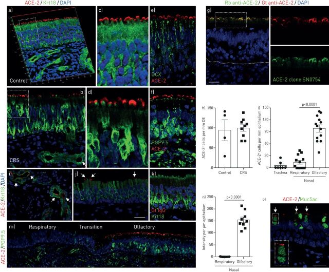
Cellular location of angiotensin-converting enzyme 2 (ACE-2) in human nasal and tracheal biopsies. a–d) Confocal image of ACE-2 (red) and Krt18 (green) immunostaining in the olfactory neuroepithelium from healthy control (a and c) and chronic rhinosinusitis (CRS) patient (b and d). The three-dimensional image (a) shows that the ACE-2 is localised to the apical surface of Krt18-positive sustentacular cells in the olfactory epithelium. Confocal images were obtained under Z stack mode which covered 8 µm in depth. The boxed area in panel (a and b) was highlighted in (c and d), respectively. e and f) The location of ACE-2- and DCX-positive immature (e) or PGP9.5-positive mature (f) olfactory sensory neurons in control. g) Confocal image verified the apical expression pattern of ACE-2 by co-staining of Goat anti-ACE-2 and another Rabbit anti-ACE-2 antibody (clone SN0754). The boxed area in (g) was highlighted in right panels. h) Quantification of ACE-2-positive cells per mm olfactory epithelium (OE). At least three images were collected from each specimen (four controls and nine CRS biopsies) using 40×objectives under the Z stack scan mode at same depth. i and j) Expression of ACE-2 in glands (i) and nasal respiratory epithelium (j). k) No detectable signal in Goat IgG isotype control. l) Quantification of ACE-2-positive cells per mm epithelium. The positive cells in seven tracheal biopsies and 13 nasal specimen that contained both respiratory and olfactory epithelium were counted. m) Representative image of respiratory-olfactory mucosa adjacent area. PGP9.5 and ACE-2 co-staining image was obtained using confocal microscope under the tile scan mode. n) Quantification of the ACE-2 fluorescence intensity per µm epithelium. Nasal specimen that contained both respiratory and olfactory epithelium were quantified using Image J. o) Co-staining of ACE-2 and secretory cell marker Muc5ac in tracheal airway epithelium. The inset represents magnification of the selected area. Dots in graph represent independent specimens (h, l and n). Data are represented as mean±sem . p-value was calculated by unpaired two-tailed Student's t-test. Differences were considered significant when p<0.05. Scale bars: 20 µm.
Update of
-
Elevated ACE2 expression in the olfactory neuroepithelium: implications for anosmia and upper respiratory SARS-CoV-2 entry and replication.bioRxiv [Preprint]. 2020 May 9:2020.05.08.084996. doi: 10.1101/2020.05.08.084996. bioRxiv. 2020. Update in: Eur Respir J. 2020 Sep 24;56(3):2001948. doi: 10.1183/13993003.01948-2020. PMID: 32511390 Free PMC article. Updated. Preprint.
References
Publication types
MeSH terms
Substances
Grants and funding
LinkOut - more resources
Full Text Sources
Other Literature Sources
Medical
Miscellaneous