Review
. 2020 Oct 8;202(21):e00398-20.
doi: 10.1128/JB.00398-20. Print 2020 Oct 8.

A Compass To Boost Navigation: Cell Biology of Bacterial Magnetotaxis

Affiliations
Review

A Compass To Boost Navigation: Cell Biology of Bacterial Magnetotaxis

Frank D Müller et al. J Bacteriol. .

Abstract

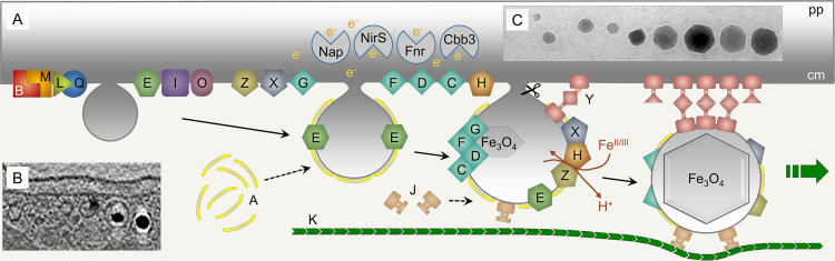
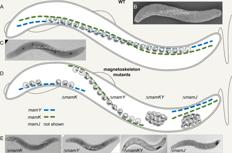
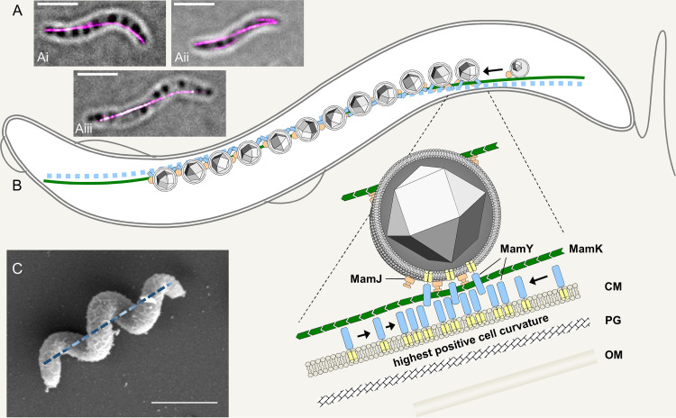
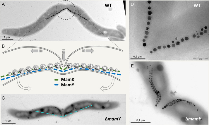
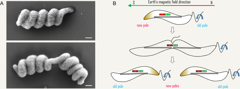
Magnetotactic bacteria are aquatic or sediment-dwelling microorganisms able to take advantage of the Earth's magnetic field for directed motility. The source of this amazing trait is magnetosomes, unique organelles used to synthesize single nanometer-sized crystals of magnetic iron minerals that are queued up to build an intracellular compass. Most of these microorganisms cannot be cultivated under controlled conditions, much less genetically engineered, with only few exceptions. However, two of the genetically amenable Magnetospirillum species have emerged as tractable model organisms to study magnetosome formation and magnetotaxis. Recently, much has been revealed about the process of magnetosome biogenesis and dedicated structures for magnetosome dynamics and positioning, which suggest an unexpected cellular intricacy of these organisms. In this minireview, we summarize new insights and place the molecular mechanisms of magnetosome formation in the context of the complex cell biology of Magnetospirillum spp. First, we provide an overview on magnetosome vesicle synthesis and magnetite biomineralization, followed by a discussion of the perceptions of dynamic organelle positioning and its biological implications, which highlight that magnetotactic bacteria have evolved sophisticated mechanisms to construct, incorporate, and inherit a unique navigational device. Finally, we discuss the impact of magnetotaxis on motility and its interconnection with chemotaxis, showing that magnetotactic bacteria are outstandingly adapted to lifestyle and habitat.

Keywords: cell shape; cytoskeleton; flagella; magnetoskeleton; magnetosome; magnetospirillum; magnetotaxis.


Figures

FIG 1
FIG 1
Steps and some key proteins of magnetosome biogenesis. (A) Essential membrane-bound Mam proteins (labeled with respective letters) (see also Table 1) are thought to tightly interact at the cytoplasmic membrane and eventually facilitate formation and growth of vesicles. Later, more proteins that function in iron transport, redox control, and magnetite precipitation are recruited. Soluble proteins are associated with the periphery of the vesicles and either play a role in magnetosome membrane assembly, e.g., MamA, or are involved in the positioning and mobility of the vesicles, e.g., MamJ and MamK. Magnetite precipitation and crystal growth require not only magnetosome vesicles equipped with Mam and Mms proteins as a “nanoreactor” but also appropriate cellular redox conditions, depending on nitrate and oxygen respiration controlled by Nap, NirS, Fnr, and Cbb3 that act in the periplasmic (pp) space or the cytoplasmic membrane (cm). In M. gryphiswaldense, vesicles eventually become pinched off the cytoplasmic membrane by an unknown mechanism. Note that only a subset of the essential and accessory factors is shown. For a synopsis of magnetosome-associated proteins in magnetospirilla, refer to Table 1. (B) Cryo-electron tomography image of nascent magnetosomes in an M. gryphiswaldense cell. (Adapted from PLoS Genetics [22].) (C) Transmission electron microscopy (TEM) image of magnetite crystals in a wild-type cell close to the end of a chain highlighting their even cuboctahedral shape.
FIG 2
FIG 2
Magnetosome organization in wild-type and phenotypes of magnetoskeleton mutants in M. gryphiswaldense. (A to C) Wild type. (A) Magnetosomes in wild-type cells are organized in extended straight chains along the magnetoskeleton formed by membrane-bound MamY, the cytoplasmic actin-like MamK, and MamJ proteins (MamJ is not shown). Due to the two-dimensional (2D) nature of the scheme, the chain seemingly detaches from the cell envelope in its central part but actually stays in close proximity to it. (B) Scanning electron microscopy image of an M. gryphiswaldense wild-type cell harboring two magnetosome chains (arrowheads, iron detected by energy dispersive X-ray spectroscopy) and indicating the twisted cell morphology. (C) TEM (2D) image of an M. gryphiswaldense wild-type cell. Arrowheads indicate the magnetosome chain. (D and E) Phenotypes of magnetoskeleton mutants in M. gryphiswaldense. (D) Schematic view of magnetosome organization in mamK, mamY, mamKY, and mamJ mutants. mamK, short, fragmented, off-center chains attached to positive membrane curvature; mamY, chain shifted to the negative inner cell curvature; mamKY, agglomerated magnetosomes or magnetic flux-closed rings; mamJ, similar to mamKY. (E) Representative TEM images are shown in the lower panel. The position of the magnetosomes is indicated by arrowheads.
FIG 3
FIG 3
Localization and function of magnetoskeleton constituents. (A) Localization of mCherry-MamK (i and ii) and mCherry-MamY (iii) in live M. gryphiswaldense cells imaged by 3D structured illumination microscopy (3D-SIM; maximum-intensity projection overlaid with a bright-field image; scale bar, 2 μm). (B) 2D model of the current view of the “magnetoskeleton” in M. gryphiswaldense, as suggested by Toro-Nahuelpan et al. (99). The actin-like MamK (green) polymerizes into cell-spanning dynamic cytoplasmic filaments that nucleate at the cell poles and “treadmill” toward midcell. Maturating magnetosomes become attached to this filament via MamJ (orange). MamY is a protein of the cytoplasmic and magnetosome membranes (membrane helices, yellow; cytoplasmic domain, blue) with high potential to self-interact. MamY assemblies are suggested to become curvature sensitive and localize to sites of highest positive inner membrane curvature coinciding with the geodetic cell axis, where they recruit magnetosome chains. CM, cytoplasmic membrane; PG, peptidoglycan; OM, outer membrane. The model is not drawn to scale. (C) SEM image of an M. gryphiswaldense cell illustrating a corkscrew-like cell morphology. The geodetic cell axis is indicated as a dashed line. Scale bar, 2 μm.
FIG 4
FIG 4
Cytokinesis in the presence of a magnetosome chain, as suggested by Katzmann et al. (94). (A) TEM image of a dividing M. gryphiswaldense wild-type cell. The center of the magnetosome chain is precisely localized at the division plane, ensuring that each daughter cell inherits exactly half of the magnetosomes (89, 93). Note the “buckling” cell center reflecting a distortion of the otherwise preserved helical cell morphology. (B) 2D scheme of the center from a dividing wild-type cell. Shaded arrows indicate direction of main cell wall growth at the division plane. Open arrows indicate the direction of cell bending. (In three dimensions, some twisting may occur as well.) Within the division plane, the septum grows by asymmetric indentation starting unilaterally from the site of negative inner cell curvature, resulting in a fracture-like appearance of the magnetosome chain. (C) In the mamY mutant, the magnetosome chain seemingly becomes disrupted by the wedge-like growing division septum. However, the buckling cell shape upon division is still apparent. The dashed line indicates the magnetosome chain position in wild-type cells. (D and E) Magnification of magnetosome chains immediately after splitting in the wild type (D) and the mamY mutant (E).
FIG 5
FIG 5
Helicity of M. gryphiswaldense cells grown on solid media and a hypothetical mechanism for preservation of cell polarity upon cytokinesis in MTB. (A) SEM images of M. gryphiswaldense cells that were recovered from a colony growing on agar-solidified medium. Note the highly spiralized cell morphology in comparison to cells grown in liquid medium (e.g., Fig. 2B). Cell division (lower image) seems to require (or to cause) some “unwinding” of the tight helix at the division plane, and yet the “buckling” appearance of the dividing cell (Fig. 4) is still visible. Scale bars, 500 nm. (B) Maintenance of polarity during cytokinesis in a monopolarly flagellated MTB. Before cytokinesis, swimming polarity is defined by the internal magnetic dipole (indicated by the bar magnet) and a gradient in redox potential (data not shown), leading to preferred swimming toward one pole of the external magnetic field. However, cell division generates daughter cells of opposite magnetic polarity with respect to the new cell pole. To ensure that both cells have the same magnetic polarity with respect to their flagellated pole, the daughter cell that does not inherit the flagellum must synthesize a new flagellum at the new cell pole (127).

References

    1. Blakemore R. 1975. Magnetotactic bacteria. Science 190:377–379. doi:10.1126/science.170679. - DOI - PubMed
    1. Bellini S. 2009. On a unique behavior of freshwater bacteria. Chin J Ocean Limnol 27:3–5. doi:10.1007/s00343-009-0003-5. - DOI
    1. Lefèvre CT, Bazylinski DA. 2013. Ecology, diversity, and evolution of magnetotactic bacteria. Microbiol Mol Biol Rev 77:497–526. doi:10.1128/MMBR.00021-13. - DOI - PMC - PubMed
    1. Lin W, Pan Y, Bazylinski DA. 2017. Diversity and ecology of and biomineralization by magnetotactic bacteria. Environ Microbiol Rep 9:345–356. doi:10.1111/1758-2229.12550. - DOI - PubMed
    1. Blakemore RP, Frankel RB, Kalmijn AJ. 1980. South-seeking magnetotactic bacteria in the southern hemisphere. Nature 286:384–385. doi:10.1038/286384a0. - DOI

Publication types

LinkOut - more resources